

Transvaginal sonographic characteristics of paraovarian borderline tumor
- PMID: 25932220
- PMCID: PMC4402867
Transvaginal sonographic characteristics of paraovarian borderline tumor
Abstract
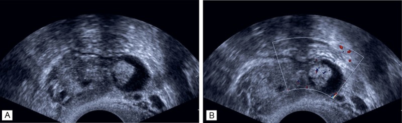
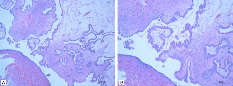
Parovarian cysts are common disorders which constitute 10-20% of adnexal masses in pathologically verified series. Most of these cysts are benign, and borderline parovarian tumors are rare and documented only as case reports in the literature. The study was aim to examine the sonographic features of parovarian borderline tumors for making an accurate preoperative diagnosis. Four patients (mean age 49 years, ranged from 35 to 75 years) with a pathological proven parovarian borderline tumor were retrospectively recruited. Preoperative transvaginal ultrasonography (TVS) and color Doppler ultrasonography were examined, and histological reports were analyzed. All tumors were correctly diagnosed as parovarian tumors at preoperative TVS. The cysts were hypoechoic and showed a variable number of papillary projections growing from the inner wall in 3 patients. Color Doppler examination of the papillae showed the presence of blood vessels in two of those three patients. In addition, histological analysis of the removed tumors demonstrated two parovarian serous borderline cystadenomas and two parovarian serous papillary borderline cystadenomas. TVS might be useful in making a preoperative diagnosis of borderline parovarian tumors.
Keywords: Parovarian cysts; borderline tumor; transvaginal sonography.
Figures
Similar articles
-
Paraovarian/paratubal cysts: comparison of transvaginal sonographic and pathological findings to establish diagnostic criteria.Ultrasound Obstet Gynecol. 2006 Sep;28(3):330-4. doi: 10.1002/uog.2829. Ultrasound Obstet Gynecol. 2006. PMID: 16823765
-
New sonographic marker of borderline ovarian tumor: microcystic pattern of papillae and solid components.Ultrasound Obstet Gynecol. 2019 Sep;54(3):395-402. doi: 10.1002/uog.20283. Epub 2019 Aug 8. Ultrasound Obstet Gynecol. 2019. PMID: 30950132
-
Preoperative sonographic features of borderline ovarian tumors.Ultrasound Obstet Gynecol. 2005 Jan;25(1):50-9. doi: 10.1002/uog.1823. Ultrasound Obstet Gynecol. 2005. PMID: 15619309
-
[Doppler ultrasonography in the diagnosis of ovarian cysts: indications, pertinence and diagnostic criteria].J Gynecol Obstet Biol Reprod (Paris). 2001 Nov;30(1 Suppl):S20-33. J Gynecol Obstet Biol Reprod (Paris). 2001. PMID: 11917373 Review. French.
-
Paratubal borderline tumor diagnosed in the adolescent period: a case report and review of the literature.J Pediatr Adolesc Gynecol. 2011 Oct;24(5):e115-6. doi: 10.1016/j.jpag.2011.05.007. Epub 2011 Jul 7. J Pediatr Adolesc Gynecol. 2011. PMID: 21737318 Review.
Cited by
-
Clinical, Imaging, Histological and Surgical Aspects Regarding Giant Paraovarian Cysts: A Systematic Review.Ther Clin Risk Manag. 2022 Apr 29;18:513-522. doi: 10.2147/TCRM.S361476. eCollection 2022. Ther Clin Risk Manag. 2022. PMID: 35516165 Free PMC article. Review.
References
-
- Alpern MB, Sandler MA, Madrazo BL. Sonographic features of parovarian cysts and their complications. AJR Am J Roentgenol. 1984;143:157–160. - PubMed
-
- Song MJ, Lee CW, Park EK, Lee AW, Park JS, Hur SY. Parovarian tumors of borderline malignancy. Eur J Gynaecol Oncol. 2011;32:445–447. - PubMed
-
- Suzuki S, Furukawa S, Kyozuka H, Watanabe T, Takahashi H, Fujimori K. Two cases of paraovarian tumor of borderline malignancy. J Obstet Gynaecol Res. 2013;39:437–441. - PubMed
-
- Seamon LG, Holt CN, Suarez A, Richardson DL, Carlson MJ, O’Malley DM. Paratubal borderline serous tumors. Gynecol Oncol. 2009;113:83–85. - PubMed
-
- Kim JS, Woo SK, Suh SJ, Morettin LB. Sonographic diagnosis of paraovarian cysts: value of detecting a separate ipsilateral ovary. AJR Am J Roentgenol. 1995;164:1441–1444. - PubMed
LinkOut - more resources
Full Text Sources