. 2015 Apr 30:4:6.
doi: 10.1186/s13630-015-0015-z. eCollection 2015.

Calcium sensors of ciliary outer arm dynein: functions and phylogenetic considerations for eukaryotic evolution

Affiliations

Calcium sensors of ciliary outer arm dynein: functions and phylogenetic considerations for eukaryotic evolution

Kazuo Inaba. Cilia. .

Abstract

The motility of eukaryotic cilia and flagella is modulated in response to several extracellular stimuli. Ca(2+) is the most critical intracellular factor for these changes in motility, directly acting on the axonemes and altering flagellar asymmetry. Calaxin is an opisthokont-specific neuronal calcium sensor protein first described in the sperm of the ascidian Ciona intestinalis. It binds to a heavy chain of two-headed outer arm dynein in a Ca(2+)-dependent manner and regulates 'asymmetric' wave propagation at high concentrations of Ca(2+). A Ca(2+)-binding subunit of outer arm dynein in Chlamydomonas reinhardtii, the light chain 4 (LC4), which is a Ca(2+)-sensor phylogenetically different from calaxin, shows Ca(2+)-dependent binding to a heavy chain of three-headed outer arm dynein. However, LC4 appears to participate in 'symmetric' wave propagation at high concentrations of Ca(2+). LC4-type dynein light chain is present in bikonts, except for some subclasses of the Excavata. Thus, flagellar asymmetry-symmetry conversion in response to Ca(2+) concentration represents a 'mirror image' relationship between Ciona and Chlamydomonas. Phylogenetic analyses indicate the duplication, divergence, and loss of heavy chain and Ca(2+)-sensors of outer arm dynein among excavate species. These features imply a divergence point with respect to Ca(2+)-dependent regulation of outer arm dynein in cilia and flagella during the evolution of eukaryotic supergroups.

Keywords: Algae; Bikont; Calaxin; Eukaryote; Evolution; Excavate; Excavates; Fertilization; Neuronal calcium sensor; Opisthokont; Sperm.


Figures
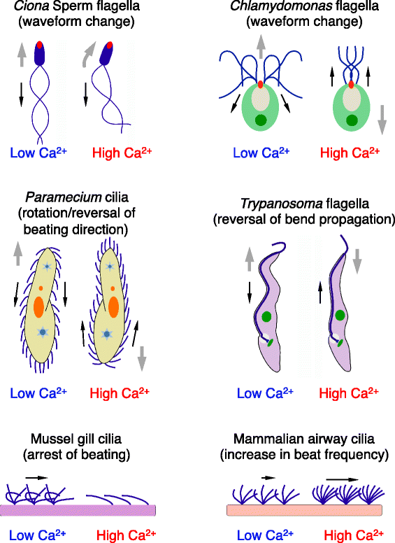
Figure 1
Figure 1
Schematic drawings of various Ca 2+ -dependent changes in wave propagation of cilia and flagella and the direction of locomotion and water flow in several organisms and tissues. Red dots in Ciona sperm and Chlamydomonas flagella indicate acrosomes and mating structure (fertilization tubules), respectively. Black and gray arrows represent the direction of wave propagation and cell locomotion, respectively.
Figure 2
Figure 2
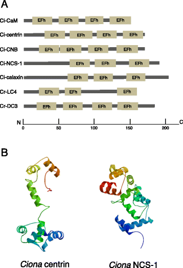
Structures of EF-hand Ca 2+ -binding proteins. (A) Domain structures of Ciona and Chlamydomonas Ca2+-sensors, drawn based on SMART searches (http://smart.embl-heidelberg.de/). The length of each protein and the positions of EF hand motifs are scaled below. (B) Molecular models of ligand-unbound Ciona centrin and NCS-1, built using SWISS-MODEL (http://swissmodel.expasy.org) [175]. Templates used are 1tnx.1 (skeletal muscle troponin) and 2d8n.1 (human recoverin) for Ciona centrin and NCS-1, respectively.
Figure 3
Figure 3
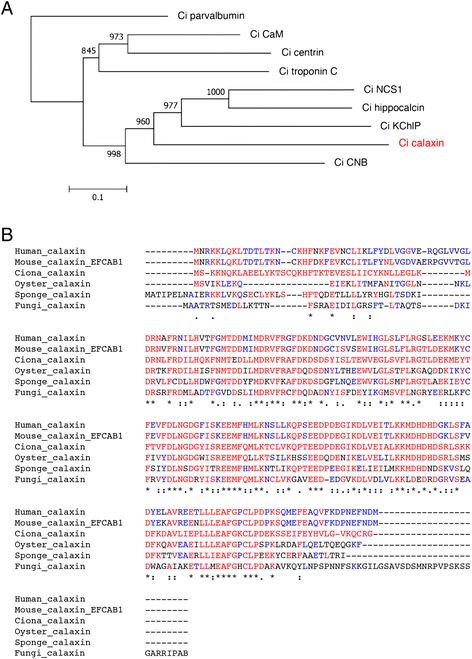
Calaxin is an opisthokont-specific Ca 2+ sensor. (A) A phylogenetic tree of Ca2+-binding proteins in the ascidian Ciona intestinalis. Proteins were aligned by CLUSTALW, and the tree was constructed by MEGA5. Ciona parvalbumin-like protein (XP_002129217) was used as the outgroup. The value shown on each branch represents the number of times that a node was supported in 1,000 bootstrap pseudo-replications. Accession numbers or NCBI reference sequence numbers of the sequence resources are as follows: calmodulin (AB076905), calaxin (AB079059), centrin (XP_004227465), troponin C (XP_002129347), NCS-1 (XP_002126443), hippocalcin (XP_002124848), KChIP (XP_004226075), calcineurin B subunit (CNB) (XP_002130765). (B) Multiple alignment of calaxin in opisthokont species. Asterisks, colons, or dots indicate identical residues in all sequences in the alignment, conserved substitutions, or semi-conserved substitutions, respectively. The amino acid residues identical to Ciona calaxin or to calaxin in other organisms are in red or blue, respectively. The sources of amino acid sequences are as follows: human calaxin (NP_078869), mouse calaxin (NP_080045), Ciona calaxin (AB079059), oyster calaxin (EKC38288), sponge calaxin (XP_003383675), and chytrid fungus calaxin (XP_006677085).
Figure 4
Figure 4
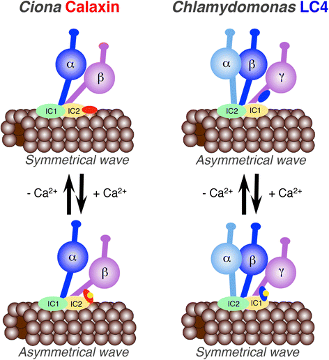
Mirror image in the function of outer arm dynein Ca 2+ sensors between Ciona and Chlamydomonas . Ciona calaxin binds to the β-heavy chain, suppresses microtubule-sliding and induces propagation of an asymmetric waveform at high concentration of Ca2+. In contrast, Chlamydomonas LC4 binds to the γ-heavy chain, becomes tethered to IC1 and induces propagation of a symmetric waveform at high concentration of Ca2+. Direct evidence for the activation of microtubule-sliding by Chlamydomonas outer arm dynein has not been obtained.
Figure 5
Figure 5
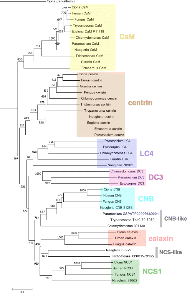
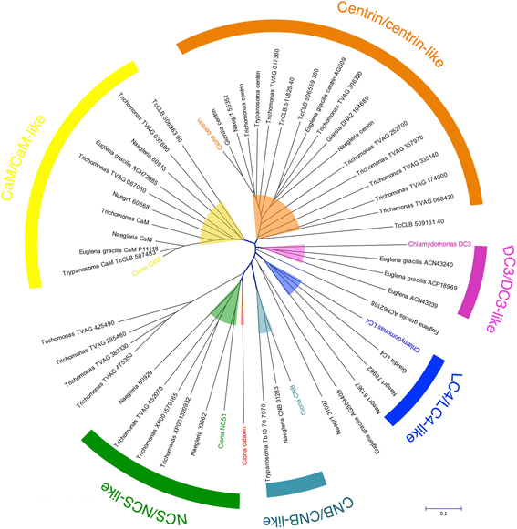
Phylogenetic analysis of Ca 2+ -binding proteins. Proteins were aligned by CLUSTALW, and the tree was constructed by MEGA5. Ciona parvalbumin-like protein (XP_002129217) was used as the outgroup. The value shown on each branch represents the number of times that a node was supported in 1,000 bootstrap pseudo-replications. Sequences were obtained from the organisms Ciona (Ciona intestinalis), human (Homo sapiens), fungus (Batrachochytrium dendrobatidis), Naegleria (Naegleria gruberi), Euglena (Euglena gracilis), Trypanosoma (Trypanosoma cruzi or T. brucei), Giardia (Giardia intestinalis or G. lamblia), Trichomonas (Trichomonas vaginalis), Chlamydomonas (Chlamydomonas reinhardtii), Paramecium (Paramecium tetraurelia), and Ectocarpus (Ectocarpus siliculosus). The sources of amino acid sequences are as follows: Ciona calmodulin (AB076905), Ciona calaxin (AB079059), Ciona centrin (XP_004227465), Ciona NCS-1 (XP_002126443), Ciona CNB (XP_002130765); human CaM (CAA36839), human calaxin (NP_078869), human NCS1 (NP_055101), human CNB (NP_000936), human centrin (NP_004057); chytrid fungus calaxin (XP_006677085), chytrid fungus CaM (XP_006678916), chytrid fungus centrin (XP_006682970), chytrid fungus NCS1 (XP_006675998), chytrid fungus CNB (XP_006677028); Naegleria CaM (XP_002683533), Naegleria centrin (XP_002678269); Trypanosoma CaM (XP_805243), Trypanosoma centrin (XP_805423), Trypanosoma calflagin (Q26680); Euglena CaM (P11118), Euglena centrin (AGS09408); Giardia CaM (XP_001705820), Giardia centrin (XP_001707577), Giardia LC4 (XP_001705117); Trichomonas CaM (XP_001326924), Trichomonas centrin (CAB55607), Trichomonas CNB (XP_002680632); Paramecium CaM (XP_001448363), Paramecium LC4 (XP_001442002), Paramecium centrin (XP_001347281), Paramecium DC3 (XP_001444482); Ectocarpus LC4 (CBN80105), Ectocarpus CaM (CBN74265), Ectocarpus centrin (CBN79657), Ectocarpus DC3 (CBJ30770). The protein sequences with specific accession numbers were obtained from DDBJ/EMBL/GenBank, or from genome browsers with the following URLs: Chlamydomonas http://genome.jgi-psf.org/Chlre4/Chlre4.home.html; Paramecium http://paramecium.cgm.cnrs-gif.fr; Naegleria http://genome.jgi-psf.org/Naegr1/Naegr1.home.html; Trichomonas http://trichdb.org; and Trypanosoma https://www.sanger.ac.uk/resources/downloads/protozoa/trypanosoma-brucei.html.
Figure 6
Figure 6
Phylogenetic analysis of homologs of Ca 2+ sensor proteins in Excavata. Proteins (EF-hand proteins, length less than 350 amino acids) were searched against genomes of each excavate by BLASTP and those with E-value <e−9 were aligned with Ciona or Chlamydomonas Ca2+-sensors by CLUSTALW. An unrooted tree was drawn by MEGA5. Branches of each Ca2+-sensor are highlighted by colors. The protein sequences (with accession numbers indicated) were obtained from DDBJ/EMBL/GenBank, or from the genome browsers shown in the legend of Figure 5.
Figure 7
Figure 7
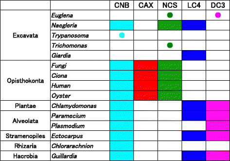
Distribution of Ca 2+ sensor proteins in eukaryotes. Based on the BLASTP search and the phylogenetic analyses in Figures 5 and 6, occurrence of each Ca2+ sensor in eukaryotic groups is summarized. Occurrence is indicated in the same colors as used in Figures 5 and 6. Closed circles in a specific color represent an occurrence of homologs with weak bootstrap support.
Figure 8
Figure 8
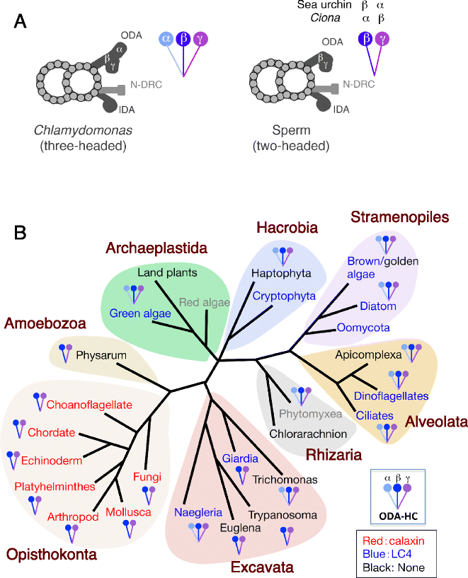
Structure of outer arm dynein and its Ca 2+ sensor across eukaryotic groups. (A) Schematic representation of the number of dynein heavy chains and the morphology of outer arm dyneins observed by electron microscopy. Chlamydomonas outer arm dynein is composed of three heavy chains, α, β, and γ. Ciona outer arm dynein has two heavy chains homologous to the Chlamydomonas β and γ chains. The α and β heavy chains in Ciona and the β and α heavy chains in sea urchin correspond to Chlamydomonas β and γ, respectively. ODA, outer arm dynein; IDA, inner arm dynein; N-DRC, nexin link/dynein regulatory complex. (B) Distribution of two-headed or three-headed outer arm dynein, and calaxin or LC4, across eukaryotic groups. The occurrence of calaxin or LC4 is indicated in red or blue, respectively, in the name of the group. A group name in black or gray indicates the lack of both calaxin and LC4, or not enough genomic information, respectively. The references for the EM images of the axonemes and the outer arm dynein are as follows: Naegleria [146]; Euglena [176,177]; Trypanosoma [66,67]; Giardia [144]; Trichomonas [147]: amoebozoan (Physarum) [101-103]; choanoflagellate (Codosiga botrytis) [178]; chordate (Ciona intestinalis and human) [62,88]; echinoderm (sea urchin: Colobocentrotus atratus) [1,3]; platyhelminthes (Dugesia tigrina) [68,179]; arthropod (Exechia seriara) [180]; Mollusca (Crassostrea gigas) [181]; chytrid fungus (Rhizophlyctis) [182]; green alga (Chlamydomonas) [137]; diatom (Biddulphia levis) [183]; golden alga (Ochromonas) [116]; ciliate (Tetrahymena pyriformis) [184]; dinoflagellate (Wolszymkia micra) [185]; apicomplexan (Plasmodium) [141]; chlorarachnion (Bigelowiella natans) [123]; haptophyte (Chrysochromulina) [186]; and phytomyxean (Plasmodiophora brassicae) [140].
Figure 9
Figure 9
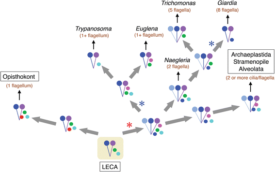
A possible model for the evolution of, and diversification in, the structures of outer arm dynein and corresponding Ca 2+ sensors during eukaryotic evolution. The model is based on analyses of the structures of outer arm dynein (two-headed, three-headed) and the types of Ca2+-sensor in each group of eukaryotes. It is assumed that the heavy chains and Ca2+-sensors of outer arm dynein of the last eukaryotic common ancestor (LECA) preceded duplication, and that duplication and divergence of Ca2+-sensors occurred at an early stage of eukaryotic diversification. The model is arranged so that the positions of eukaryotic groups match with widely accepted phylogenetic relationships [128,158]. The number of cilia/flagella per cell is also indicated in parenthesis (brown letters). Note that the numbers of cilia/flagella in Euglena and Trypanosoma are indicated as ‘1+,’ since these organisms are considered to have been biflagellates but lost or largely degenerated one of the two flagella during evolution. In this model, duplication of dynein heavy chain occurred at the root of the bikont lineage. Duplication and divergence of Ca2+-sensors would have already occurred in the ancestral organisms that contained three-headed dynein. An ancestral organism containing three-headed dynein might have recruited LC4-like sensors or CNB/NCS-like sensors and then branched into the Metamonadan (Trichomonas + Giardia) and Discoban lineages. Loss of dynein heavy chains would have occurred in Giardia and the Euglenozoa. Red or blue asterisks represent duplication or loss of a dynein heavy chain, respectively. Colored dots next to the two- or three-headed dyneins represent Ca2+-sensors (red, calaxin; blue, LC4; magenta, DC3; green, NCS; cyan, CNB). In the lineage of opisthokonts or Archaeplastida/Stramenopile/Alveolata, calaxin, LC4 or DC3 is demonstrated to be bound to the dynein heavy chain, although it is not known whether Ca2+-sensors in Excavates or any of the hypothetical ancestors could bind to the dynein or not.

Similar articles

Cited by

References

    1. Gibbons IR. Cilia and flagella of eukaryotes. J Cell Biol. 1981;91:107s–124s. - PMC - PubMed
    1. Brokaw CJ. Calcium-induced asymmetrical beating of triton-demembranated sea urchin sperm flagella. J Cell Biol. 1979;82:401–411. - PMC - PubMed
    1. Gibbons BH, Gibbons IR. The effect of partial extraction of dynein arms on the movement of reactivated sea-urchin sperm. J Cell Sci. 1973;13:337–357. - PubMed
    1. Brokaw CL, Kamiya R. Bending patterns of Chlamydomonas flagella: IV. Mutants with defects in inner and outer dynein arms indicate differences in dynein arm function. Cell Motil Cytoskeleton. 1987;8:68–75. - PubMed
    1. Lindemann CB. Testing the geometric clutch hypothesis. Biol Cell. 2004;96:681–690. - PubMed

LinkOut - more resources