. 2015 Sep;156(9):1647-1659.
doi: 10.1097/j.pain.0000000000000206.

The lidocaine metabolite N-ethylglycine has antinociceptive effects in experimental inflammatory and neuropathic pain

Affiliations

The lidocaine metabolite N-ethylglycine has antinociceptive effects in experimental inflammatory and neuropathic pain

Robert Werdehausen et al. Pain. 2015 Sep.

Abstract

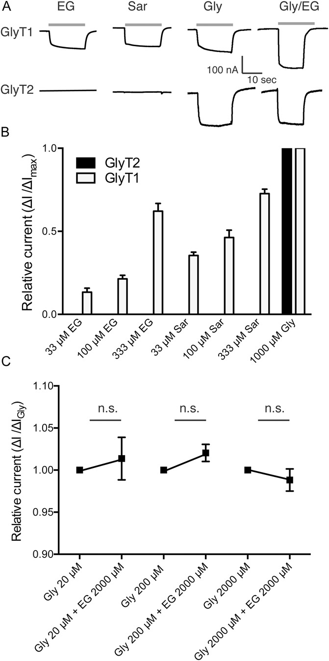
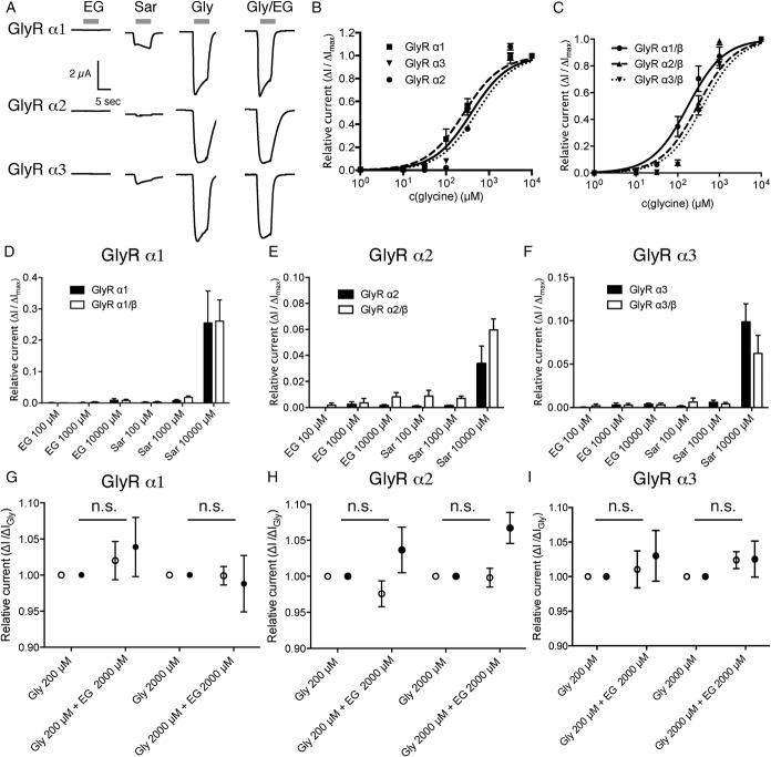
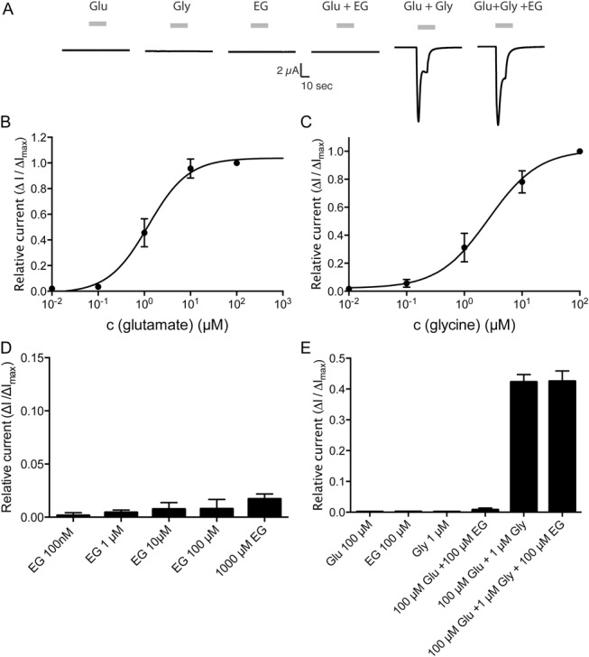
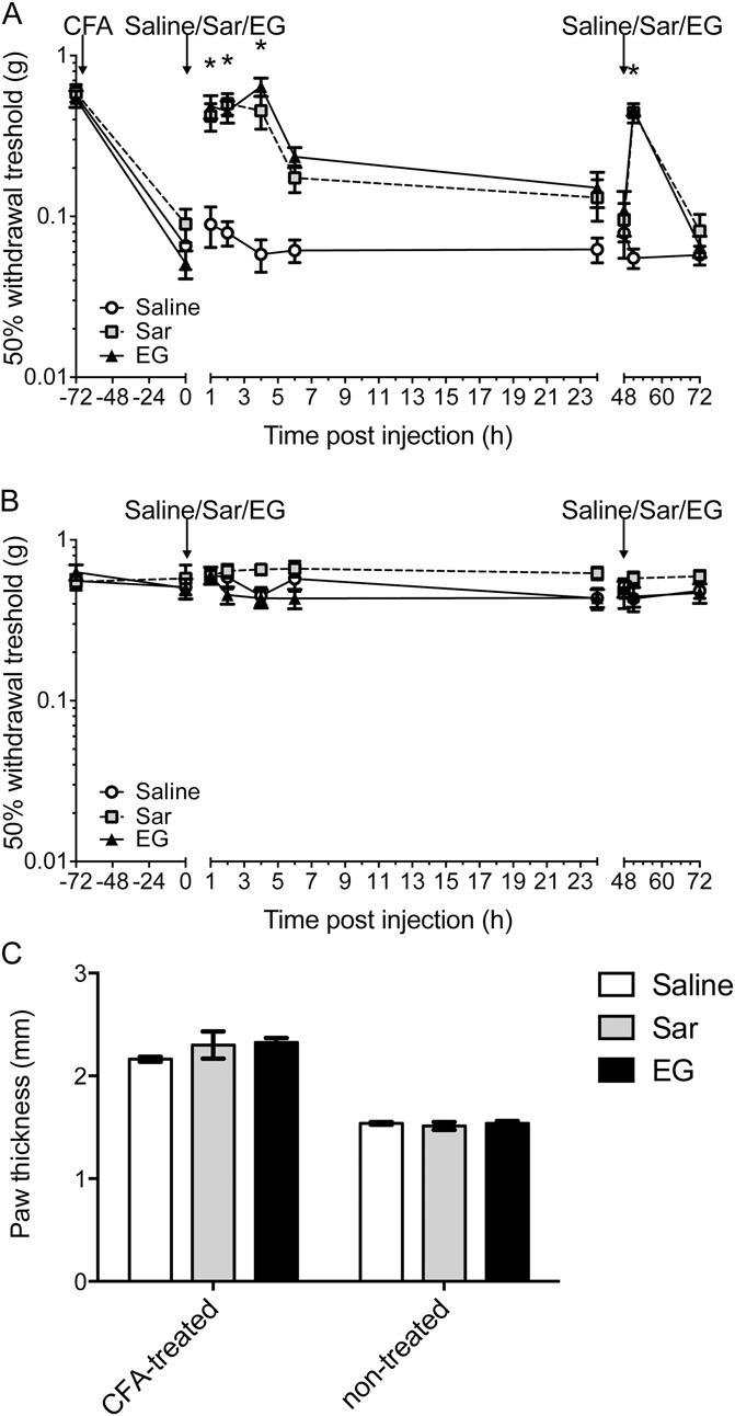
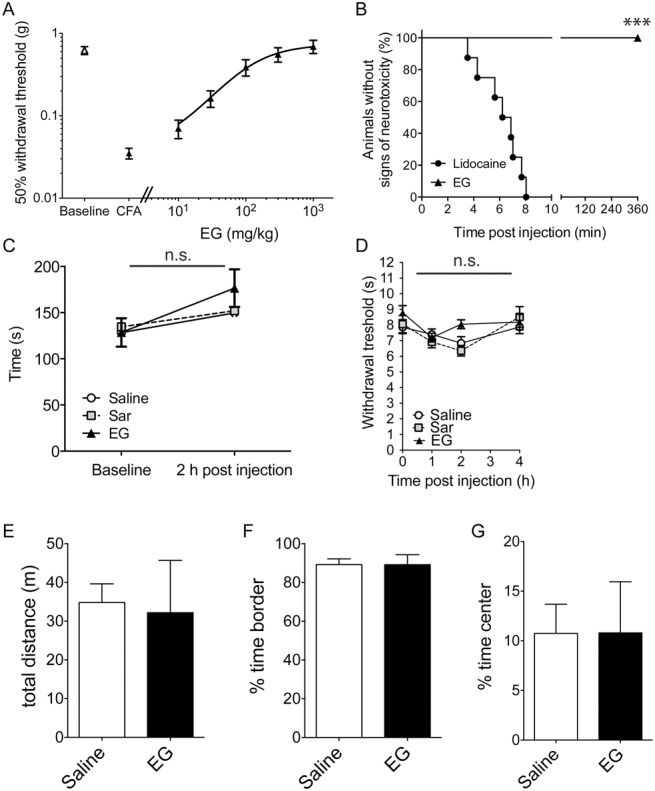
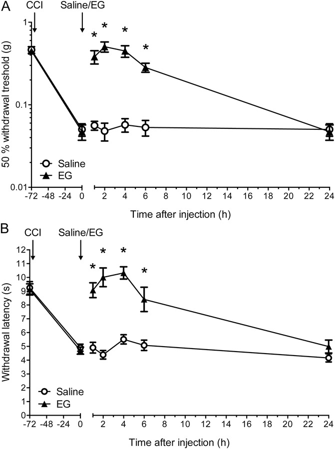
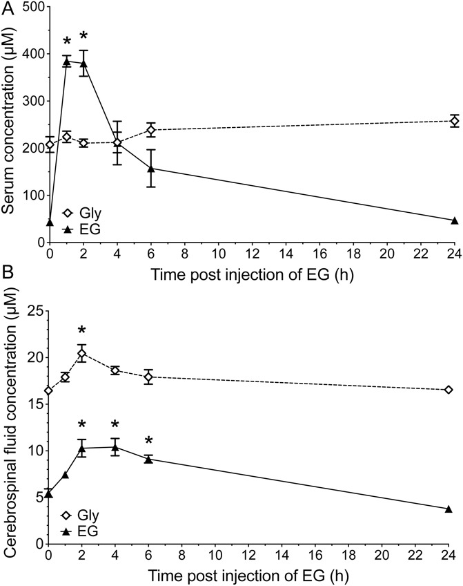
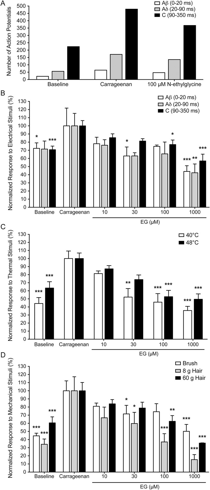
Glycine transporter 1 (GlyT1) plays a crucial role in regulating extracellular glycine concentrations and might thereby constitute a new drug target for the modulation of glycinergic inhibition in pain signaling. Consistent with this view, inhibition of GlyT1 has been found to induce antinociceptive effects in various animal pain models. We have shown previously that the lidocaine metabolite N-ethylglycine (EG) reduces GlyT1-dependent glycine uptake by functioning as an artificial substrate for this transporter. Here, we show that EG is specific for GlyT1 and that in rodent models of inflammatory and neuropathic pain, systemic treatment with EG results in an efficient amelioration of hyperalgesia and allodynia without affecting acute pain. There was no effect on motor coordination or the development of inflammatory edema. No adverse neurological effects were observed after repeated high-dose application of EG. EG concentrations both in blood and spinal fluid correlated with an increase of glycine concentration in spinal fluid. The time courses of the EG and glycine concentrations corresponded well with the antinociceptive effect. Additionally, we found that EG reduced the increase in neuronal firing of wide-dynamic-range neurons caused by inflammatory pain induction. These findings suggest that systemically applied lidocaine exerts antihyperalgesic effects through its metabolite EG in vivo, by enhancing spinal inhibition of pain processing through GlyT1 modulation and subsequent increase of glycine concentrations at glycinergic inhibitory synapses. EG and other substrates of GlyT1, therefore, may be a useful therapeutic agent in chronic pain states involving spinal disinhibition.


Conflict of interest statement

Sponsorships or competing interests that may be relevant to content are disclosed at the end of this article.

Figures

Figure 1
Figure 1
N-ethylglycine (EG) is an artificial substrate for glycine transporter 1 (GlyT1) but has no effect on glycine transporter 2 (GlyT2) function. Xenopus laevis oocytes were injected with complementary RNA encoding for mouse GlyT1 or GlyT2. Substance-induced currents were recorded at a membrane potential held at −50 mV. The respective substances were applied for 20 seconds each followed by a washout of 30 seconds. (A), Representative substance-induced currents for 30 μM glycine (Gly), 1 mM sarcosine (Sar), 1 mM EG, and Gly/EG coapplication were recorded from oocytes expressing GlyT1 or GlyT2, respectively. (B), Average amplitude of the substance-induced currents (EG or sarcosine in the respective concentration) in recordings from GlyT1- or GlyT2-expressing oocytes in relation to the maximal glycine-induced current. Note that EG and sarcosine did not induce currents in GlyT2-expressing oocytes, while glycine led to robust GlyT2-mediated currents. Data are expressed as mean ± SEM (n = 4-6). (C), Comparison of substance-induced currents recorded from GlyT2-expressing oocytes induced by glycine at indicated concentrations or after coapplication of glycine and EG at indicated concentrations. EG did not alter the glycine-induced currents. Data are expressed as mean ± SEM (n = 4-6).
Figure 2
Figure 2
N-ethylglycine (EG) does not induce significant glycine receptor (GlyR)–mediated currents in Xenopus laevis oocytes. Xenopus laevis oocytes were injected with complementary RNA encoding for mouse GlyR α1, GlyR α2, or GlyR α3 alone or coinjected with complementary RNA encoding for GlyR β. Substance-induced currents were recorded with the membrane potential held at −50 mV. The respective substances were applied for 15 seconds each followed by a washout of 30 seconds. (A), Representative substance-induced currents for 200 μM glycine (Gly), 1 mM sarcosine (Sar), and 1 mM EG were recorded from oocytes expressing the indicated GlyR subunit. (B and C), Concentration/response curve of currents elicited by glycine normalized to maximal glycine inducible currents as determined on oocytes expressing GlyR α1, GlyR α2, or GlyR α3 (depicted in B) or GlyR α1/β, GlyR α2/β, or GlyR α3/β (depicted in C), respectively. Data are expressed as mean ± SEM (n = 4-6). (D-F), Oocytes expressing GlyR α1 (D), GlyR α2 (E), or GlyR α3 (F) or the respective heteropentameric GlyR α/β receptors were superfused with EG- or sarcosine-containing solutions. Substance-induced currents were given in relation to the maximal glycine-induced current. Note that although small sarcosine-induced currents were observed in recordings from oocytes expressing any of the GlyR subunits, no or negligible EG-induced currents were observed. Data are expressed as mean ± SEM (n = 4-6). (G-I), Oocytes expressing the respective GlyR subunit (GlyR α1 in [G], GlyR α2 in [H], or GlyR α3 in [I], depicted with open circles) or the respective heteropentameric GlyR α/β receptors (depicted as filled circles) were superfused with glycine at the indicated concentration alone or in combination with 2 mM EG. Currents were determined as fraction of the current elicited by glycine alone. Data are expressed as mean ± SEM (n = 4-6).
Figure 3
Figure 3
N-ethylglycine (EG) does not influence NMDAR-mediated currents in Xenopus laevis oocytes. Xenopus laevis oocytes were injected with complementary RNA encoding for mouse. Substance-induced currents were recorded with the membrane potential held at −50 mV. The respective substances were applied for 15 seconds each followed by a washout of 30 seconds. (A), Representative substance-induced currents for 1 μM glutamate (Glu), 2 μM glycine (Gly), 100 μM EG, or the indicated coapplications EG were recorded from oocytes expressing the NMDAR (NR1/NR2a). (B), Concentration/response curve of currents elicited by glutamate in the presence of saturating concentrations of glycine (100 μM) normalized to maximal glutamate inducible currents as determined on oocytes expressing the NMDAR (NR1/NR2a). Data are expressed as mean ± SEM (n = 4-6). (C), Concentration/response curve of currents elicited by glycine in the presence of saturating concentrations of glutamate (100 μM) normalized to maximal glycine inducible currents as determined on oocytes expressing the NMDAR (NR1/NR2a). Data are expressed as mean ± SEM (n = 4-6). (D), Oocytes expressing NMDAR superfused with EG at the indicated concentrations in the presence of saturating concentrations of glutamate (100 μM). Substance-induced currents were given in relation to the maximal glycine-induced current (determined after superfusion with 100 μM glutamate and 100 μM glycine). Data are expressed as mean ± SEM (n = 4-6). (E), Oocytes expressing the NMDAR (NR1/NR2a) were superfused with glutamate, glycine, or EG at the indicated concentration alone or in the indicated combinations. Currents were determined as fraction of the current elicited by glycine and glutamate (100 μM each) alone. Data are expressed as mean ± SEM (n = 4-6).
Figure 4
Figure 4
N-ethylglycine (EG) reduces nociceptive behavior in inflammatory pain. (A), Behavioral data in adult mice after induction of inflammatory pain by injection of complete Freund's adjuvant (5 μL) in the left hind paw expressed as 50% withdrawal threshold (in g). EG (200 mg/kg), the known GlyT1 substrate sarcosine (Sar, 200 mg/kg), or saline was applied subcutaneously after allodynia had developed 3 days after induction (marked with arrow). Assessment of mechanical allodynia was performed with von Frey hairs at indicated time points. Please note that the treatment was repeated at 48 hours after initial treatment (marked with arrow) to test whether the effects can be achieved again. Data are expressed as mean ± SEM; n = 8 per group; *P < 0.05 vs control group (saline); 2-way repeated-measures analysis of variance and Bonferroni post hoc test. (B), Corresponding data from complete Freund's adjuvant–untreated right hind paws. (C), Inflammatory edema was assessed by means of paw thickness (mm) 24 hours after first treatment with EG. Data are expressed as mean ± SEM; n = 8 per group; no significant differences were detected between treatment groups; 1-way repeated-measures analysis of variance and Bonferroni post hoc test.
Figure 5
Figure 5
Dose–response relationship of N-ethylglycine (EG) for treatment of inflammatory pain and screening for secondary and acute analgesic effects. (A), Dose–response for the effect of EG on thermal hyperalgesia (withdrawal thresholds using the Hargreaves method) 3 days after the induction of inflammatory pain by complete Freund's adjuvant (5 μL) injection. Data are expressed as mean ± SEM; n = 8 per group. (B), High-dose treatment with EG does not induce adverse neurological side effects; time until occurrence of signs of adverse effects was recorded in adult naive mice after subcutaneous injection of lidocaine (40 mg/kg) or EG (400 mg/kg). Data are expressed as the fraction (in %) of symptom-free animals (no irregular movements or signs of distress); n = 8 per group; ***P < 0.001 between groups; Mantel–Cox log-rank test. (C), EG (200 mg/kg), the known GlyT1 substrate sarcosine (Sar, 200 mg/kg), or saline was applied subcutaneously in adult mice after allodynia had developed 3 days after induction. Motor coordination was tested using the rotarod performance test before and 2 hours after treatment with EG. Riding times (in seconds) were measured. Data are expressed as mean ± SEM; n = 8 per group; no significant differences were detected between treatment groups; 1-way repeated-measures analysis of variance and Bonferroni post hoc test. (D), Behavioral data in naive adult mice in the left hind paw expressed as paw withdrawal threshold (in seconds) after subcutaneous application of EG (200 mg/kg), sarcosine (200 mg/kg), or saline. Assessment of analgesic effects was performed using the Hargreaves method at indicated time points. Data are expressed as mean ± SEM; n = 8 per group; no significant differences vs control group (saline) were detected; 2-way repeated-measures analysis of variance and Bonferroni post hoc test. (E-G), Open-field experiments were performed 2 hours after receiving a single dose of EG (200 mg/kg, subcutaneously). Total distance (E) and the time spent in the periphery (F) or the central quadrant (G) were determined. Data are expressed as mean ± SEM, n = 6 to 7 per group; no significant differences vs control group (saline) were detected (Student t test, P > 0.6).
Figure 6
Figure 6
N-ethylglycine (EG) reduces nociceptive behavior in neuropathic pain. (A), Behavioral data in adult mice after induction of neuropathic pain by chronic constriction injury in the left hind paw expressed as 50% withdrawal threshold (in g). EG (200 mg/kg) or saline was applied subcutaneously after mechanical allodynia had developed 3 days after chronic constriction injury. Assessment of mechanical allodynia was performed with von Frey filaments at indicated time points. (B), Thermal hyperalgesia was assessed by means of paw withdrawal thresholds (in seconds) in response to a heat source applied to the left hind paw (Hargreaves method). Data are expressed as mean ± SEM; n = 8 per group; *P < 0.05 vs control group (saline); 2-way repeated-measures analysis of variance and Bonferroni post hoc test.
Figure 7
Figure 7
Subcutaneous injection of N-ethylglycine (EG) in rats leads to an increased EG concentration in blood serum and to increased EG and glycine (Gly) concentrations in cerebrospinal fluid. Concentrations of EG and glycine were determined using high-pressure liquid chromatography in (A) blood serum and (B) cerebrospinal fluid samples from adult rats before and at indicated time points after subcutaneous injection of EG (200 mg/kg). Data are expressed as mean ± SEM; n = 4 per group; *P < 0.05 vs before injection of EG; 1-way analysis of variance and Bonferroni post hoc test.
Figure 8
Figure 8
N-ethylglycine (EG) reduces wide-dynamic-range neuron response in inflammatory pain. Action potentials of single neurons in response to electrical stimulation (A/B), thermal stimulation (C) or mechanical stimulation (D) of the receptive field were recorded. Responses evoked by Aβ, Aδ, and C fibers after electrical stimuli were superimposed and separated according to latency (0-20, 20-90, and 90-350 milliseconds, respectively). Representative data from one individual neuron recording (A) demonstrating that the majority of action potentials are elicited by C-fiber input after electrical stimulation. (B-D), Relative number of action potentials elicited by the indicated fiber inputs, with the respective number recorded after carrageenan treatment set to 100%. Thermal stimuli were applied by means of a constant water jet at 40°C and 48°C. Mechanical stimuli were applied with a fine brush or von Frey hairs (8 and 60g force). Inflammatory pain was induced in adult rats by injection of carrageenan (1%, 20 μL) at the left hind paw. EG was applied intrathecally after hyperexcitability had developed 3 hours after carrageenan application. Effect of EG on neuronal response to stimuli was tested 30 minutes after application. (B-D), Data are expressed as mean ± SEM; n = 8 for controls and n = 4 for each concentration of EG; *P < 0.05, **P < 0.01 and ***P < 0.001 vs neuronal response after carrageenan application; 1-way repeated-measures analysis of variance and Bonferroni post hoc test.

References

    1. Ahmadi S, Lippross S, Neuhuber WL, Zeilhofer HU. PGE(2) selectively blocks inhibitory glycinergic neurotransmission onto rat superficial dorsal horn neurons. Nat Neurosci 2002;5:34–40. - PubMed
    1. Attal N, Cruccu G, Baron R, Haanpaa M, Hansson P, Jensen TS, Nurmikko T; European Federation of Neurological S. EFNS guidelines on the pharmacological treatment of neuropathic pain: 2010 revision. Eur J Neurol 2010;17:1113–e88. - PubMed
    1. Backonja MM. Neuropathic pain therapy: from bench to bedside. Semin Neurol 2012;32:264–8. - PubMed
    1. Baron R, Binder A, Wasner G. Neuropathic pain: diagnosis, pathophysiological mechanisms, and treatment. Lancet Neurol 2010;9:807–19. - PubMed
    1. Barthel F, Urban A, Schlosser L, Eulenburg V, Werdehausen R, Brandenburger T, Aragon C, Bauer I, Hermanns H. Long-term application of glycine transporter inhibitors acts antineuropathic and modulates spinal N-methyl-D-aspartate receptor subunit NR-1 expression in rats. Anesthesiology 2014;121:160–9. - PubMed

Publication types

MeSH terms