. 2015 May 1;10(5):e0123187.
doi: 10.1371/journal.pone.0123187. eCollection 2015.

Plasminogen activator inhibitor-1 in cigarette smoke exposure and influenza A virus infection-induced lung injury

Affiliations

Plasminogen activator inhibitor-1 in cigarette smoke exposure and influenza A virus infection-induced lung injury

Yashodhar P Bhandary et al. PLoS One. .

Abstract

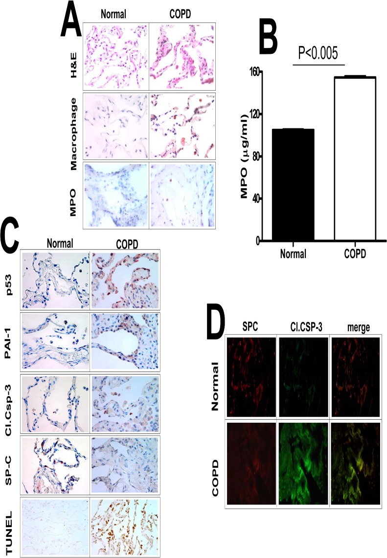
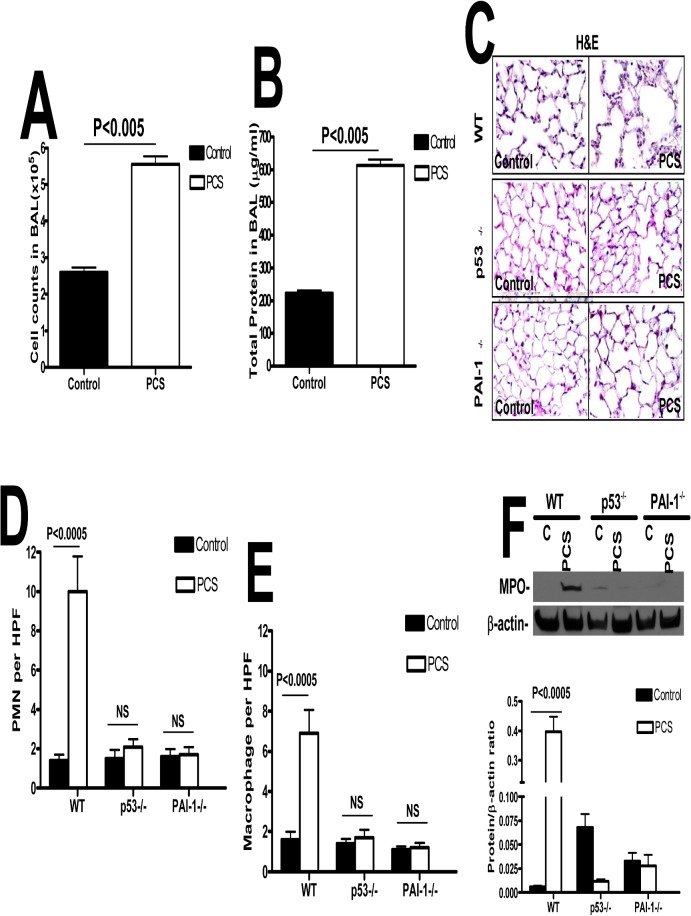
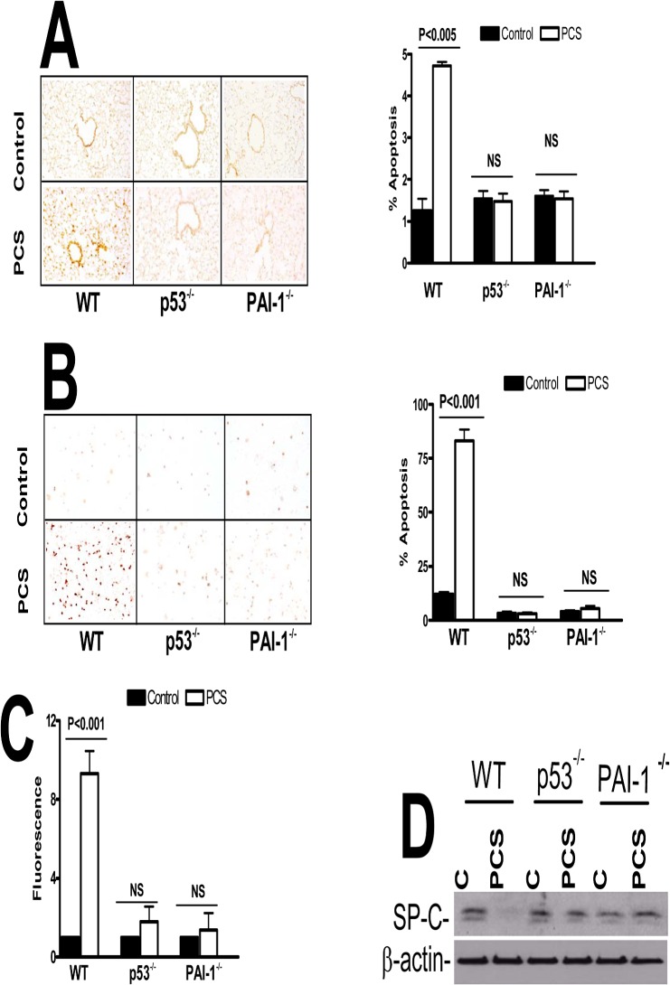
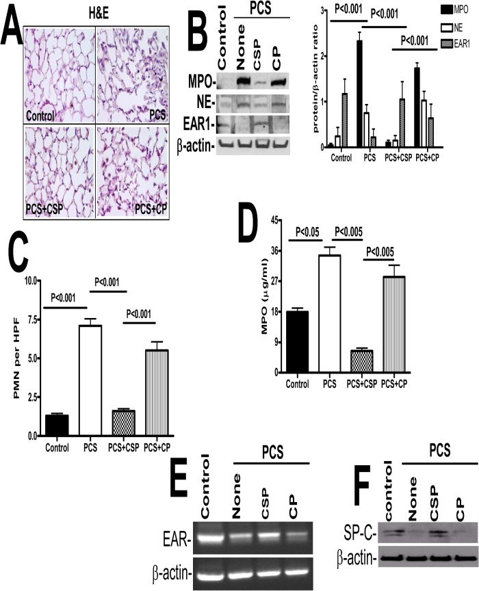
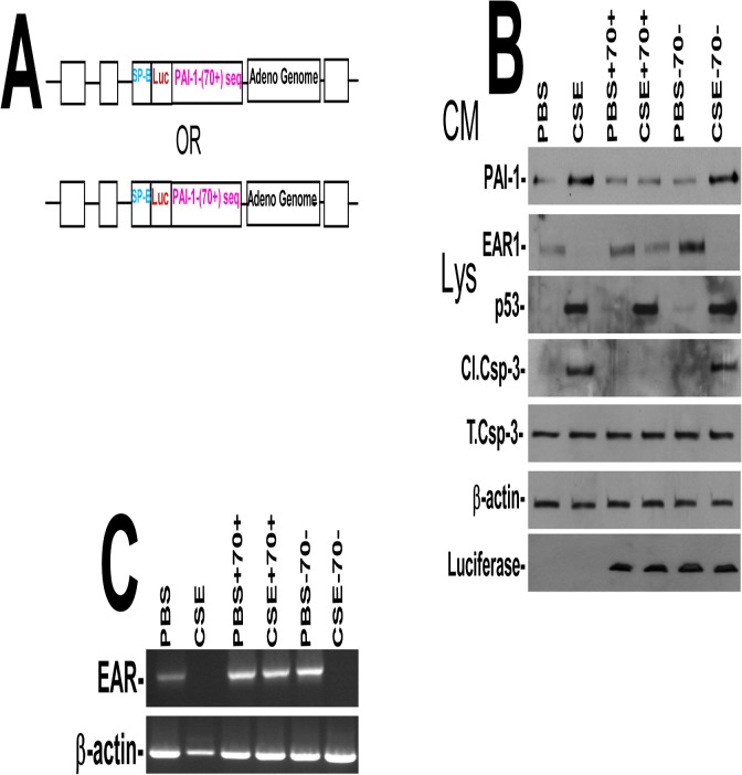
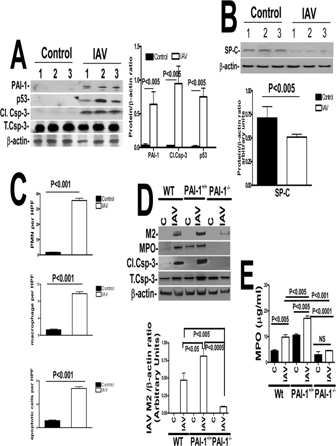
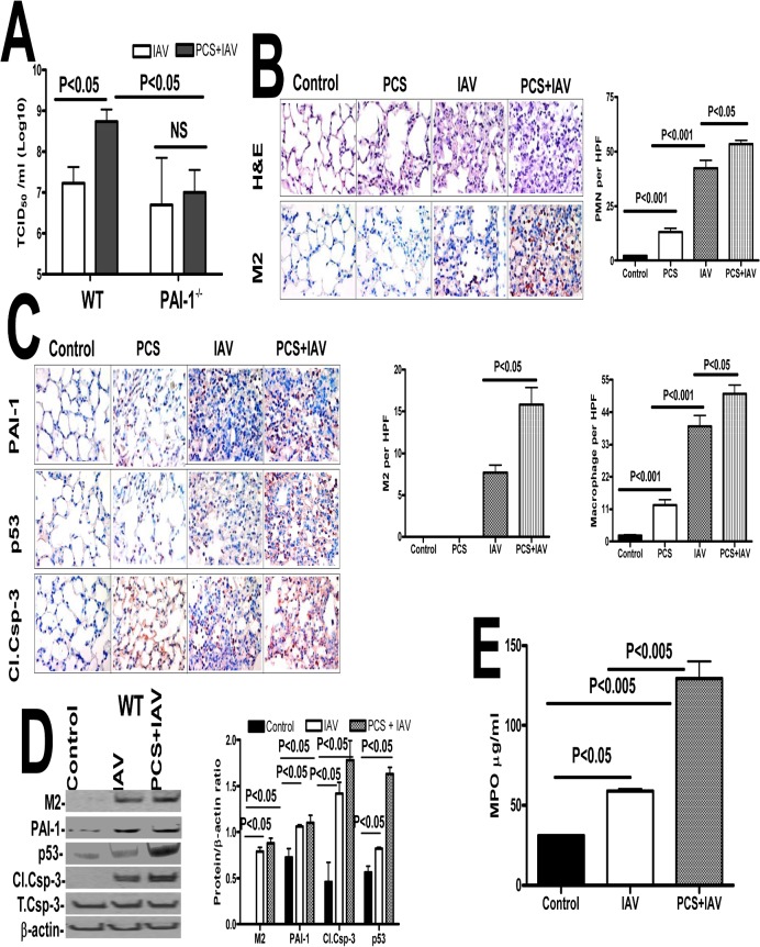
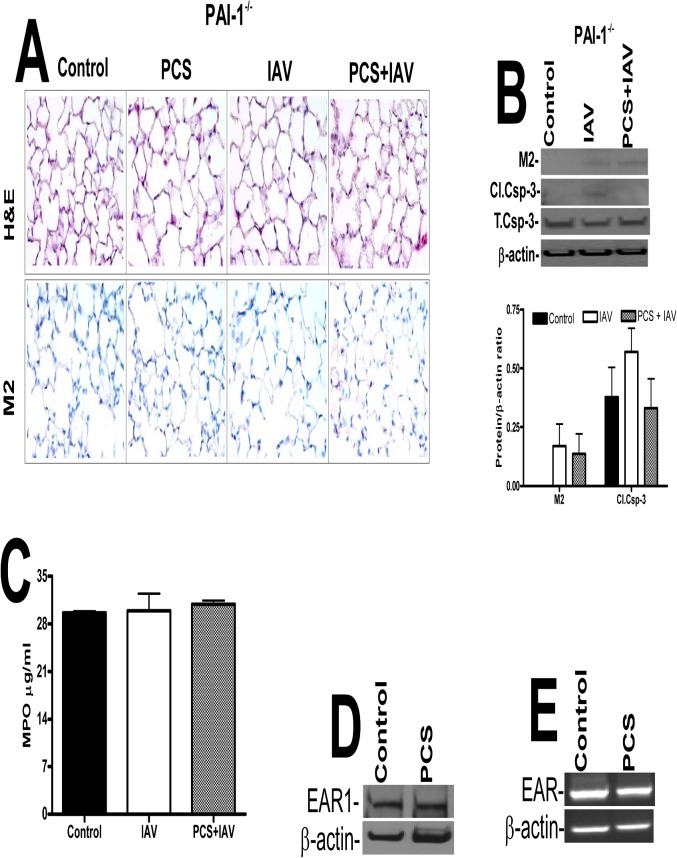
Parenchymal lung inflammation and airway and alveolar epithelial cell apoptosis are associated with cigarette smoke exposure (CSE), which contributes to chronic obstructive pulmonary disease (COPD). Epidemiological studies indicate that people exposed to chronic cigarette smoke with or without COPD are more susceptible to influenza A virus (IAV) infection. We found increased p53, PAI-1 and apoptosis in AECs, with accumulation of macrophages and neutrophils in the lungs of patients with COPD. In Wild-type (WT) mice with passive CSE (PCSE), p53 and PAI-1 expression and apoptosis were increased in AECs as was lung inflammation, while those lacking p53 or PAI-1 resisted AEC apoptosis and lung inflammation. Further, inhibition of p53-mediated induction of PAI-1 by treatment of WT mice with caveolin-1 scaffolding domain peptide (CSP) reduced PCSE-induced lung inflammation and reversed PCSE-induced suppression of eosinophil-associated RNase1 (EAR1). Competitive inhibition of the p53-PAI-1 mRNA interaction by expressing p53-binding 3'UTR sequences of PAI-1 mRNA likewise suppressed CS-induced PAI-1 and AEC apoptosis and restored EAR1 expression. Consistent with PCSE-induced lung injury, IAV infection increased p53, PAI-1 and apoptosis in AECs in association with pulmonary inflammation. Lung inflammation induced by PCSE was worsened by subsequent exposure to IAV. Mice lacking PAI-1 that were exposed to IAV showed minimal viral burden based on M2 antigen and hemagglutination analyses, whereas transgenic mice that overexpress PAI-1 without PCSE showed increased M2 antigen and inflammation after IAV infection. These observations indicate that increased PAI-1 expression promotes AEC apoptosis and exacerbates lung inflammation induced by IAV following PCSE.


Conflict of interest statement

Competing Interests: The authors have declared that no competing interests exist.

Figures

Fig 1
Fig 1. Increased p53 and PAI-1 antigen levels, and AEC apoptosis in the lung tissues of patients with COPD.
(A) Paraffin embedded sections from COPD and histologically “normal” donor lung tissues were subjected to H & E and IHC staining for macrophages and myeloperoxidase (MPO) using specific antibodies. (B) Lung homogenates from COPD (n = 3) and histologically “normal” donor lung tissues from control patients were also tested for MPO activity by colorimetric assay. Data shown in bar graphs are mean ± SD of two independent experiments. (C) The lung sections were also subjected to IHC analysis using anti-p53, anti-PAI-1, anti-active caspase-3 and anti-SP-C antibodies to assess their expression and apoptosis in AECs. Lung sections were also subjected to TUNEL staining to assess apoptosis. (D) Immunofluorescence staining was performed for the above lung sections using anti-SP-C and anti-active caspase-3 primary antibodies and fluorescently labeled secondary antibodies to assess apoptosis of type II AECs. Representative fields from 1 of 3 sections per subject are shown at X 400 magnification.
Fig 2
Fig 2. p53-mediated induction of PAI-1 expression contributes to increased pulmonary MPO levels in mice with PCSE.
(A) WT mice were exposed to ambient air (control) or PCS (n = 5/group) for 20 weeks. LL collected from these mice was subjected to total cell counting. (B) Total protein in the lavage was quantified from the above exposed mice. (C) Paraffin embedded lung sections from WT, p53- and PAI-1-deficient mice (n = 5mice/group) exposed to ambient air (control) or PCS for 20 weeks were subjected to H&E staining. Representative fields from 1 of 3 sections per subject are shown at X 400 magnification. Lung sections were subjected to IHC analysis for neutrophils using anti-MPO antibody and for macrophages using anti-F4/80 antibody. Neutrophils (D) and macrophages (E) were counted in 10 high-power fields (hpf) are shown as bar graph. (F) Lung homogenate from WT, p53- and PAI-1-deficient mice exposed to ambient air or PCS for 20 weeks were immunoblotted for changes in the levels of MPO using anti-MPO antibody. These membranes were later stripped and analyzed for β-actin to assess loading. Data shown in bar graphs are mean ± SD of two independent experiments (n = 5 mice/group). Differences between treatments are statistically significant *(P<0.05).
Fig 3
Fig 3. p53 and PAI-1 are prominently linked to PCSE-induced type II AEC apoptosis.
(A) WT, p53- and PAI-1-deficient mice were exposed to ambient air (control) or PCS for 20 weeks. Lung section obtained from these mice were subjected to TUNEL staining and the bar graph represents percent apoptosis in these groups with error bars and significance *(p<0.005) (n = 5 mice/group). (B) Type II AECs isolated from WT, p53- and PAI-1-deficient mice as described in methods were subjected to TUNEL staining and the bar graph represents percent apoptosis in these groups with error bars and significance *(p<0.005) (n = 5 mice/group). (C) Type II AECs isolated from WT, p53- and PAI-1-deficient mice were subjected to flow cytometric analysis after staining with anti-annexin-v antibody and PI to assess apoptosis. NS = the differences are not statistically significant (n = 5 mice/group). (D) Type II AECs isolated from WT and p53- and PAI-1-deficient mice as described above were immunoblotted for SP-C and β-actin as a loading control.
Fig 4
Fig 4. CSP inhibits PCSE-induced induced MPO and neutrophil elastase in mice.
WT mice were exposed to ambient air or PCS as described in the Methods for 5 days per week. After 4 weeks of PCS exposure, mice exposed to PCS were IP injected with or without 18.75 mg/kg body weight of CSP or CP once every week for 4 more weeks. After 20 weeks of PCS exposure, mice were euthanized. (A) Paraffin embedded lung sections from WT mice were subjected to H & E staining. Representative fields from 1 of 3 sections per subject are shown at X 400 magnification. (B) Lung homogenates from these mice were tested for changes in MPO, neutrophil elastase (NE), EAR1 and β-actin by Western blotting. Densities of individual bands normalized against β-actin are shown in a bar graph of two independent experiments (n = 5 mice/group). (C) Lung sections of the mice were subjected to IHC analysis using anti-MPO antibodies. Neutrophils were quantified by counting positive cells in 10 high-power fields (hpf) are shown as bar graph. (D) Lung homogenates from WT mice exposed to ambient air or PCS treated with or without CSP or CP were tested for MPO activity by colorimetric assay. (E) Total RNA obtained from the lungs of these mice were tested for changes in the expression of EAR and β-actin mRNA by RT-PCR. Experiments were repeated at least two times (n = 5 mice/group). (F) Type II AECs isolated from the mice as described above were immunoblotted for SP-C with β-actin as the loading control.
Fig 5
Fig 5. (A) Map showing the Ad-vector harboring SP-B promoter plus chimeric luciferase cDNA having either p53-binding (Ad-PAI-170+) or corresponding control (Ad-PAI-170-) PAI-1 3’UTR sequence.
(B) Type II AECs isolated from WT mice were transduced with Ad-vector alone or Ad-PAI-170+ or Ad-PAI-170- in vitro. One day after transduction, these cells were either treated with PBS or 1.5% of CS extract (CSE, O.D. = 1.00 at 260 nm = 100%) for additional 24h. Conditioned media (CM) were tested for changes in PAI-1 and EAR1, and the cell lysates (CL) were immunoblotted for p53, luciferase and active caspase-3. (C) Total RNA obtained from WT AECs as described above was analyzed for EAR mRNA by RT-PCR. Experiments were repeated at least two times.
Fig 6
Fig 6. Increased PAI-1 expression sensitizes mice to IAV infection and alveolar injury.
(A) Mice (n = 3) treated with saline or 0.5 LD50 of purified mouse-adapted IAV (strain A/PR/8/34) in 50 μl by intranasal instillation. LL fluids were analyzed for PAI-1, and isolated type II AECs lysates were immunoblotted for p53, activation of caspase-3 and β-actin. Data shown in bar graphs are means ± SD of two independent experiments. Differences between treatments are statistically significant *(P<0.05) (n = 3 mice/group). (B) Mice exposed to saline or IAV as described above were analyzed for SP-C and β-actin. Data shown in bar graphs are means ± SD of two independent experiments. Differences between treatments are statistically significant *(P<0.05) (n = 3 mice/group). (C) Lung sections from the mice treated as described above were subjected to IHC analysis for MPO and macrophage antigens, and TUNEL staining to assess inflammation and apoptosis. Neutrophils, macrophages and apoptotic (TUNEL-positive) cells were quantified by counting positive cells in 10 high-powered fields (hpf) are shown as bar graph. (D) WT mice or transgenic mice that over express PAI-1 (PAI-1+/+) or PAI-1-deficient mice (PAI-1-/-) were exposed to 50 μl saline or IAV in saline. Lung homogenates were immunoblotted for changes in IAV M2, MPO and active caspase-3 antigen levels to assess severity of IAV infection, inflammation and lung injury. β-actin was tested to gauge similar loading. Bar represents fold changes in the densities of bands (IAV M2) normalized against β-actin levels in the same sample (n = 3 mice/group). (E) Lung homogenates from WT mice or transgenic mice that overexpress PAI-1 (PAI-1+/+) or PAI-1-deficient mice (PAI-1-/-) were also tested for MPO activity by colorimetric assay. Data shown in bar graphs are means ± SD of two independent experiments (n = 3 mice/group).
Fig 7
Fig 7. Increased IAV infection in mice with PCSE is associated with augmented p53 and PAI-1 expression, and type II AEC apoptosis.
(A) Mice exposed to ambient air (AIR) or PCS were treated with 50 μl saline or IAV in saline via intranasal instillation. One week after IAV infection, these mice were euthanized. Lungs homogenates were quantified for viral titers using hemagglutination assay. (B) Mice exposed to ambient air (AIR) or PCS were treated with 50 μl saline or IAV in saline via intranasal instillation. One week after IAV infection, these mice were sacrificed. Sections (5 μM) from the inflated lungs were subjected to H & E staining, IHC analysis to detect viral protein M2, neutrophil and macrophage staining using specific antibodies. Representative fields from 1 of 3 sections per subject are shown at X 400 magnification (n = 5 mice/group). The changes in neutrophils, macrophages and M2 levels in the lung sections were quantified by counting positive cells in 10 high-powered fields (hpf) are shown as bar graph. (C) Mice exposed to ambient air or PCS for 19 weeks were treated with 50 μl saline or 0.5 LD50 of purified IAV in saline through intranasal instillation. One week after IAV infection these mice were sacrificed. Lung sections were analyzed for changes in p53 and PAI-1 and active caspase-3 antigen levels by IHC. Representative fields from 1 of 3 sections per subject are shown at X 400 magnification. (D) Lung homogenates were immunoblotted for changes in IAV M2 antigens to assess severity of IAV infection and also for changes in p53 and PAI-1 expression and active caspase-3 for apoptosis. Bar represents ratios in the densities of bands normalized against β-actin levels in the same sample (n = 5 mice/group). (E) Lung homogenates were tested for MPO activity by colorimetric assay which are represented as bar a graph of two independent experiments (n = 5 mice/group).
Fig 8
Fig 8. Mice lacking PAI-1 expression resist type II AEC apoptosis.
Mice in ambient AIR or exposed to PCS were treated with 50 μl saline or purified IAV in saline via intranasal instillation. One week after IAV infection, the mice were sacrificed. (A) Lung sections were subjected to H & E and IHC staining for M2 antigen. Representative fields from 1 of 3 sections per subject are shown at X 400 magnification (n = 5 mice/group). (B) Lung homogenates were immunoblotted for changes in IAV M2 and active caspase-3antigens to assess severity of IAV infection and apoptosis respectively. The plot represents ratios in the densities of bands normalized against β-actin levels in the same sample (n = 5 mice/group). (C) Lung homogenates were tested for MPO activity by colorimetric assay and represented as a bar graph of two independent experiments (n = 5 mice/group). (D) Lung homogenates from the mice in ambient AIR or exposed to PCS for 20 weeks were immunoblotted for changes in EAR1 with β-actin antibody as a loading control. (E) Total RNA from the mice exposed to AIR or PCS was analyzed for EAR and β-actin mRNA (n = 5 mice/group).

Similar articles

Cited by

References

    1. Doll R, Peto R. The causes of cancer: quantitative estimates of avoidable risks of cancer in the United States today. J Natl Cancer Inst. 1981; 66:1191–1308. - PubMed
    1. Jinot J, Bayard S. Respiratory health effects of passive smoking: EPA's weight-of-evidence analysis. J Clin Epidemiol. 1994; 47:339–349. - PubMed
    1. Heffner JE, Mularski RA, Calverley PM. COPD performance measures: missing opportunities for improving care. Chest. 2010; 137(5):1181–9. 10.1378/chest.09-2306 - DOI - PubMed
    1. Sopori M. Effects of cigarette smoke on the immune system. Nat. Rev. Immunol. 2002; 2:372–377. - PubMed
    1. Kotani N, Kushikata T, Hashimoto H, Sessler DI, Muraoka M, Matsuki A. Recovery of intraoperative microbicidal and inflammatory functions of alveolar immune cells after a tobacco smoke-free period. Anesthesiology. 2001; 94(6):999–1006. - PubMed

Publication types

MeSH terms