. 2015 May 1;10(5):e0125345.
doi: 10.1371/journal.pone.0125345. eCollection 2015.

The Mycobacterium tuberculosis ClpP1P2 Protease Interacts Asymmetrically with Its ATPase Partners ClpX and ClpC1

Affiliations

The Mycobacterium tuberculosis ClpP1P2 Protease Interacts Asymmetrically with Its ATPase Partners ClpX and ClpC1

Julia Leodolter et al. PLoS One. .

Erratum in

Abstract

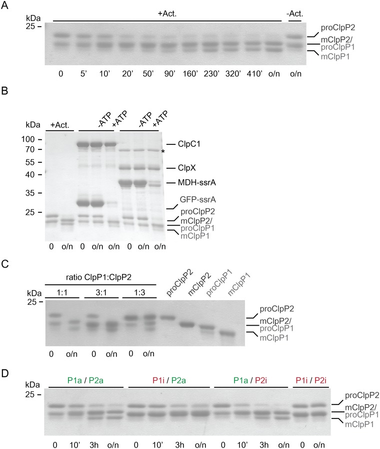
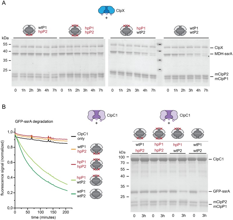
Clp chaperone-proteases are cylindrical complexes built from ATP-dependent chaperonerings that stack onto a proteolytic ClpP double-ring core to carry out substrate protein degradation.Interaction of the ClpP particle with the chaperone is mediated by an N-terminal loop and a hydrophobic surface patch on the ClpP ring surface. In contrast to E. coli, Myco bacterium tuberculosis harbors not only one but two ClpP protease subunits, ClpP1 and ClpP2,and a homo-heptameric ring of each assembles to form the ClpP1P2 double-ring core. Consequently,this hetero double-ring presents two different potential binding surfaces for the interaction with the chaperones ClpX and ClpC1. To investigate whether ClpX or ClpC1 might preferentially interact with one or the other double-ring face, we mutated the hydrophobicchaperone-interaction patch on either ClpP1 or ClpP2, generating ClpP1P2 particles that are defective in one of the two binding patches and thereby in their ability to interact with their chaperone partners. Using chaperone-mediated degradation of ssrA-tagged model substrates, we show that both Mycobacterium tuberculosis Clp chaperones require the intact interaction face of ClpP2 to support degradation, resulting in an asymmetric complex where chaperones only bind to the ClpP2 side of the proteolytic core. This sets the Clpproteases of Mycobacterium tuberculosis, and probably other Actinobacteria, apart from the well-studied E. coli system, where chaperones bind to both sides of the protease core,and it frees the ClpP1 interaction interface for putative new binding partners [corrected].


Conflict of interest statement

Competing Interests: The authors have declared that no competing interests exist.

Figures

Fig 1
Fig 1. Assembly of ClpP1 and ClpP2 to the ClpP1P2 double-ring particle.
Analytical gel filtration runs were performed on a Superdex 200 gel filtration column (24 ml). Unless noted otherwise, all runs were performed at room temperature. The assembly state of the proteins is indicated by a cartoon depiction in the topmost graph of either single rings in light grey for ClpP1 and dark grey for ClpP2, or the assembled double-ring particle. Molecular size markers are indicated as arrow tips above the elution profiles in the first graph. A marker for E. coli ClpP (EcClpP) is included as a reference for the double-ring assembly. The concentration of ClpP1 and ClpP2 is always 25 μM protomer. A. ClpP1 and ClpP2 containing the N-terminal propeptide (proClpP1, proClpP2) assemble to double-ring complexes in the absence of the activator. When proClpP1 (dashed light grey) and proClpP2 (dashed dark grey) are incubated together at room temperature overnight they assemble into the double-ring complex (dark brown). SDS-PAGE shows a 1:1 ratio of ClpP1 to ClpP2 in the peak fraction. Incubation of the assembled complex at 4°C for 3 hours leads to disassembly (blue). Reincubation of the complex at room temperature for 3 hours leads to reassembly into the double-ring complex (light brown). B. Analytical gel filtration of individually loaded mature ClpP1 (mClpP1, dashed light grey), mature ClpP2 (mClpP2, dashed dark grey) and Δ15NClpP2 (dashed orange). mClpP2 shows its main elution peak at 10 ml corresponding to a size of around 450 kDa. Elution profiles were also recorded after overnight incubation of mClpP1 and mClpP2 (black), and of mClpP1 and Δ15NClpP2 (orange). C. Elution profiles of in vitro processed mClpP1P2. To produce the mature complex proClpP1 and proClpP2 were incubated in the presence of 1 mM activator overnight at room temperature. The activator was then removed by buffer exchange and the mClpP1P2 complex was incubated at 4°C for 3 hours leading to disassembly of the complex (blue). Reincubation of the complex at room temperature for 3 hours leads to reassembly into the double-ring complex (light brown).
Fig 2
Fig 2. Propeptide processing of ClpP1 and ClpP2 in the ClpP1P2 double-ring assembly.
Maturation of double-ring assembled ClpP1 and ClpP2 carrying the N-terminal propeptide (proClpP1, proClp2) to the mature proteins (mClpP1, mClpP2) was performed at room temperature and followed by SDS-PAGE analysis. Samples were taken at the indicated time points. A. Propeptide processing of ClpP1 and ClpP2 (25 μM protomer each), in the presence of 1 mM activator (+Act.), or without activator (−Act.). B. Propeptide processing of ClpP1 and ClpP2 (0.5 μM double-ring particle) in the absence of activator and the presence of 1 μM ClpC1 hexamer, 10 μM GFP-ssrA or 1 μM ClpX hexamer and 10 μM MDH-ssrA, and 5mM ATP (+ATP) or without ATP (−ATP). For comparison, overnight processing of ClpP1 and ClpP2 in the presence of 1 mM activator is shown. The asterisk (*) indicates a protein contaminant that was identified by mass spectrometry to contain DnaK from E. coli. C. Propeptide processing of ClpP1 and ClpP2 at different molar ratios of ClpP1:ClpP2 with 25 and 75 μM, respectively, ClpP1 or ClpP2 protomer concentration in the presence of 1 mM activator. D. Propeptide processing of double-ring complexes assembled from wild-type (active) ClpP1 and ClpP2 (P1a, P2a, 25 μM protomer each) and protease-inactive ClpP1S98A, ClpP2S110A (P1i, P2i) in the presence of 1 mM activator.
Fig 3
Fig 3. Generation of hydrophobic patch variants of ClpP1 and ClpP2.
A. Cartoon representation of the LGF-loops (dark blue) of the chaperone binding to hydrophobic surface patches (green) on the protease core (grey). B. Top view of a heptameric protease ring. The hydrophobic patches (green) are formed by residues of two adjacent protease subunits (grey). C. Mutations introduced in ClpP1 and ClpP2 to create the hydrophobic patch variants hpClpP1 (ClpP1S61A, Y63V, L83A, Y91V) and hpClpP2 (ClpP2Y75V, Y95V). D. Alignment of Mtb ClpP1 and ClpP2 with EcClpP. Conservation is colored from white (not conserved) to black (identical). The identity between ClpP1/ClpP2 is 39.5%, between EcClpP/ClpP1 46% and EcClpP/ClpP2 44.4%. Red arrows highlight the residue positions of the EcClpP hydrophobic patch residues. Residues depicted in green in panel E are marked with a green box. E. Surface representation of ClpP1, ClpP2 (4U0G.pdb) and EcClpP (3MT6.pdb) ring faces. Individual subunits are colored alternatingly in light and dark grey. Hydrophobic patch residues are colored in green and labelled accordingly. For ClpP1 and ClpP2 the hydrophobic patch residues used for mutation are shown, for EcClpP reference residues are shown as described in the literature [14]. F. Creation of a set of mature mixed wild-type ClpP1 and ClpP2 (wtP1, wtP2) and hydrophobic patch variants (hpP1, hpP2). Large-scale processing of ClpP1 and ClpP2 (70 μM protomer each) containing the N-terminal propeptide (proClpP1, proClpP2) to mature ClpP1 and ClpP2 (mClpP1, mClpP2) in the presence of 1 mM activator. The reaction was performed in Buffer A. All samples were run on the same gel. The lane containing the size marker was removed for better visual representation (white line). G. Analytical gel filtration was performed with the mature ClpP1P2 complexes created in panel F. The peak at 11 ml shows that all complexes have assembled into double-rings.
Fig 4
Fig 4. Chaperone-mediated degradation of ssrA-tagged substrates by ClpP1P2 requires the hydrophobic patch on ClpP2.
ClpX and ClpC1-dependent degradation of model substrates was assayed with a set of mature ClpP1P2 particles, created from mixed wild-type (wtP1, wtP2) and hydrophobic patch variants (hpP1, hpP2) of ClpP1 and ClpP2. A. Degradation of MDH-ssrA (2 μM) mediated by ClpX (1 μM hexamer) and wt, hp or mixed mature ClpP1P2 particles (0.5 μM double-ring particle), was followed by the disappearance of the MDH-ssrA band in SDS-PAGE. The band just below MDH-ssrA that is not degraded (*) was confirmed by MS/MS to be composed of MDH, most probably lacking the ssrA tag. B. Degradation of GFP-ssrA (2 μM) mediated by ClpC1 (1 μM hexamer) and by wt, hp and mixed mature ClpP1P2 particles (0.5 μM double-ring particle) was monitored by the loss of the intrinsic GFP fluorescence signal. The signal was globally normalized. Additionally, time points were taken at the beginning and the end of the reaction and degradation of GFP-ssrA was confirmed by SDS-PAGE.

Similar articles

Cited by

References

    1. Gandhi NR, Nunn P, Dheda K, Schaaf HS, Zignol M, van Soolingen D, et al. Multidrug-resistant and extensively drug-resistant tuberculosis: a threat to global control of tuberculosis. The Lancet. 2010;375(9728):1830–43. 10.1016/S0140-6736(10)60410-2 - DOI - PubMed
    1. Jassal M, Bishai WR. Extensively drug-resistant tuberculosis. The Lancet Infectious Diseases. 2009;9(1):19–30. 10.1016/S1473-3099(08)70260-3 - DOI - PubMed
    1. Brotz-Oesterhelt H, Beyer D, Kroll HP, Endermann R, Ladel C, Schroeder W, et al. Dysregulation of bacterial proteolytic machinery by a new class of antibiotics. Nature medicine. 2005;11(10):1082–7. 10.1038/nm1306 - DOI - PubMed
    1. Kirstein J, Hoffmann A, Lilie H, Schmidt R, Rubsamen-Waigmann H, Brotz-Oesterhelt H, et al. The antibiotic ADEP reprogrammes ClpP, switching it from a regulated to an uncontrolled protease. EMBO molecular medicine. 2009;1(1):37–49. 10.1002/emmm.200900002 - DOI - PMC - PubMed
    1. Ollinger J, O'Malley T, Kesicki EA, Odingo J, Parish T. Validation of the essential ClpP protease in Mycobacterium tuberculosis as a novel drug target. Journal of bacteriology. 2012;194(3):663–8. Epub 2011/11/30. 10.1128/JB.06142-11 - DOI - PMC - PubMed

Publication types

MeSH terms