. 2015 May 1;10(5):e0125617.
doi: 10.1371/journal.pone.0125617. eCollection 2015.

OXPHOS-Mediated Induction of NAD+ Promotes Complete Oxidation of Fatty Acids and Interdicts Non-Alcoholic Fatty Liver Disease

Affiliations

OXPHOS-Mediated Induction of NAD+ Promotes Complete Oxidation of Fatty Acids and Interdicts Non-Alcoholic Fatty Liver Disease

Thomas E Akie et al. PLoS One. .

Abstract

OXPHOS is believed to play an important role in non-alcoholic fatty liver disease (NAFLD), however, precise mechanisms whereby OXPHOS influences lipid homeostasis are incompletely understood. We previously reported that ectopic expression of LRPPRC, a protein that increases cristae density and OXPHOS, promoted fatty acid oxidation in cultured primary hepatocytes. To determine the biological significance of that observation and define underlying mechanisms, we have ectopically expressed LRPPRC in mouse liver in the setting of NAFLD. Interestingly, ectopic expression of LRPPRC in mouse liver completely interdicted NAFLD, including inflammation. Consistent with mitigation of NAFLD, two markers of hepatic insulin resistance--ROS and PKCε activity--were both modestly reduced. As reported by others, improvement of NAFLD was associated with improved whole-body insulin sensitivity. Regarding hepatic lipid homeostasis, the ratio of NAD+ to NADH was dramatically increased in mouse liver replete with LRPPRC. Pharmacological activators and inhibitors of the cellular respiration respectively increased and decreased the [NAD+]/[NADH] ratio, indicating respiration-mediated control of the [NAD+]/[NADH] ratio. Supporting a prominent role for NAD+, increasing the concentration of NAD+ stimulated complete oxidation of fatty acids. Importantly, NAD+ rescued impaired fatty acid oxidation in hepatocytes deficient for either OXPHOS or SIRT3. These data are consistent with a model whereby augmented hepatic OXPHOS increases NAD+, which in turn promotes complete oxidation of fatty acids and protects against NAFLD.


Conflict of interest statement

Competing Interests: The authors have declared that no competing interests exist.

Figures

Fig 1
Fig 1. LRPPRC promotes mitochondrially encoded gene expression in liver.
(A) Protein expression and (B) quantification in livers of wild-type littermate controls (WT), hemizygous (Tg/0), and double hemizygous (Tg/Tg) LRPPRC transgenic mice. (C) Protein expression in heart, spleen, brown adipose tissue (BAT) and liver in WT and Tg/Tg mice. (D) Protein expression in WT and Tg/Tg cultured primary hepatocytes. (E) Nuclear Lrpprc, mitochondrial polymerase (Polrmt), mitochondrial transcription factor A (Tfam), and mitochondrial transcription factor B2 (Tfb2m) and (F) mitochondrially encoded respiratory complex subunit gene expression in Tg/Tg and control livers. (G) Genetic determination of mitochondrially encoded DNA content in the samples. For all experiments, n = 3 or 4. Data are mean ± SEM * p<0.05, ** p<0.01, *** p<0.001 vs. WT by two-tailed unpaired Student’s t-test (B, E, G) or 2-way ANOVA with Bonferroni’s post-test (F).
Fig 2
Fig 2. Liver-specific LRPPRC augments hepatic OXPHOS.
(A) Representative plot, (B) absolute and (C) relative changes in oxygen consumption rate (OCR) in isolated mitochondria from WT and LRPPRC Tg/Tg livers measured on a Clarke-type electrode. (D) FCCP induced maximal OCR vs. LRPPRC protein levels in isolated mitochondria from WT and Tg/Tg livers. (E) P:O ratio calculated from Clarke-electrode data. For all experiments, n = 3 or 4. Data are mean ± SEM (B—E), * p<0.05, ** p<0.01, *** p<0.001 by two-tailed unpaired Student’s t-test (B, C) or linear regression (D).
Fig 3
Fig 3. Hepatic OXPHOS enhances whole body insulin sensitivity.
(A) Glucose and (B) insulin tolerance tests and area-under-the-curve (insets) and (C) weights in wild-type littermate control mice (WT), LRPPRC single hemizygous (Tg/0), and double hemizygous (Tg/Tg) mice fed a high-fat diet for 12 weeks, n = 10. (D) Hourly activity, (E) daily food intake, and (F) diurnal energy expenditure (7AM—7PM) in WT and Tg/Tg mice fed a high-fat diet > 20 weeks n = 3–6. (G) ER stress and (H) antioxidant gene expression in high-fat fed WT and Tg/Tg mouse liver, n = 9 or 10. (I) Mitochondrial superoxide production and (J) mitochondrial membrane potential (ΔΨm) in HepG2 cells ectopically expressing LacZ (control) or LRPPRC n = 24. (K) Immunoblot and quantification of phospho-AKT in cultured primary hepatocytes from chow-fed WT and Tg/Tg mice treated with 25nM insulin, n = 3. (L) Immunoblot and (M) quantification of membrane or (N) membrane: cytosolic PKCε in high-fat fed WT and Tg/Tg mouse livers, n = 5 or 6. Data are mean ± SEM, *p<0.05, **p<0.01, ***p<0.001 vs. WT by two-way ANOVA (A, B), two-tailed unpaired Student’s t-test (insets, F, H, J, K), mixed model with Bonferroni correction (C) or one-tailed unpaired Student’s t-test (M, N).
Fig 4
Fig 4. Augmented hepatic OXPHOS interdicts NAFLD.
(A) Representative histological sections of livers from wild-type littermate control mice (WT) and LRPPRC double hemizygous (Tg/Tg) mice. (B) Pathologic classification of inflammation in livers of the same mice. (C) Expression of inflammatory genes in livers of WT and Tg/Tg mice fed a high-fat diet for 12 weeks. (D) Pathologic classification of steatosis in high-fat fed WT and Tg/Tg mouse livers. (E) Biochemical determination of triglyceride content in the same samples. (F) Determination of Fsp27 expression in high-fat fed WT and Tg/Tg livers. (G) Serum triglyceride content in the same mice. Expression of genes involved in (H) lipid export, (I) lipid uptake, (J) Lipogenesis, and (K) fatty acid β-oxidation in high-fat fed WT and Tg/Tg mouse livers. For all experiments, n = 9 or 10. Data are mean ± SEM *p<0.05, **p<0.01, ***p<0.001 vs. WT by chi-squared analysis (B, D), 2-way ANOVA with Bonferroni’s post-test (C), or by two-tailed, unpaired Student’s t-test (D-K).
Fig 5
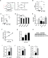
Fig 5. OXPHOS dictates NAD+, promoting complete oxidation of fatty acids (A) Fatty acid metabolic pathway in liver: NAD+ dependent β-oxidation catalyzes lipids to acetyl-CoA and ketone bodies (incomplete oxidation).
Acetyl-CoA is further metabolized via the NAD+ dependent TCA cycle, yielding CO2 (complete oxidation). NAD+ allosterically activates TCA cycling, while NADH inhibits this process. (B) NAD+ and (C) [NAD+]/[NADH] levels in chow fed wild-type littermate control mice (WT), LRPPRC single hemizygous (Tg/0), and double hemizygous (Tg/Tg) mice.(D) NAD+ synthetic gene expression in WT and Tg/Tg mice. (E) [NAD+]/[NADH] levels in cultured primary hepatocytes treated with 250 μM palmitic acid, 1 μM rotenone, or 4 μM FCCP. (F) NAD+ levels in nicotinamide (NAM) treated cultured primary hepatocytes. (G) Palmitate oxidation to CO2 in cultured primary hepatocytes treated with NAM. (H) Immunoblot of citrate synthase (CS) expression in cultured primary hepatocytes treated with NAM. (I) Palmitate oxidation in Sirt3 -/- (Sirt3 KO) primary hepatocytes treated with NAM. Palmitate oxidation in primary hepatocytes treated with (J) rotenone or (K) FCCP and NAM. For all experiments, n = 3 or 4 Data are mean ± SEM *p<0.05, **p<0.01, ***p<0.001 vs. vehicle unless otherwise indicated and ###p<0.001 by two-tailed unpaired Student’s t-test (B, C, E, G, I, J, K) or linear regression (F).
Fig 6
Fig 6. Activation of hepatic OXPHOS increases cellular NAD+ and complete fatty acid oxidation, mitigating NAFLD.
In obesity and nutrient excess, increased ratio of reduced NADH to oxidized NAD+ inhibits complete oxidation of fatty acids in the liver, leading to lipid accumulation, PKCε activation, and insulin resistance. Augmented hepatic OXPHOS, through enhanced redox cycling and/or increased ATP dependent NAD+ synthesis, increases the ratio of [NAD+]/[NADH], thereby driving complete oxidation of fatty acids, reducing steatosis, and promoting insulin sensitivity. Additionally, enhanced OXPHOS reduces reactive oxygen species, which may contribute to increased insulin sensitivity (not shown).

References

    1. Bellentani S, Scaglioni F, Marino M, Bedogni G. Epidemiology of non-alcoholic fatty liver disease. Dig Dis. 2010;28(1):155–61. doi: 000282080 [pii]; 10.1159/000282080 - DOI - PubMed
    1. Angulo P. Nonalcoholic fatty liver disease. N Engl J Med. 2002. April 18;346(16):1221–31. 10.1056/NEJMra011775 [doi];346/16/1221 [pii]. - DOI - PubMed
    1. Adams LA, Lymp JF, St SJ, Sanderson SO, Lindor KD, Feldstein A, et al. The natural history of nonalcoholic fatty liver disease: a population-based cohort study. Gastroenterology. 2005. July;129(1):113–21. doi: S0016508505006943 [pii]. - PubMed
    1. White DL, Kanwal F, El-Serag HB. Association between nonalcoholic fatty liver disease and risk for hepatocellular cancer, based on systematic review. Clin Gastroenterol Hepatol. 2012. December;10(12):1342–59. doi: S1542-3565(12)01153-6 [pii]; 10.1016/j.cgh.2012.10.001 . - DOI - PMC - PubMed
    1. Bellentani S, Saccoccio G, Masutti F, Croce LS, Brandi G, Sasso F, et al. Prevalence of and risk factors for hepatic steatosis in Northern Italy. Ann Intern Med. 2000. January 18;132(2):112–7. doi: 200001180–00004 [pii]. - PubMed

Publication types

MeSH terms