. 2015 Jul;156(7):2503-17.
doi: 10.1210/en.2014-1815. Epub 2015 May 1.

Adrenal Development in Mice Requires GATA4 and GATA6 Transcription Factors

Affiliations

Adrenal Development in Mice Requires GATA4 and GATA6 Transcription Factors

Sergei G Tevosian et al. Endocrinology. 2015 Jul.

Abstract

The adrenal glands consist of an outer cortex and an inner medulla, and their primary purposes include hormone synthesis and secretion. The adrenal cortex produces a complex array of steroid hormones, whereas the medulla is part of the sympathetic nervous system and produces the catecholamines epinephrine and norepinephrine. In the mouse, GATA binding protein (GATA) 4 and GATA6 transcription factors are coexpressed in several embryonic tissues, including the adrenal cortex. To explore the roles of GATA4 and GATA6 in mouse adrenal development, we conditionally deleted these genes in adrenocortical cells using the Sf1Cre strain of animals. We report here that mice with Sf1Cre-mediated double deletion of Gata4 and Gata6 genes lack identifiable adrenal glands, steroidogenic factor 1-positive cortical cells and steroidogenic gene expression in the adrenal location. The inactivation of the Gata6 gene alone (Sf1Cre;Gata6(flox/flox)) drastically reduced the adrenal size and corticosterone production in the adult animals. Adrenocortical aplasia is expected to result in the demise of the animal within 2 weeks after birth unless glucocorticoids are provided. In accordance, Sf1Cre;Gata4(flox/flox)Gata6(flox/flox) females depend on steroid supplementation to survive after weaning. Surprisingly, Sf1Cre;Gata4(flox/flox)Gata6(flox/flox) males appear to live normal lifespans as vital steroidogenic synthesis shifts to their testes. Our results reveal a requirement for GATA factors in adrenal development and provide a novel tool to characterize the transcriptional network controlling adrenocortical cell fates.


Figures

Figure 1.
Figure 1.
Adrenal gland aplasia in Sf1Cre;Gata4flox/flox Gata6flox/flox mice. Representative pictures of torsos from the control (A and B) and Sf1Cre;Gata4flox/floxGata6flox/flox (C and D) of female (XX; A and C) and male (XY; B and D) mice at PND9. A and B, Adrenal glands are encircled in dashed orange lines. Note the absence of adrenal glands in Sf1Cre;Gata4flox/floxGata6flox/flox (C and D) animals. RK, right kidney; LK, left kidney; A, anterior; P, posterior. E–M, Histological appearance of the abdominal area of the control (E, H, and K), Sf1Cre;Gata4flox/+Gata6flox/flox (F, I, and L), and Sf1Cre;Gata4flox/floxGata6flox/flox (G, J, and M) fetuses at E15.5 (E–J) and 19.5 (K–M). Sections were stained with hematoxylin and eosin (H&E). Arrows in panels E–G point to the adrenal gland. Panels H–J are higher magnification of the adrenal gland showed in E–G, respectively. Scale bars, 200 μm (E–G), 100 μm (K–M), and 50 μm (H–J).
Figure 2.
Figure 2.
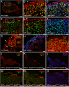
Loss of steroidogenic cells in Sf1Cre;Gata4flox/floxGata6flox/flox adrenals. Representative sections from controls (A–I) and Sf1Cre;Gata4flox/floxGata6flox/flox (J–O) adrenals at E15.5 were stained for GATA6 (red) and SF1 (green) (A–C, J, and M); GATA4 (red) and SF1 (green) (D–F, K, and N); GATA6 (red) and GATA4 (green) (G); and 3βHSD (red) (I, L, and O) and SF1 (green) (I). DAPI (blue) was used as nuclear staining. In the control adrenals, most SF1-positive cells express GATA6 but not GATA4 (compare B and E). Only anterior capsular cells consistently express GATA4 (arrow in E), whereas most the capsular cells express GATA6 (arrows in B and G). G, A small subset of subcapsular cells (arrowheads) and rare capsular cells (asterisk) coexpresses both GATA factors. In J–O, note that steroidogenic (SF1- or 3βHSD-positive) cells are absent in the Sf1Cre;Gata4flox/floxGata6flox/flox adrenals. B and C, and E and F, are higher magnifications of A and D, respectively, and M–O are higher magnifications of J–L, respectively. Scale bars, 100 μm (A and J–L), 50 μm (D and M–O), and 20 μm (B, C, and E–I).
Figure 3.
Figure 3.
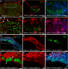
The adrenal area in Sf1Cre;Gata4flox/floxGata6flox/flox fetuses is occupied by medullar cells. Torso sections of control (A–D) and Sf1Cre;Gata6flox/flox (E–K) fetuses at E15.5 were stained for TH (green) and NF (NF68) (red) (A, B, E, and F), 3βHSD (red) and TH (green) (I). In A and E, adrenals are encircled by a blue dashed line, and B–D and F–H are higher magnifications of A and E, respectively. In the control, the medullar (THhigh/NFlow) and the nervous (THlow/NFhigh) zones are separated by the negative adrenocortical mass (A and B). Note that the Sf1Cre;Gata4flox/floxGata6flox/flox animals lack of adrenocortical cells, these 2 populations are directly juxtaposed (E and F). Also note in I and K the absence of steroidogenic cells (defined by 3βHSD staining) in the adrenal glands of Sf1Cre;Gata4flox/floxGata6flox/flox fetuses. cx, cortex; m, medulla; sg, sympathetic ganglion. Scale bars, 200 μm (A and E), 100 μm (B–D), and 50 μm (F–K).
Figure 4.
Figure 4.
Adrenocortical development is severely impaired in the absence of GATA6. A–F, Representative sections from control (A and B) and ROSAmT/mG; Sf1Cre;Gata4flox/+Gata6flox/flox (C–F) adrenals at E15.5 were stained for SF1 (bright green) (A and B) and GATA4 (red) (A–D and F). Cells that underwent Sf1Cre-mediated recombination are traced by membrane EGFP (EGFPm) (dark green). In B note that GATA4 expression is mostly limited to capsular cells (arrowhead), with rare cortical cells expressing the protein (arrows). In contrast, numerous cortical cells are GATA4-positive in the ROSAmT/mG; Sf1Cre;Gata4flox/+Gata6flox/flox adrenal (C, D, and F; encircled in C). In C–E only some EGFP-positive cells express GATA4 (see also Supplemental Figure 4). G–L, A limited number of steroidogenic cells is present in the ROSAmT/mG; Sf1Cre;Gata6flox/flox adrenals. Adrenal sections of ROSAmT/mG; Sf1Cre;Gata6flox/flox at E15.5 were stained with antibodies against GATA6 (red; G, H, and J–L) and SF1 (bright green) (G, I, J, and L). EGFPm expression (dark green) traces SF1Cre-mediated excision (G, I, J, and L). In J, arrows point to SF1-positive nuclei that are strictly confined to the EGFPm-positive/GATA6-negative cells. J–L, Higher magnifications of a rectangular area in G. Scale bars, 50 μm (A, C, and G–I) and 20 μm (B, D–F, and J–L).
Figure 5.
Figure 5.
The sole functional allele of the Gata4 gene is sufficient to support adrenocortical development and steroidogenesis. Adrenal sections from control (A and B) and ROSAmT/mG; Sf1Cre;Gata4flox/+Gata6flox/flox (C–J) fetuses at E15.5 were stained for 3βHSD (red) (A–F) and TH (bright green) (A–H); SF1 (bright green) (I and J) and 3βHSD (red) (I and J). In the ROSAmT/mG; Sf1Cre;Gata4flox/+Gata6flox/flox adrenals, EGFP expression traces SF1Cre-mediated gene excision only by displaying membrane staining (EGFPm, dark green) (C–J). DAPI was used as nuclear staining (blue). Scale bars, 200 μm (C, E, G, and I), 100 μm (A, D, F, and H), 50 μm (D, F, and H), and 20 μm (B and J).
Figure 6.
Figure 6.
Gene expression analysis of adrenal glands. RNA expression analysis via qPCR for the steroidogenic enzymes Star, Cyp11a1, Cyp11b1, Cyp21a2, and Cyp17a1 and the medullar-specific genes Th and Nefl. Adrenal glands were obtained from controls (at least n = 5), Sf1Cre;Gata4flox/+ Gata6flox/flox (at least n = 4), and Sf1Cre;Gata4flox/flox Gata6flox/flox (n = 3) at E15.5. Bars represent mean ± SEM of fold change of Sf1Cre;Gata4flox/+Gata6flox/flox (black bar) and Sf1Cre;Gata4flox/floxGata6flox/flox (gray bar) relative to the controls. Data were analyzed by ANOVA (one-way) followed by Tukey's multiple comparisons test, with significance at *, P < .05; **, P < .01; and ***, P < .001. Statistical difference between Sf1Cre;Gata4flox/+Gata6flox/flox and Sf1Cre;Gata4flox/floxGata6flox/flox is shown by the respective P value.
Figure 7.
Figure 7.
Presence of adrenal-like cells in the embryonic Sf1Cre;Gata4flox/floxGata6flox/flox testes. Representative testicular sections from controls (A and B) and Sf1Cre;Gata4flox/floxGata6flox/flox (C and D) males at E18.5 were stained for the adrenal enzyme CYP21A2. B and D, Higher magnification of A and C, respectively. In B and D, arrowheads point to CYP21A2-positive cells. Scale bars, 100 μm (A and C) and 50 μm (B and D). E and F, Quantitative changes in the expression of Insl3, Star, Cyp11a1, Hsd3b6, Mc2r, Cyp21a1, Cyp11b1, Cyp11b2, Gata4, and Gata6 in Sf1Cre;Gata4flox/floxGata6flox/flox testes at E15.5 (E) and E18.5 (F). The results are plotted as the mean ± SEM of the fold change relative to the controls from n = 3 biological replicates of each genotype. G, Glucose concentration (mg/dL) from whole blood of the controls (n = 3; black bar) and Sf1Cre;Gata4flox/floxGata6flox/flox (n = 3; gray bar) males at PND60. H and I, Corticosterone concentration in plasma (ng/mL) from the controls (black bar) and Sf1Cre;Gata4flox/flox Gata6flox/flox (gray bar) male mice at PND4–PND10 (n = 4) (L) and 60 (n = 3) (M). J, Intratesticular corticosterone concentration (ng/mL) of the controls (n = 3; black bar) and Sf1Cre;Gata4flox/flox Gata6flox/flox (n = 3; gray bar) mice at PND45. Results are shown as the mean ± SEM, and datasets were analyzed by Student's t test, with significance considered at *, P < .05; **, P < .01; and ***, P < .001.
Figure 8.
Figure 8.
Rescue of Sf1Cre;Gata4flox/floxGata6flox/flox female mice with GC treatment. A, Plasma corticosterone concentration (ng/mL) of the controls (n = 3; black bar) and Sf1Cre;Gata4flox/floxGata6flox/flox (n = 3; gray bar) females between PND4 and PND9. Results are shown as the mean ± SEM, and the data were analyzed by Student's t test, with significance considered at *, P < .05. B, Percent survival of the controls treated with DPBS vehicle (n = 7; dashed line), Sf1Cre;Gata4flox/floxGata6flox/flox females that were injected daily with a cocktail of GCs (fludrocortisone acetate + dexamethasone 21-phosphate) (n = 7; dotted line), and nontreated Sf1Cre;Gata4flox/floxGata6flox/flox females (n = 6; solid line). C, Glucose concentration (mg/dL) in whole blood from the controls treated with DPBS vehicle (black bar), treated Sf1Cre;Gata4flox/floxGata6flox/flox females (light gray bar), and noninjected Sf1Cre;Gata4flox/flox Gata6flox/flox females (dark gray bar). The data are presented as the mean ± SEM, and datasets were analyzed with ANOVA (one-way) followed by Tukey's test for multiple comparisons. Bars with different superscripts differ significantly (***, P < .001). D–G, Representative ovarian sections from the controls (D and E) and Sf1Cre;Gata4flox/floxGata6flox/flox (F and G) at PND4. Sections were stained for the adrenal enzyme CYP21A2. E and G, Higher magnification of D and F, respectively. Note the lack of CYP21A2-positive cells in the ovaries of both genotypes. Pr, primordial; P, primary; PA, preantral. Scale bars, 50 μm (D and F) and 20 μm (E and G).

Similar articles

Cited by

References

    1. Muglia L, Jacobson L, Dikkes P, Majzoub JA. Corticotropin-releasing hormone deficiency reveals major fetal but not adult glucocorticoid need. Nature. 1995;373(6513):427–432. - PubMed
    1. Parker KL, Schimmer BP. Steroidogenic factor 1: a key determinant of endocrine development and function. Endocr Rev. 1997;18(3):361–377. - PubMed
    1. Caron KM, Soo SC, Wetsel WC, Stocco DM, Clark BJ, Parker KL. Targeted disruption of the mouse gene encoding steroidogenic acute regulatory protein provides insights into congenital lipoid adrenal hyperplasia. Proc Natl Acad Sci USA. 1997;94(21):11540–11545. - PMC - PubMed
    1. Barik J, Marti F, Morel C, et al. Chronic stress triggers social aversion via glucocorticoid receptor in dopaminoceptive neurons. Science. 2013;339(6117):332–335. - PubMed
    1. Niwa M, Jaaro-Peled H, Tankou S, et al. Adolescent stress-induced epigenetic control of dopaminergic neurons via glucocorticoids. Science. 2013;339(6117):335–339. - PMC - PubMed

Publication types

MeSH terms