. 2015 May 1;10(5):e0124888.
doi: 10.1371/journal.pone.0124888. eCollection 2015.

The PGE2/IL-10 Axis Determines Susceptibility of B-1 Cell-Derived Phagocytes (B-1CDP) to Leishmania major Infection

Affiliations

The PGE2/IL-10 Axis Determines Susceptibility of B-1 Cell-Derived Phagocytes (B-1CDP) to Leishmania major Infection

Angélica F Arcanjo et al. PLoS One. .

Abstract

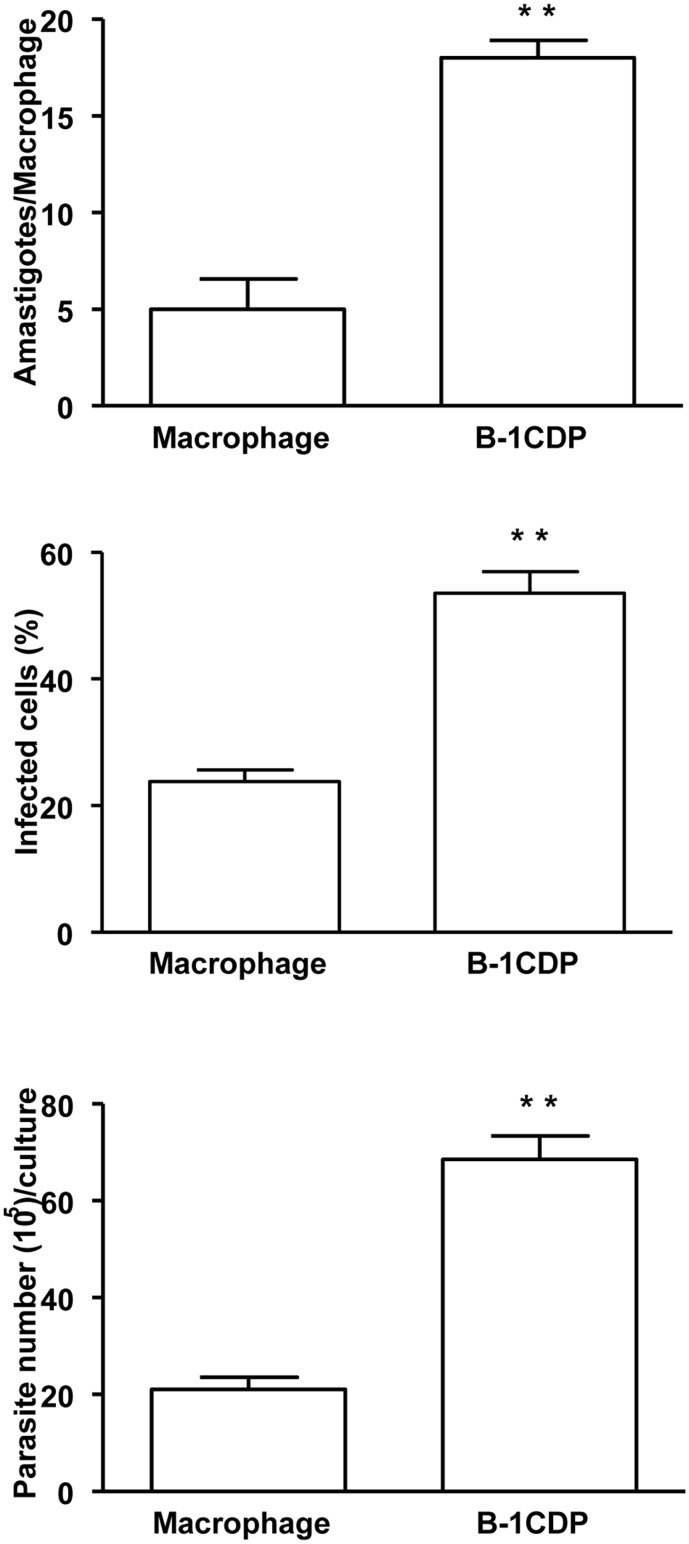
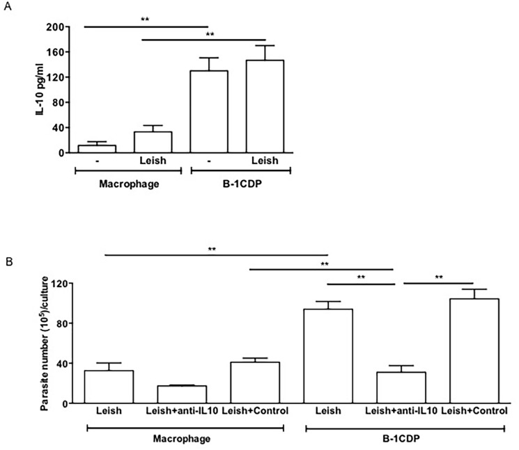
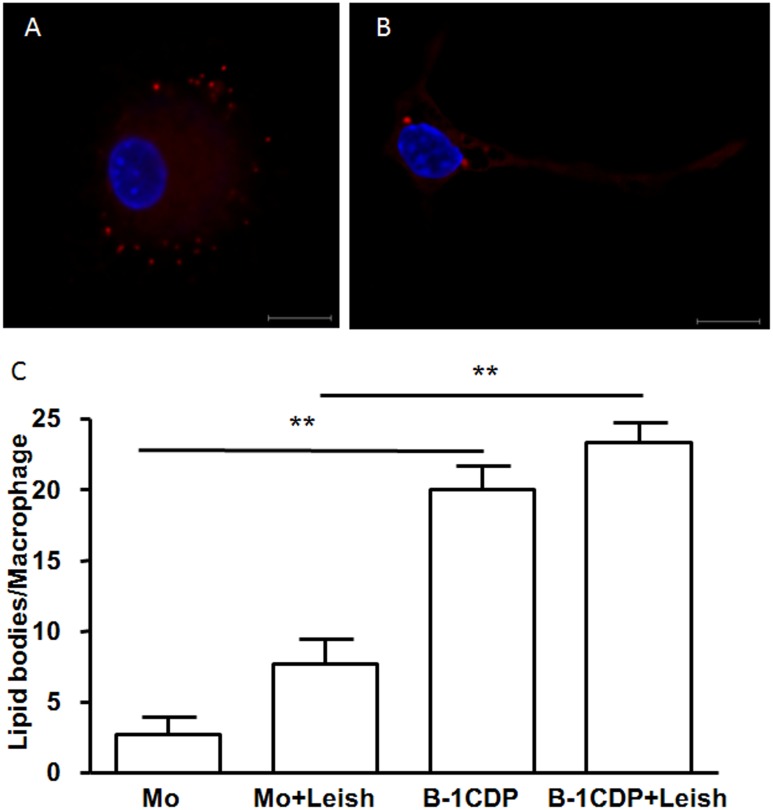
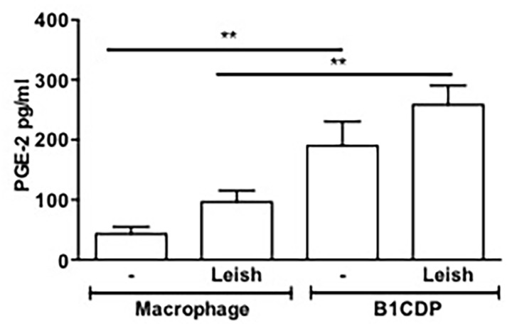
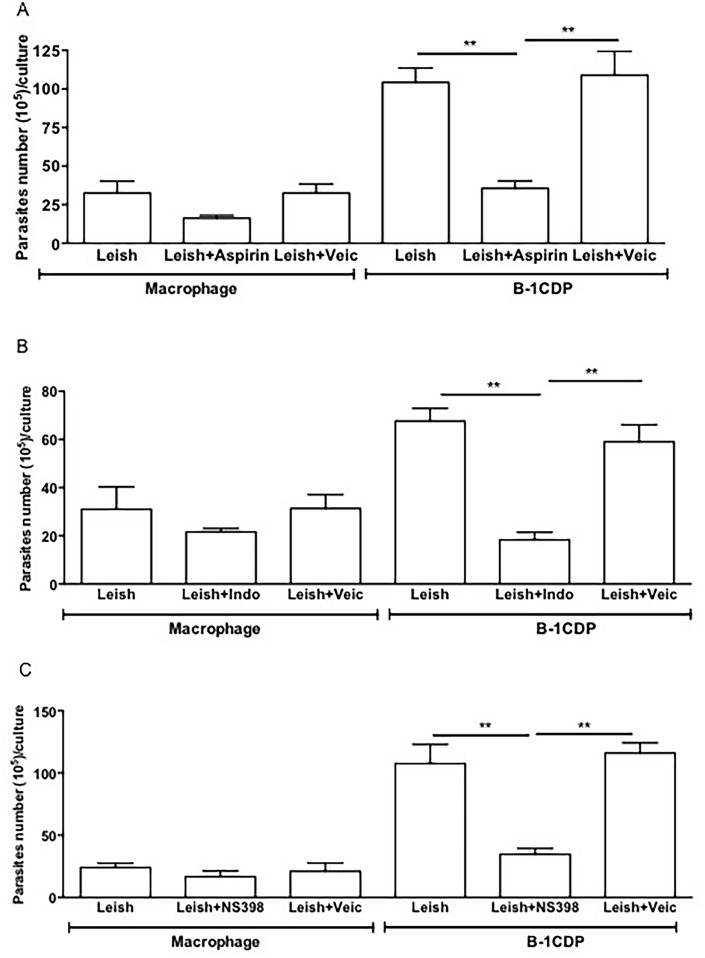
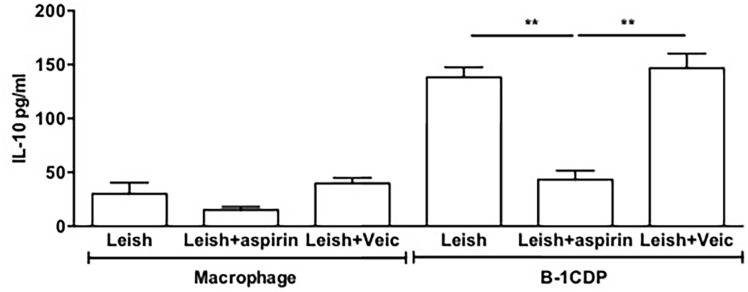
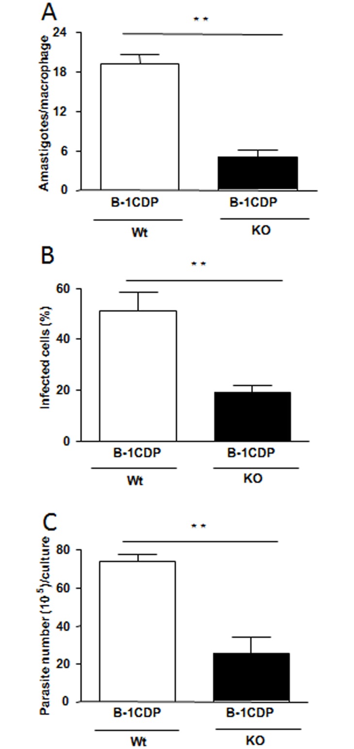
B-1 cells can be differentiated from B-2 cells because they are predominantly located in the peritoneal and pleural cavities and have distinct phenotypic patterns and activation properties. A mononuclear phagocyte derived from B-1 cells (B-1CDP) has been described. As the B-1CDP cells migrate to inflammatory/infectious sites and exhibit phagocytic capacity, the microbicidal ability of these cells was investigated using the Leishmania major infection model in vitro. The data obtained in this study demonstrate that B-1CDP cells are more susceptible to infection than peritoneal macrophages, since B-1CDP cells have a higher number of intracellular amastigotes forms and consequently release a larger number of promastigotes. Exacerbated infection by L. major required lipid bodies/PGE2 and IL-10 by B-1CDP cells. Both infection and the production of IL-10 were decreased when PGE2 production was blocked by NSAIDs. The involvement of IL-10 in this mechanism was confirmed, since B-1CDP cells from IL-10 KO mice are more competent to control L. major infection than cells from wild type mice. These findings further characterize the B-1CDP cells as an important mononuclear phagocyte that plays a previously unrecognized role in host responses to L. major infection, most likely via PGE2-driven production of IL-10.


Conflict of interest statement

Competing Interests: The authors have declared that no competing interests exist.

Figures

Fig 1
Fig 1. B-1CDP cells exhibit a susceptible phenotype to infection with L. major in vitro when compared to peritoneal macrophages.
B-1CDP cells and peritoneal macrophages were cultured (105/ml) and infected with metacyclic promastigotes of L. major. After 24 hours, the cell culture was washed and phagocytes were cultured for another 3 days with DMEM supplemented with 10% FBS at 37°C. After this period, cells were stained and amastigotes inside the phagocytes were counted under the light microscope (A) and set the percentage of infected cells (B). To quantify promastigotes forms in the supernatants, the cells were infected with L. major. After 24 hours of infection, cells were washed and cultured in Schneider medium for 5 days at a temperature of 27°C. After this period, the promastigotes were quantified in the supernatant of the cultures of infected phagocytes (C). All cultures were performed in triplicate and bars show the mean + SD. Statistical analysis were performed by T-Test from representative results of three similar experiments. **p ≤ 0.05.
Fig 2
Fig 2. IL-10 is determinant for the increased parasite load in B-1CDP.
B-1CDP cells and peritoneal macrophages were cultured in the presence or absence of L. major (MOI 10:1). After 24 hours, the supernatant was collected and IL-10 (A) was measured by ELISA. All cultures were performed in triplicate and bars show the mean ± SD. Representative result of three similar experiments **p <0.05. B-1CDP cells and peritoneal macrophages were treated or not with doses of monoclonal neutralizing anti-IL-10 or control isotype. Once were infected with L. major, after 24 hours, the cell cultures were washed with DMEM and incubated 3 days and then passed to Schneider medium. After 5 days in medium Schneider, promastigotes were counted in the supernatant (B). Statistical analysis were performed by Student’ t test from representative results of three similar experiments and bars show the mean +SD. **p ≤ 0.05.
Fig 3
Fig 3. B-1CDP cells naturally express increased numbers of lipid bodies as compared to peritoneal macrophages.
B-1CDP cells (A) and peritoneal macrophages (B) were incubated with glass coverslips, some cultures were infected with L. major (3C). Stained with Nile red (Sigma), the slides were washed and stained with DAPI (Sigma). The morphology of fixed cells was observed, and Nile red LBs were counted by light microscopy with a 100× objective lens in 50 consecutively scanned leukocytes. Statistical analysis were performed by Student’ t test from representative results of three similar experiments and bars show the mean +SD. **p ≤ 0.05. Bar, 10 μm. Representative of two experiments with identical results.
Fig 4
Fig 4. B-1CDP cells secrete high levels of PGE2 regardless the infection.
B-1CDP cells and macrophages were incubated in the presence or absence of L. major (MOI 10:1). After 24 hours of infection the supernatant was collected, and PGE2 was measured by EIA. All cultures were performed in triplicate and bars show the mean ± SD. Statistical analysis were performed by Student’ t test from representative results of three similar experiments and bars show the mean +SD. **p ≤ 0.05.
Fig 5
Fig 5. Blockage of the cyclooxygenase pathway shift the B-1CDP cells to protective phenotype in the L major infection.
Macrophages and B-1CDP were incubated in the presence or absence of L. major treated or not with (A) aspirin (10 mg/mL), (B) indomethacin (1 mg/mL) or (C) NS-398 (1 mM). After 24 hours of incubation the cells were washed and incubated again for 5 days. After this time, the promastigotes were counted in the culture supernatant. All cultures were performed in triplicate and bars show the mean ± SD. Statistical analysis were performed by Student’ t test from representative results of three similar experiments and bars show the mean +SD. **p ≤ 0.05.
Fig 6
Fig 6. Effect of inhibition of PGE2 on IL-10 releases.
Macrophages and B-1CDP cells were incubated with or without aspirin (10μg/mL) for 1 hour. They were then incubated in the presence or absence of L. major (MOI 10:1). After 24 hours of incubation, the supernatant was collected and IL-10 measured by ELISA. All cultures were performed in triplicate and bars show the mean ± SD. Statistical analysis were performed by Student’ t test from representative results of three similar experiments and bars show the mean +SD. **p ≤ 0.05.
Fig 7
Fig 7. B-1CDP cells from IL-10 deficient mice are more competent to control L. major infection.
To confirm the production of IL-10 is involved in the susceptibility to infection by L. major, we used B-1CDP cells from IL-10 KO mice. When compared to B-1CDP from wild type mice, the cells from IL-10 KO mice are more resistant to infection. Our data demonstrate decreased in the number of intracellular amastigotes (A) and percentage of infected cells (B). We also observed the significant decrease in the liberated promastigotes forms by B-1CDP from IL-10 KO mice (C). Statistical analysis were performed by Student’ t test from representative results of three similar experiments and bars show the mean +SD. **p ≤ 0.05.

References

    1. van Furth R, Cohn ZA. The origin and kinetics of mononuclear phagocytes. J Exp Med. 1968;128(3):415–35. - PMC - PubMed
    1. Okabe Y, Medzhitov R. Tissue-specific signals control reversible program of localization and functional polarization of macrophages. Cell. 2014;157(4):832–44. 10.1016/j.cell.2014.04.016 - DOI - PMC - PubMed
    1. Ginhoux F, Jung S. Monocytes and macrophages: developmental pathways and tissue homeostasis. Nat Rev Immunol. 2014;14(6):392–404. 10.1038/nri3671 - DOI - PubMed
    1. Lopes JD, Mariano M. B-1 cell: the precursor of a novel mononuclear phagocyte with immuno-regulatory properties. An Acad Bras Cienc. 2009;81(3):489–96. - PubMed
    1. Popi AF, Osugui L, Perez KR, Longo-Maugeri IM, Mariano M. Could a B-1 cell derived phagocyte "be one" of the peritoneal macrophages during LPS-driven inflammation? PLoS One. 2012;7(3):e34570 10.1371/journal.pone.0034570 - DOI - PMC - PubMed

Publication types

MeSH terms