Case Reports
. 2015 May 2:8:181.
doi: 10.1186/s13104-015-1149-1.

Acute systemic embolism due to an idiopathic floating thrombus of the thoracic aorta: success of medical management: a case report

Affiliations
Case Reports

Acute systemic embolism due to an idiopathic floating thrombus of the thoracic aorta: success of medical management: a case report

Yves Ghislain Abissegue et al. BMC Res Notes. .

Abstract

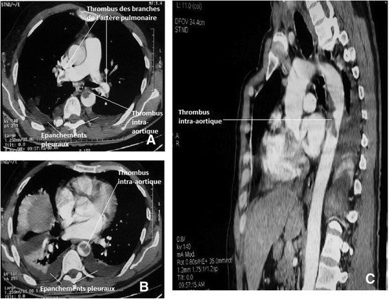
Background: Idiopathic thoracic aortic mural thrombi are rare. They can be responsible for dramatic systemic embolization. Early treatment is imperative because of their high morbidity and mortality rate.

Case presentation: A 55-year-old previously healthy Moroccan male came in an array of acute right lower limbs pain and abdominal sensibility. Severe systemic embolism involving the lower extremities, spleen, kidney, and digestive tract, due to an idiopathic mural thrombus of the thoracic aorta was diagnosed. He received medical treatment leading to the complete disappearance of the thrombus and the effects caused by the latter.

Conclusions: When faced unexplained peripheral embolization, research for a thrombus of the thoracic aorta should be performed. Medical treatment should be considered for its management, especially in patients with high surgical risk.


Figures

Figure 1
Figure 1
CT angiography showing the thoracic aorta of normal caliber without parietal atherosclerotic lesion, with an intra-aortic thrombus. Axial section; A, B. Sagittal reconstruction; E. Kidney infarct and peri-splenic hematoma, axial section C, D.
Figure 2
Figure 2
CT angiography of the lower limbs showing. Right; poor patency of the operated arterial axis, and Left; a change in contrast at the beginning of the popliteal artery.
Figure 3
Figure 3
CT angiography showing a thrombus in the pulmonary artery branches. Axial section A; bilateral pleural effusions, Axial A and B; persistence of intra aortic thrombus. Axial section A and B and sagittal reconstruction C.
Figure 4
Figure 4
CT angiography showing a complete reversal of the thoracic aorta with disappearance of thrombus. Axial section A and sagittal reconstruction C. Notch sequelae of kidney infarct, axial section B.

Similar articles

Cited by

References

    1. Tsilimparis N, Hanack U, Pisimisis G, Yousefi S, Wintzer C, Rückert RI. Thrombus in the non- aneurysmal, non atherosclerotic descending thoracic aorta—An unusual source of arterial embolism. Eur J Vasc Endovasc Surg. 2011;41:450–7. doi: 10.1016/j.ejvs.2010.11.004. - DOI - PubMed
    1. Hahn T, Dalsing MC, Sawchuk AP, Cikrit DL, Lalka SG. Primary aortic mural thrombus: presentation and treatment. Ann Vasc Surg. 1999;13(I):52–9. doi: 10.1007/s100169900220. - DOI - PubMed
    1. Blangetti I, Fenoglio L, Avallato C, Bertora M, Novali C, Peinetti F, et al. Transesophageal ecocardiography: the correct intraoperative Way to detect the source of peripheral embolism in an emergency. Ann Vasc Surg. 2013;27(8):e13–1185. doi: 10.1016/j.avsg.2012.10.029. - DOI - PubMed
    1. Yoshikawa Y, Kamikub Y, Sonod H, Yamakage M. Unexpectedly resolved multiple mobile thrombi in a normal thoracic aorta associated with colorectal cancer and protein C deficiency. J Cardiothorac Vasc Anesth. 2014;28(3):714–7. doi: 10.1053/j.jvca.2012.11.006. - DOI - PubMed
    1. Turley SR, Unger J, Cox MW, Lawson J, McCann RL, Shortell CK. Atypical aortic thrombus: should nonoperative management Be first line? Ann Vasc Surg. 2014;28(7):1610–7. doi: 10.1016/j.avsg.2014.03.028. - DOI - PubMed

Publication types