. 2015 Jul 9:298:467-74.
doi: 10.1016/j.neuroscience.2015.04.049. Epub 2015 Apr 28.

Hindbrain lactate regulates preoptic gonadotropin-releasing hormone (GnRH) neuron GnRH-I protein but not AMPK responses to hypoglycemia in the steroid-primed ovariectomized female rat

Affiliations

Hindbrain lactate regulates preoptic gonadotropin-releasing hormone (GnRH) neuron GnRH-I protein but not AMPK responses to hypoglycemia in the steroid-primed ovariectomized female rat

P K Shrestha et al. Neuroscience. .

Abstract

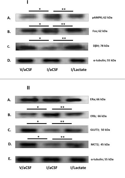
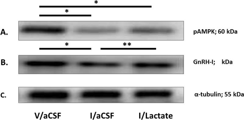
Steroid positive-feedback activation of the gonadotropin-releasing hormone (GnRH)-pituitary luteinizing hormone (LH) neuroendocrine axis propagates the pre ovulatory LH surge, a crucial component of female reproduction. Our work shows that this key event is restrained by inhibitory metabolic input from hindbrain A2 noradrenergic neurons. GnRH neurons express the ultra-sensitive energy sensor adenosine 5'-monophosphate-activated protein kinase (AMPK); here, we investigated the hypothesis that GnRH nerve cell AMPK and peptide neurotransmitter responses to insulin-induced hypoglycemia are controlled by hindbrain lack of the oxidizable glycolytic end-product L-lactate. Data show that hypoglycemic inhibition of LH release in steroid-primed ovariectomized female rats was reversed by coincident caudal hindbrain lactate infusion. Western blot analyses of laser-microdissected A2 neurons demonstrate hypoglycemic augmentation [Fos, estrogen receptor-beta (ER-β), phosphoAMPK (pAMPK)] and inhibition (dopamine-beta-hydroxylase, GLUT3, MCT2) of protein expression in these cells, responses that were normalized by insulin plus lactate treatment. Hypoglycemia diminished rostral preoptic GnRH nerve cell GnRH-I protein and pAMPK content; the former, but not the latter response was reversed by lactate. Results implicate caudal hindbrain lactoprivic signaling in hypoglycemia-induced suppression of the LH surge, demonstrating that lactate repletion of that site reverses decrements in A2 catecholamine biosynthetic enzyme and GnRH neuropeptide precursor protein expression. Lack of effect of lactate on hypoglycemic patterns of GnRH AMPK activity suggests that this sensor is uninvolved in metabolic-inhibition of positive-feedback-stimulated hypophysiotropic signaling to pituitary gonadotropes.

Keywords: A2 noradrenergic neurons; Western blot; gonadotropin-releasing hormone; laser-microdissection; luteinizing hormone surge; phosphoAMPK.


Figures

Figure 1
Figure 1. Effects of Hypoglycemia on Hindbrain Dorsal Vagal Complex A2 Noradrenergic Nerve Cell Phospho-Adenosine 5’-Monophosphate-Activated Protein Kinase (pAMPK), Fos, Dopamine-β-Hydroxylase (DH), Steroid Hormone Receptor, and Substrate Fuel Transporter Protein Expression in Steroid-Primed Ovariectomized (OVX) Female Rats: Impact of Caudal Fourth Ventricular L-Lactate Infusion
Individual tyrosine hydroxylase (TH)-immunoreactive (-ir) A2 neurons were laser-microdissected from 10 μm-thick sections of the caudal dorsal vagal complex of steroid-primed OVX animals 2 hr after sc injection of vehicle (V; group 1; n=5) or neutral protamine Hagedorn Insulin (I; 12.5 U/kg bw; groups 2 and 3; n=6/group). Animals were continuously infused with artificial cerebrospinal fluid (aCSF; groups 1 and 2) or aCSF containing L-lactate (25μM/2.0μL/hr; group 3) into the caudal fourth ventricle between −10 and +120 min after injections. For each protein of interest, separate lysates of pooled n=50 A2 cells (n=8-10/rat) were analyzed by Western blot in triplicate at minimum. Figure 1.I depicts representative immunoblots of A2 phosphoAMPK (Row A), Fos (Row B), DβH (Row C); and α-tubulin (Row D) for each treatment group. Figure 1.II shows typical Western blots of A2 estrogen receptor-alpha (ER ; Row A), estrogen receptor-beta (ER ; Row B), glucose transporter-3 (GLUT3; Row C), monocarboxylate transporter-2 (MCT2; Row D), and α-tubulin (Row E). *p < 0.05, versus V/aCSF; **p < 0.05, versus I/aCSF; these symbols denote outcomes of statistical analyses shown in Figure 2.
Figure 2
Figure 2. Effects of Hindbrain Lactate Repletion on Normalized A2 Nerve Cell Protein Expression in Hypoglycemic Steroid-Primed OVX Animals
Protein band optical densities (O.D.) of immunoblots of n=50 A2 neurons/treatment group from V/aCSF- (white bars), I/aCSF- (gray bars), and I/Lactate (diagonal-striped gray bars) - treated rats were quantified with Syngene Genetool 4.01 software and expressed relative to α-tubulin. Panels depict mean normalized O.D. values ± S.E.M. for A2 nerve cell AMPK (Figure 2.A), pAMPK (Figure 2.B), Fos (Figure 2.C), DβH (Figure 2.D), ER (Figure 2.E), ERβ (Figure 2.F), progesterone receptor (PR; Figure 2.G), GLUT3 (Figure 2.H), glucose transporter-4 (GLUT4; Figure 2.I), and MCT2 (Figure 2.J) protein levels 2 hr after sc injections. *p < 0.05, versus V/aCSF; **p < 0.05, versus I/aCSF.
Figure 3
Figure 3. Effects of Hypoglycemia on Rostral Preoptic Gonadotropin-Releasing Hormone (GnRH) pAMPK and GnRH-I Protein Expression in Steroid-Primed OVX Female Rats: Impact of Caudal Fourth Ventricular L-Lactate Infusion
Individual GnRH-ir neurons were laser-microdissected from sections of the rostral preoptic area of steroid-primed OVX animals 2 hr after sc injection of V (group 1) or I (groups 2 and 3). Animals were continuously infused with aCSF (groups 1 and 2) or aCSF containing L-lactate (25μM/2.0μL/hr; group 3) into the caudal fourth ventricle between −10 and +120 min after injections. For each protein of interest, separate lysates of pooled n=50 GnRH neurons (n=8-10/rat) were analyzed by Western blot in triplicate at minimum. Figure 1.I depicts representative immunoblots of GnRH phosphoAMPK (Row A), GnRH-I (Row B), and α-tubulin (Row C). *p < 0.05, versus V/aCSF; **p < 0.05, versus I/aCSF; these symbols denote outcomes of statistical analyses shown in Figure 4.
Figure 4
Figure 4. Effects of Hindbrain Lactate Repletion on Normalized GnRH Nerve Cell Protein Expression in Hypoglycemic Steroid-Primed OVX Rats
Protein band optical densities (O.D.) of immunoblots of n=50 GnRH neurons/treatment group from V/aCSF- (white bars), I/aCSF- (gray bars), and I/Lactate (diagonal-striped gray bars) - treated rats were quantified with Syngene Genetool 4.01 software and expressed relative to α-tubulin. Panels depict mean normalized O.D. values ± S.E.M. for GnRH nerve cell pAMPK (Figure 2.A) and GnRH-I (Figure 2.B) protein content 2 hr after treatments. *p < 0.05, versus V/aCSF; **p < 0.05, versus I/aCSF

References

    1. Adam CL, Findlay PA. (Inhibition of luteinizing hormone secretion and expression of c-fos and corticotrophin-releasing factor genes in the paraventricular nucleus during insulin-induced hypoglycaemia in sheep. J. Neuroendocrinol. 1998;10:777–783. - PubMed
    1. Barros LF. (Metabolic signaling by lactate in the brain. Trends Neurosci. 2013;36:396–404. - PubMed
    1. Briski KP, Koshy Cherian A, Genabai NK, Vavaiya KV. (In situ coexpression of glucose and monocarboxylate transporter mRNAs in metabolic-sensitive dorsal vagal complex catecholaminergic neurons: transcriptional reactivity to insulin-induced hypoglycemia (IIH) and caudal hindbrain glucose or lactate repletion during IIH. Neuroscience. 2009;164:1152–1160. - PubMed
    1. Briski KP, Sylvester PW. (Effects of the glucose antimetabolite, 2-deoxy-D-glucose (2-DG), on the LH surge and Fos expression by preoptic GnRH neurons in ovariectomized, steroid-primed rats. J. Neuroendocrinol. 1998;10:769–776. - PubMed
    1. Cagampang FR, Cates PS, Sandhu S, Strutton PH, McGarvey C, Coen CW, O'Byrne KT. (Hypoglycaemia-induced inhibition of pulsatile luteinizing hormone secretion in female rats: role of oestradiol, endogenous opioids and the adrenal medulla. J. Neuroendocrinol. 1997;9:867–872. - PubMed

MeSH terms

LinkOut - more resources