. 2015 Jul:56:154-64.
doi: 10.1016/j.biomaterials.2015.03.048. Epub 2015 Apr 17.

Deoxycholate bile acid directed synthesis of branched Au nanostructures for near infrared photothermal ablation

Affiliations

Deoxycholate bile acid directed synthesis of branched Au nanostructures for near infrared photothermal ablation

Dong-Hyun Kim et al. Biomaterials. 2015 Jul.

Abstract

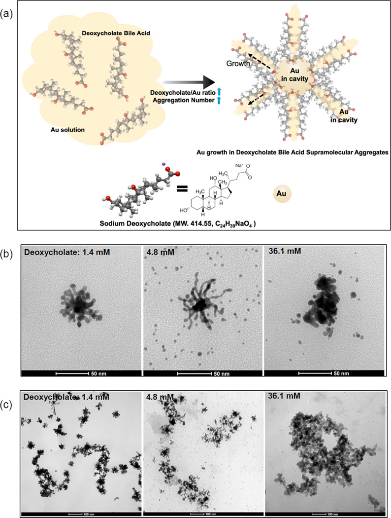
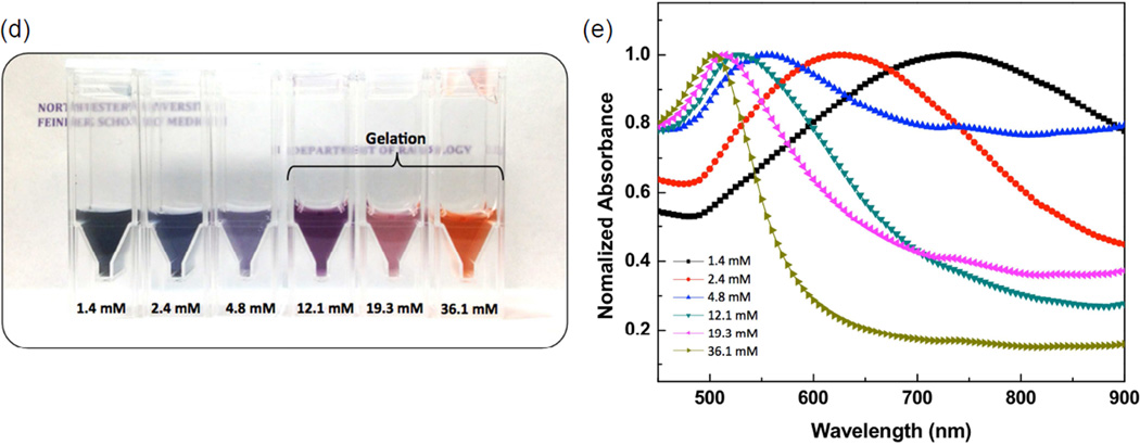
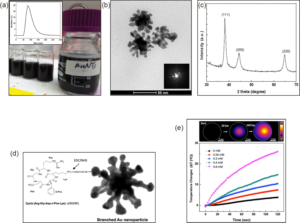
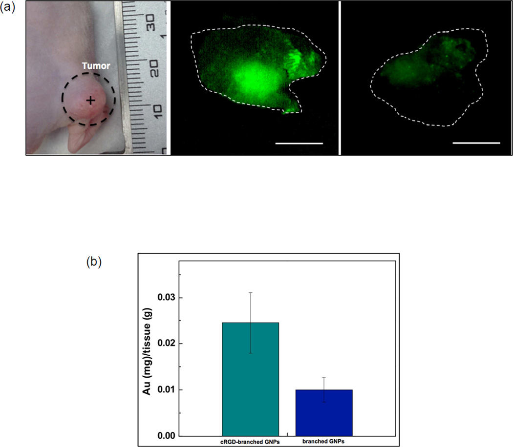
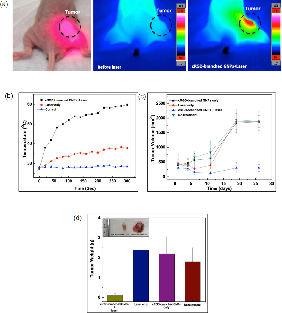
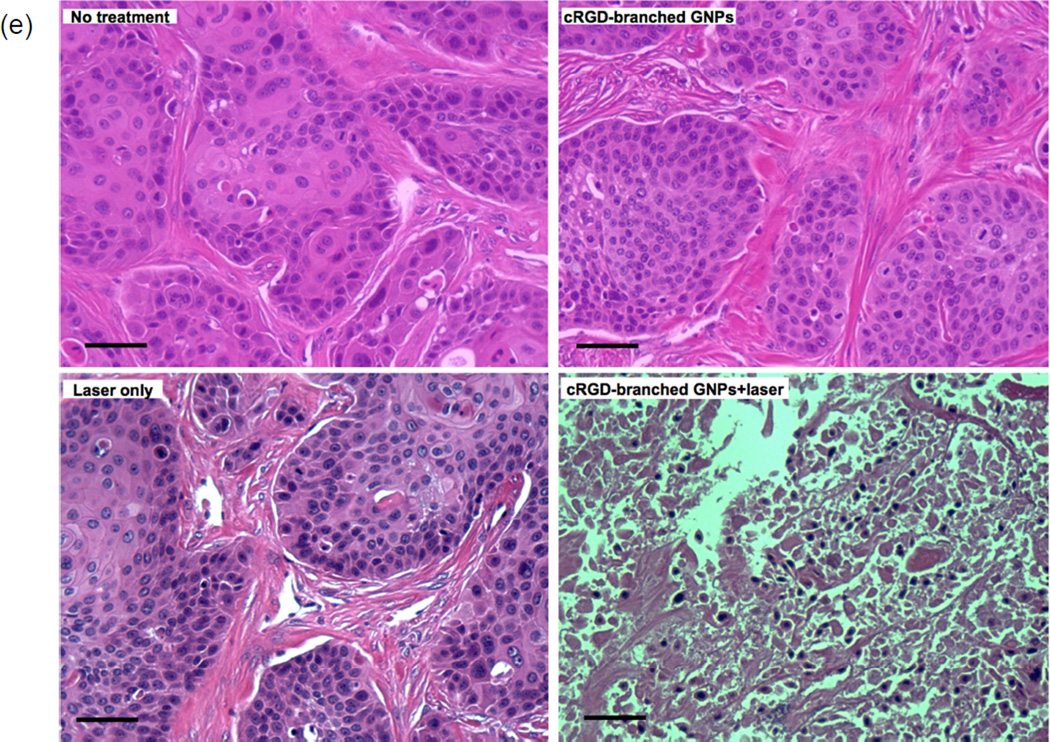
We report an approach for simple, reproducible and high-yield synthesis of branched GNPs directed by deoxycholate bile acid supramolecular aggregates in Au solution. A growth process involving stepwise trapping of the GNP seeds and Au ions in the deoxycholate bile acid solution yields multiple-branched GNPs. Upon NIR laser irradiation strong NIR absorption for branched GNPs induced photothermal-heating to destroy tumor cells. Subsequently, these branched GNPs were biofunctionalized with cRGD cell penetrating-targeting peptides for photothermal cancer treatment applications. Branched GNPs conjugated with cRGD peptides enhanced internalization of the branched GNPs in BxPC3 human pancreatic adenocarcinoma cells and effectively ablated BxPC3 cells when irradiated with a NIR laser (808 nm). Their potential use as photothermal transducing agents was demonstrated in in vivo settings using a pancreatic cancer xenograft model. The tumors were effectively ablated with cRGD-branched GNPs injection and laser exposure without any observation of tumor recurrence. This firstly reported method for deoxycholate bile acid directed synthesis of branched GNPs opens new possibilities for the production of strong NIR absorbing nanostructures for selective nano-photothermolysis of cancer cells and the further design of novel materials with customized spectral and structural properties for broader applications.

Keywords: Bile acid; Gold nanoparticles; Green chemistry; Nanomedicine; Photothermal treatment.


Figures

Figure 1
Figure 1
(a) Schematic of the formation of branched GNPs directed by sodium deoxycholate (MW. 414.55, C24H39NaO4) aggregations (increased aggregation number by increasing the concentration of deoxycholate molecules) and (b and c) TEM images of the synthesized branched GNPs (1.4 mM (left)) and GNPs synthesized with different concentrations of sodium deoxycholate solution (4.8 (middle) and 36.1 mM (right)), (d) a photograph and (e) absorption spectra of the synthesized GNP solutions in various concentrations of sodium deoxycholate solution (1.4~36.1 mM).
Figure 1
Figure 1
(a) Schematic of the formation of branched GNPs directed by sodium deoxycholate (MW. 414.55, C24H39NaO4) aggregations (increased aggregation number by increasing the concentration of deoxycholate molecules) and (b and c) TEM images of the synthesized branched GNPs (1.4 mM (left)) and GNPs synthesized with different concentrations of sodium deoxycholate solution (4.8 (middle) and 36.1 mM (right)), (d) a photograph and (e) absorption spectra of the synthesized GNP solutions in various concentrations of sodium deoxycholate solution (1.4~36.1 mM).
Figure 2
Figure 2
(a) A photograph image of the branched GNPs solution in 10 ml and 100 ml batches and (inset) their hydrodynamic size, (b) TEM image of the synthesized branched GNPs with a 1.4 mM sodium deoxycholate solution and (inset) their electron diffraction, (c) XRD pattern of the synthesized branched GNPs, (d) schematics for biofunctionalized branched GNPs with carboxyl-PEG-thiol and cRGD peptides to target integrin ανβ3, and (e) IR thermal image of agar phantom including the branched GNPs (0.6 mM) and heating profiles of the branched GNPs at various concentrations (0~0.6 mM) under irradiation with 808 nm laser (0.47 W/cm2).
Figure 3
Figure 3
(a) Cellular targeting and uptake of cRGD-branched GNPs incubated with BxPC-3 cells for 24 h. Fluorescent images (red: cRGD-branched GNPs and blue: nucleus) and a TEM image of the cRGD-branched GNPs uptaken by BxPC3 cells, (b) Viability of BxPC3 cells after incubation with increasing concentrations of branched GNPs and cRGDbranched GNPs for 24 h without laser irradiation, and (c) photothermal destruction of BxPC-3 cells exposed to increasing concentrations of cRGD-branched GNPs (0~0.8 mM) and power densities (0.47 and 0.96 W/cm2) of NIR laser (808 nm) for 3 mins.
Figure 4
Figure 4
(a) A photograph of BxPC3 pancreatic tumor model in nude mouse and fluorescent images of the excised tumors at 24 h after respective intra-tumoral injections of cyto780 labeled cRGD-branched GNPs and non-conjugated branched GNPs (without cRGD). Scale bars= 5 mm (Cyto780 labeled cRGD-branched GNPs (green) and dotted line: border of tumors) and (b) quantified amounts of targeted branched GNPs in the tumor tissues at 24 h post-injection (P < 0.05).
Figure 5
Figure 5
(a) A photograph and IR thermal images of a BxPC3 pancreatic tumor bearing mouse after cRGD-branched GNPs injection during NIR irradiation (808 nm; 1.4 W/cm2) for 5 mins, (b) Temperature changes measured in control tumors (without laser irradiation) and during NIR laser irradiation with and without injection of cRGD-branched GNPs, (c) tumor growth curves as a function of time after the different treatment protocols (control, laser irradiation only, cRGDbranched GNPs injection-only and cRGD-branched GNPs injection+laser irradiation) in BxPC3 pancreatic xenograft tumors; (d) average tumor weight and (inset) a digital picture of tumors excised at day 26 after treatment in each group, (e) representative photographs of hematoxylin and eosin-stained slides of tumor tissues from each group (scale bars=50 um).
Figure 5
Figure 5
(a) A photograph and IR thermal images of a BxPC3 pancreatic tumor bearing mouse after cRGD-branched GNPs injection during NIR irradiation (808 nm; 1.4 W/cm2) for 5 mins, (b) Temperature changes measured in control tumors (without laser irradiation) and during NIR laser irradiation with and without injection of cRGD-branched GNPs, (c) tumor growth curves as a function of time after the different treatment protocols (control, laser irradiation only, cRGDbranched GNPs injection-only and cRGD-branched GNPs injection+laser irradiation) in BxPC3 pancreatic xenograft tumors; (d) average tumor weight and (inset) a digital picture of tumors excised at day 26 after treatment in each group, (e) representative photographs of hematoxylin and eosin-stained slides of tumor tissues from each group (scale bars=50 um).

References

    1. Lv WP, Wang Y, Feng WQ, Qi JJ, Zhang GL, Zhang FB, et al. Robust and smart gold nanoparticles: one-step synthesis, tunable optical property, and switchable catalytic activity. J Mater Chem. 2011;21:6173–6178.
    1. Turner M, Golovko VB, Vaughan OPH, Abdulkin P, Berenguer-Murcia A, Tikhov MS, et al. Selective oxidation with dioxygen by gold nanoparticle catalysts derived from 55-atom clusters. Nature. 2008;454:981-U31. - PubMed
    1. Thacker VV, Herrmann LO, Sigle DO, Zhang T, Liedl T, Baumberg JJ, et al. DNA origami based assembly of gold nanoparticle dimers for surface-enhanced Raman scattering. Nat Commun. 2014;5 - PubMed
    1. Hutter E, Maysinger D. Gold-nanoparticle-based biosensors for detection of enzyme activity. Trends Pharmacol Sci. 2013;34:497–507. - PubMed
    1. Peng G, Tisch U, Adams O, Hakim M, Shehada N, Broza YY, et al. Diagnosing lung cancer in exhaled breath using gold nanoparticles. Nat Nanotechnol. 2009;4:669–673. - PubMed

Publication types

MeSH terms

LinkOut - more resources