. 2015 Jun 26;290(26):16215-25.
doi: 10.1074/jbc.M115.653303. Epub 2015 May 1.

Combined Crystal Structure of a Type I Cohesin: MUTATION AND AFFINITY BINDING STUDIES REVEAL STRUCTURAL DETERMINANTS OF COHESIN-DOCKERIN SPECIFICITIES

Affiliations

Combined Crystal Structure of a Type I Cohesin: MUTATION AND AFFINITY BINDING STUDIES REVEAL STRUCTURAL DETERMINANTS OF COHESIN-DOCKERIN SPECIFICITIES

Kate Cameron et al. J Biol Chem. .

Abstract

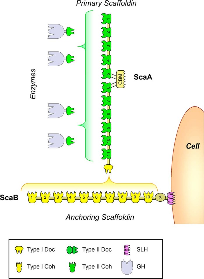
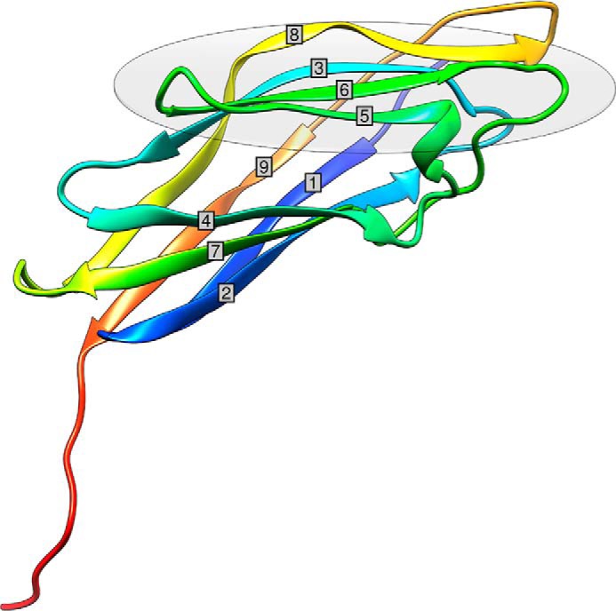
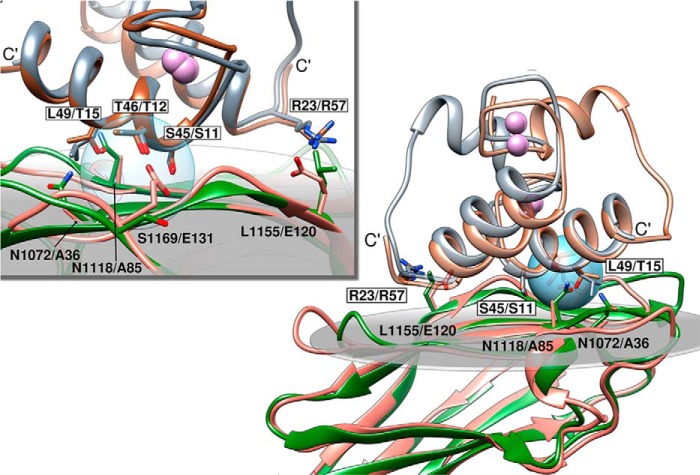
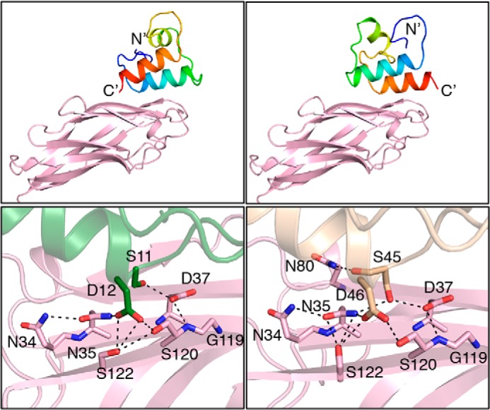
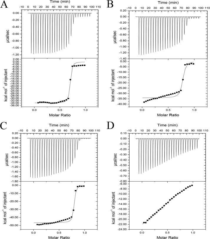
Cohesin-dockerin interactions orchestrate the assembly of one of nature's most elaborate multienzyme complexes, the cellulosome. Cellulosomes are produced exclusively by anaerobic microbes and mediate highly efficient hydrolysis of plant structural polysaccharides, such as cellulose and hemicellulose. In the canonical model of cellulosome assembly, type I dockerin modules of the enzymes bind to reiterated type I cohesin modules of a primary scaffoldin. Each type I dockerin contains two highly conserved cohesin-binding sites, which confer quaternary flexibility to the multienzyme complex. The scaffoldin also bears a type II dockerin that anchors the entire complex to the cell surface by binding type II cohesins of anchoring scaffoldins. In Bacteroides cellulosolvens, however, the organization of the cohesin-dockerin types is reversed, whereby type II cohesin-dockerin pairs integrate the enzymes into the primary scaffoldin, and type I modules mediate cellulosome attachment to an anchoring scaffoldin. Here, we report the crystal structure of a type I cohesin from B. cellulosolvens anchoring scaffoldin ScaB to 1.84-Å resolution. The structure resembles other type I cohesins, and the putative dockerin-binding site, centered at β-strands 3, 5, and 6, is likely to be conserved in other B. cellulosolvens type I cohesins. Combined computational modeling, mutagenesis, and affinity-based binding studies revealed similar hydrogen-bonding networks between putative Ser/Asp recognition residues in the dockerin at positions 11/12 and 45/46, suggesting that a dual-binding mode is not exclusive to the integration of enzymes into primary cellulosomes but can also characterize polycellulosome assembly and cell-surface attachment. This general approach may provide valuable structural information of the cohesin-dockerin interface, in lieu of a definitive crystal structure.

Keywords: cellulase; cellulose; cellulosome; computational biology; computer modeling; protein-protein interaction.


Figures

FIGURE 1.
FIGURE 1.
Schematic representation of the B. cellulosolvens cellulosome. The primary scaffoldin, ScaA, binds up to 11 cellulosomal enzymes through type II cohesin dockerin interactions (in green). ScaA contains a C-terminal type I dockerin (in yellow) that binds to one of the 10 type I cohesins of anchoring scaffoldin ScaB. ScaB binds to the cell surface through a C-terminal S-layer homology (SLH) domain (in violet).
FIGURE 2.
FIGURE 2.
Three-dimensional structure of BcCohI color-ramped from N (blue) to C (red) terminus. BcCohI has a β-sandwich fold, and the nine cohesin β-strands are labeled. The gray oval represents the putative dockerin-interacting cohesin face.
FIGURE 3.
FIGURE 3.
Interaction of the type I Bc-DocA with a cohesin microarray. CBM-Coh fusion proteins, as listed in the key, were applied in triplicate in successive 2-fold dilution, starting from a maximum concentration of 8 μm (∼5 nl). The XynDoc fusion protein (10 ml per slide) was then applied at a final concentration of 2 nm, and the amount of dockerin bound to the cohesin samples was visualized by immunofluorescence. The triplet of spots at the base of each sample area denotes a marker, composed of a Xyn-CBM conjugate, which indicates the location of the samples on the cellulose slide. GenBankTM accession numbers for sequences described in this figure are as follows: A. cellulolyticus ScaA (AF155197), ScaB (AY221112), ScaC (AY221113), and ScaD (AY221114); A. fulgidus Orfs 2375 and 2376 (AE000782); B. cellulosolvens ScaA and ScaB (AF224509) and Cel48A (AAR23324); C. acetobutylicum CipA (AE001437); C. cellulolyticum CipC (U40345) and OrfX (Cc-OX, AF081458); C. thermocellum primary scaffoldin, CipA (Q06851), and anchoring scaffoldins, OlpA (Ct-OA, Q06848), SdbA (U49980), OlpB (Q06852), and Orf2p (Q06853); R. flavefaciens ScaA, ScaB, ScaC, and ScaE (AJ278969).
FIGURE 4.
FIGURE 4.
Interaction of dockerins from various sources with a type I cohesin from B. cellulosolvens ScaB (Bc-B3). Results are shown for four enzyme-borne dockerins (from C. thermocellum, C. cellulolyticum, B. cellulosolvens, and R. flavefaciens), the lone dockerin from A. fulgidus, and five scaffoldin-borne dockerins from C. thermocellum, A. cellulolyticus, and R. flavefaciens. None of these dockerins interacted with Bc-B3, but all of them interacted selectively with the target cohesins from their own species. Experiments were carried out as described in the legend to Fig. 3, with the designated XynDoc fusion proteins. See key and legend in Fig. 3 for the nomenclature of the different cohesins identified in this figure. GenBankTM accession numbers for dockerin sequences described in this figure are as follows: Ct-Doc48A (WP_003516749), Cc-Doc5A (WP_015924614), Bc-Doc48A (AAR23324), Rf-Doc44B (WP_026053020), Af-Doc (WP_010879862), Ct-CipA-Doc (Q06851), Ac-ScaA-XDoc (AF155197), Ac-ScaB-Doc (AY221112), Rf-ScaA-Doc, and Rf-ScaB-XDoc (AJ278969).
FIGURE 5.
FIGURE 5.
Superposition of BcCohI with the type I cohesin-dockerin complexes from C. thermocellum. Structural overlay of C. thermocellum type I cohesin-dockerin complexes in two binding orientations (PDB codes 1ohz and 2ccl) superposed with BcCohI. Residues involved in C. thermocellum cohesin-dockerin binding are highlighted in stick representation. Dockerin residue numbers are boxed and indicate equivalent residue positions on CtCoh-1ohz (gray colored structure) and CtCoh-2ccl (brown color), respectively. The cohesin residue numbers refer to structurally equivalent positions on BcCohI (green) and CtCoh-1ohz-A (salmon), respectively. Calcium ions are represented as spheres colored in purple. The top inset is a magnified view from the opposite side, around the blue spherical surface that highlights the canonical C. thermocellum Ser/Thr interface residue pair. Figures were prepared using UCSF Chimera (38).
FIGURE 6.
FIGURE 6.
Computational model of the type I B. cellulosolvens cohesin-dockerin complex. Left and right panels represent two different model structures related to one another by a pseudo 2-fold symmetry axis about the interaction between the monomers. Hydrogen bonds mediating the interactions are represented by dashed lines. Panels were prepared using PyMOL (39). Top panel shows the dockerin in rainbow hues (blue to red) from the N to C terminus, respectively. The cohesin structure is colored lavender. In the bottom panels, the interface is magnified, and the major interacting residues are shown.
FIGURE 7.
FIGURE 7.
Binding of ScaA dockerin and its mutant derivatives to BcCohI evaluated by isothermal titration calorimetry. ScaA dockerin is highly symmetric and potentially contains two identical cohesin-binding surfaces. The key residues that participate in cohesin recognition at each cohesin-binding surface were changed to alanine. ScaA dockerins DocS23A/D24A and DocS60A/D61A only have one functional binding face, whereas in the quadruple mutant, DocS23A/D24A/S60A/D61A, the two cohesin-binding faces were inactivated. The upper part of each panel shows the raw heats of binding, and the lower parts comprise the integrated heats after correction for heat dilution. The curve represents the best fit to a single-site binding model. BcCohI-DocScaA; B, BcCohI-DocS23A/D24A; C, BcCohI-Doc/S60A/D61A; and D, BcCohI-DocS23A/D24A/S60A/D61A.

References

    1. Gilbert H. J. (2010) The biochemistry and structural biology of plant cell wall deconstruction. Plant Physiol. 153, 444–455 - PMC - PubMed
    1. Bayer E. A., Belaich J.-P., Shoham Y., Lamed R. (2004) The cellulosomes: multienzyme machines for degradation of plant cell wall polysaccharides. Annu. Rev. Microbiol. 58, 521–554 - PubMed
    1. Fontes C. M., Gilbert H. J. (2010) Cellulosomes: highly efficient nanomachines designed to deconstruct plant cell wall complex carbohydrates. Annu. Rev. Biochem. 79, 655–681 - PubMed
    1. Carvalho A. L., Dias F. M., Prates J. A., Nagy T., Gilbert H. J., Davies G. J., Ferreira L. M., Romão M. J., Fontes C. M. (2003) Cellulosome assembly revealed by the crystal structure of the cohesin-dockerin complex. Proc. Natl. Acad. Sci. U.S.A. 100, 13809–13814 - PMC - PubMed
    1. Artzi L., Dassa B., Borovok I., Shamshoum M., Lamed R., Bayer E. A. (2014) Cellulosomics of the cellulolytic thermophile Clostridium clariflavum. Biotechnol. Biofuels 7, 100 - PMC - PubMed

Publication types

MeSH terms

Substances