Review
. 2015 Oct;67(2 Pt A):142-52.
doi: 10.1016/j.molimm.2015.04.001. Epub 2015 Apr 28.

In vivo imaging with antibodies and engineered fragments

Affiliations
Review

In vivo imaging with antibodies and engineered fragments

Amanda C Freise et al. Mol Immunol. 2015 Oct.

Abstract

Antibodies have clearly demonstrated their utility as therapeutics, providing highly selective and effective drugs to treat diseases in oncology, hematology, cardiology, immunology and autoimmunity, and infectious diseases. More recently, a pressing need for equally specific and targeted imaging agents for assessing disease in vivo, in preclinical models and patients, has emerged. This review summarizes strategies for developing and optimizing antibodies as targeted probes for use in non-invasive imaging using radioactive, optical, magnetic resonance, and ultrasound approaches. Recent advances in engineered antibody fragments and scaffolds, conjugation and labeling methods, and multimodality probes are highlighted. Importantly, antibody-based imaging probes are seeing new applications in detection and quantitation of cell surface biomarkers, imaging specific responses to targeted therapies, and monitoring immune responses in oncology and other diseases. Antibody-based imaging will provide essential tools to facilitate the transition to truly precision medicine.

Keywords: Antibody fragments; Image-guided surgery; ImmunoPET; Molecular imaging.


Figures

Figure 1
Figure 1. Development of antibodies for in vivo imaging: a multifaceted endeavor
Numerous characteristics must be optimized in order to employ antibodies for targeting and imaging in vivo. A typical imaging probe will consist of a targeting moiety, linker, and signaling agent.
Figure 2
Figure 2. Desired characteristics of imaging vs. therapeutic antibodies
Properties such as effector function, half-life, and conjugation sites are modified to suit either therapy or imaging applications.
Figure 3
Figure 3. Antibody fragments
Summary of salient properties of intact antibodies, enzymatic fragments, recombinant fragments and smaller scaffolds.
Figure 4
Figure 4. Fluorescence-guided resection of tumors
By probing with a fluorescently-labeled antibody for a tumor marker, surgeons can immediately determine whether a tumor has been fully resected, and identify local metastases that may not be visible by eye. Here, a far-red fluorescent dye (Cy5)-labeled anti-PSCA diabody was used to guide resection of an intramuscular 22rv1-PSCA prostate cancer xenograft in a murine model. Cy5-anti-PSCA diabody was injected intravenously and surgery was performed 6 hours post-injection. (A) A white-light image of the tumor bed post-resection, with residual tumor purposely left unresected. (B) Fluorescence imaging shows residual tumor clearly. (Behesnilian et al., 2015)
Figure 5
Figure 5. Dual-modality antibody imaging
Non-invasive staging of lymph nodes (LN) followed by image-guided resection of only cancer-positive LNs offers more precision in identifying and removing metastases. Hall et al. used a dual-labeled anti-EpCAM mAb, conjugated with both IRDye 800CW and 64Cu-DOTA, to image prostate cancer LN metastases with PET/CT (A) and NIR fluorescence (B) imaging. PC3 cells were implanted in the prostate and imaging was performed 10–12 weeks later. Lumbar LNs (LLNs); renal LNs (RLNs); sciatic LN (SLN). This research was originally published in JNM. Hall MA et al. J Nucl Med. 2012;53:1427–37. © by the Society of Nuclear Medicine and Molecular Imaging, Inc.
Figure 6
Figure 6. Applications of antibody imaging in oncology
Imaging of specific biomarkers can be used to accurately locate lesions, aid in choosing a treatment, and monitor response to therapy. (A) PET imaging using the 124I-anti-PSCA minibody A11 was able to detect intratibial LAPC-9 prostate cancer xenografts with higher sensitivity and specificity than a 18F-Fluoride bone scan. (B) Response to anti-androgen treatment was monitored by visualizing reduced 124I-A11 activity in the tumors of enzalutamide-treated mice, reflecting a therapy-induced downregulation of PSCA expression. (C) The Hcc827-GR6 non-small cell lung cancer line developed resistance to gefitinib through upregulation of MET expression. PET imaging MET using a 89Zr-DFO-labeled MET-specific human minibody clearly visualized the overexpression of MET in the gefitinib-resistant tumors. (Knowles et al., 2014a; Li et al., 2014)
Figure 7
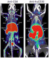
Figure 7. Imaging immune cell subsets
Tracking CD8+ T cells could help detect and stage CD8+ lymphomas and aid in monitoring T cell immunotherapy. Here, imaging of CD8+ T cells with a 64Cu-NOTA-anti-CD8 minibody in wild type mice visualizes the spleen and lymph nodes (left panel). 89Zr-DFO-anti-huCD20 cys-minibody imaging of a transgenic mouse expressing huCD20 reveals B cells in the spleen and lymph nodes (right panel); this could also be applied to for the detection of B-cell lymphomas. (Tavaré et al., 2014a; Zettlitz et al., 2013)

Similar articles

Cited by

References

    1. Agarwal P, Bertozzi CR. Site-specific antibody-drug conjugates: the nexus of bioorthogonal chemistry, protein engineering, and drug development. Bioconjug Chem. 2015 doi: 10.1021/bc5004982. - DOI - PMC - PubMed
    1. Ahrens ET, Bulte JWM. Tracking immune cells in vivo using magnetic resonance imaging. Nat Rev Immunol. 2013;13:755–63. doi: 10.1038/nri3531. - DOI - PMC - PubMed
    1. Al Faraj A, Shaik AS, Afzal S, Al Sayed B, Halwani R. MR imaging and targeting of a specific alveolar macrophage subpopulation in LPS-induced COPD animal model using antibody-conjugated magnetic nanoparticles. Int J Nanomedicine. 2014;9:1491–503. doi: 10.2147/IJN.S59394. - DOI - PMC - PubMed
    1. Atreya R, Neumann H, Neufert C, Waldner MJ, Billmeier U, Zopf Y, Willma M, App C, Münster T, Kessler H, Maas S, Gebhardt B, Heimke-Brinck R, Reuter E, Dörje F, Rau TT, Uter W, Wang TD, Kiesslich R, Vieth M, Hannappel E, Neurath MF. In vivo imaging using fluorescent antibodies to tumor necrosis factor predicts therapeutic response in Crohn’s disease. Nat Med. 2014;20:313–8. doi: 10.1038/nm.3462. - DOI - PMC - PubMed
    1. Axup JY, Bajjuri KM, Ritland M, Hutchins BM, Kim CH, Kazane SA, Halder R, Forsyth JS, Santidrian AF, Stafin K, Lu Y, Tran H, Seller AJ, Biroc SL, Szydlik A, Pinkstaff JK, Tian F, Sinha SC, Felding-Habermann B, Smider VV, Schultz PG. Synthesis of site-specific antibody-drug conjugates using unnatural amino acids. Proc Natl Acad Sci USA. 2012;109:16101–6. doi: 10.1073/pnas.1211023109. - DOI - PMC - PubMed

Publication types