. 2015 May 2:15:71.
doi: 10.1186/s12883-015-0327-z.

Non-neuronal and neuronal BACE1 elevation in association with angiopathic and leptomeningeal β-amyloid deposition in the human brain

Affiliations

Non-neuronal and neuronal BACE1 elevation in association with angiopathic and leptomeningeal β-amyloid deposition in the human brain

Zhi-Qin Xue et al. BMC Neurol. .

Abstract

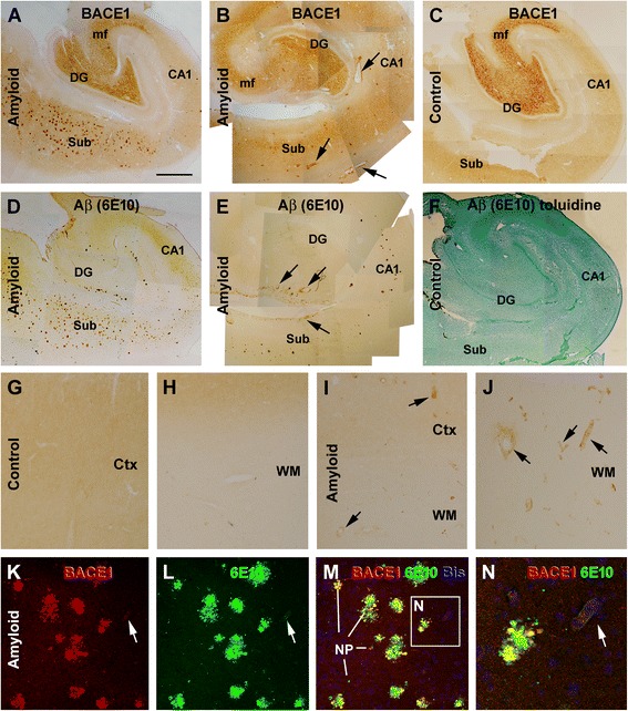
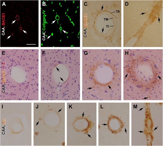
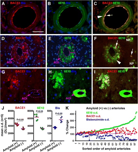
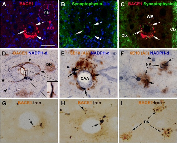
Background: Cerebral amyloid angiopathy (CAA) refers to the deposition of β-amyloid (Aβ) peptides in the wall of brain vasculature, commonly involving capillaries and arterioles. Also being considered a part of CAA is the Aβ deposition in leptomeninge. The cellular origin of angiopathic Aβ and the pathogenic course of CAA remain incompletely understood.

Methods: The present study was aimed to explore the pathogenic course of CAA in the human cerebrum via examination of changes in β-secretase-1 (BACE1), the obligatory Aβ producing enzyme, relative to Aβ and other cellular markers, by neuroanatomical and biochemical characterizations with postmortem brain samples and primary cell cultures.

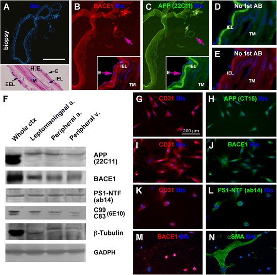
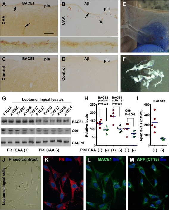
Results: Immunoreactivity (IR) for BACE1 was essentially not visible at vasculature in cases without cerebral amyloidosis (control group, n = 15, age = 86.1 ± 10.3 year). In cases with brain amyloid pathology (n = 15, age = 78.7 ± 12.7 year), increased BACE1 IR was identified locally at capillaries, arterioles and along the pia, localizing to endothelia, perivascular dystrophic neurites and meningeal cells, and often coexisting with vascular iron deposition. Double immunofluorescence with densitometric analysis confirmed a site-specific BACE1 elevation at cerebral arterioles in the development of vascular Aβ deposition. Levels of BACE1 protein, activity and its immediate product (C99) were elevated in leptomeningeal lysates from cases with CAA relative to controls. The expression of BACE1 and other amyloidogenic proteins in the endothelial and meningeal cells was confirmed in primary cultures prepared from human leptomeningeal and arteriolar biopsies.

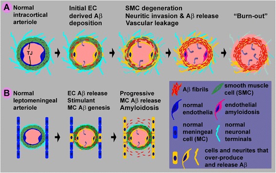
Conclusion: These results suggest that BACE1 elevation in the endothelia and perivascular neurites may be involved in angiopathic Aβ deposition, while BACE1 elevation in meningeal cells might contribute Aβ to leptomeningeal amyloidosis.


Figures

Figure 1
Figure 1
Representative images showing increased β-secretase (BACE1) immunoreactivity (IR) at selected vascular sites, in addition to neuritic plaques, in the human brains with amyloid deposition relative to controls. Panels (A-F) are montages of low magnification images showing BACE1 (A-C) and 6E10 [reactive to Aβ, potentially to β-amyloid precursor protein (APP) and APP β-C-terminal as well] (D-F) IR over the subiculum (Sub), hippocampus (CA sectors) and dentate gyrus (DG) between adjacent sections from two cases with brain amyloid pathology (A,B,D,E) and one control (C,F). In the second case (B,E), vascular BACE1 and 6E10 labeling (arrows) is evident at low magnification. In the control case, no staining is seen with 6E10 labeling (F, with tissue lamination illustrated by toluidine counterstain). Note that the BACE1 IR at the mossy fiber terminal field is comparable in (A-C). Panels (G-J) show low magnification views of BACE1 IR in the temporal neocortical grey and white matter from two additional brains, with a diffuse neuropil pattern in the control (G,H) and increased labeling in the amyloid case at selected vascular profiles (I.J). Confocal double immunofluorescence shows a partial colocalization of BACE1/6E10 IR among typical neuritic plaques (K-N), with the structures exhibiting overlapped labeling (appearing yellow) representing dystrophic neurites (M,N). A capillary profile exhibiting weak BACE1/6E10 IR is also seen in the field (M, N). Scale bar in (A) = 2.5 mm, applying to (B-F), equivalent to 500 μm for (G-J), 200 μm for (K-M) and 50 μm for (N).
Figure 2
Figure 2
Morphological characterization of vascular (arteriolar) BACE1 and 6E10 immunoreactivity (IR) in postmortem human temporal neocortex. Panels (A,B) show selective BACE1 labeling at cortical vasculature visualized by collagen IV. Panels (C) illustrates BACE1 IR at a cross-sectioned arteriole, localizing to the tunica interna (TI) and around the tunica adventitia (TA) or the perivascular area, but not in the tunica media (TM). Panel (D) shows a feather-like pattern of BACE1 IR along a longitudinally cut intracortical arteriole. Panels (E-H) illustrate variable patterns of BACE1 IR among labeled (F-H) relative to unlabeled (E) arterioles in immunohistochemical preparation with hematoxylin and eosin stain (H.E.) counterstain. Arrows points to BACE1 IR in the TI (C,F), TA (C,D) and TM (G,H). Loss of cells in the vascular wall is noticeable in (G) and (H) relative to (E) and (F). The BACE1 IR in the TA and TM appears process-like (C,D,G,H). Panels (I-L) illustrate variable patterns of Aβ labeling at arterioles, with Aβ IR occurs primarily at the TI (I) or in the TM (J-L). Perivascular Aβ IR (arrows) often co-exists among the profiles with Aβ deposition in the TM with varying intensity (J-L), and appears segmentally in longitudinal view of the labeled vessel (M). Scale bar = 500 μm in (A) applying for (B); equal to 200 μm for (M) and 100 μm for other panels.
Figure 3
Figure 3
Correlative morphometric characterization of BACE1 and 6E10 immunoreactivity (IR) in intracortical arterioles in confocal fluorescent preparation. Panels (A-C) show colocalized BACE1/6E10 IR in the endothelia (arrows), with a ring-like Aβ deposition (arrowhead) in the smooth muscle layer that is not colocalized with BACE1 IR. Panels (D-F) demonstrate an arteriole containing BACE1 labeled neurites and local Aβ deposition (arrowheads) in the vascular wall, with BACE1 IR colocalizing with 6E10 IR inside but not outside swollen process-like elements (arrows). Panels (H-J) show a vessel with densely packed Aβ products in the wall, with BACE1 IR over the same area comparable to background. Fusiform bisbenzimide (Bis)-labeled nuclei appear lost in the middle layer of arterioles in (D/E, G/H) relative to (A/B). A typical neuritic plaque (NP) consisted of BACE1 labeled dystrophic neurites and Aβ deposits is also seen in (G-I). Panels (J) plots the mean optic densities (o.d.) of BACE1, 6E10 and bisbenzimide fluorescence measured over the wall of individual normal [i.e., amyloid negative (-), 10/brain)] and amyloid [i.e., amyloid positive (+), 20/brain] arterioles from five individual brains, with the area of interest (AOI) illustrated as the purple-line circled areas in (D/E and G/H, and also in Figure 4A to demonstrate the method to measure densities at normal control vessels). Note the increase of BACE1/6E10 o.d. and reduction of bisbenzimide o.d. in the amyloid (+) relative to (-) arterioles. Panel (K) shows a correlative densitometric analysis by sorting the order of individual vessels (n = 100 from 5 brains) according to their elevated levels of 6E10 densities relative to baseline (defined as 100%, mean of densities reported from all non-amyloid vessels). BACE1 density increases initially in parallel with that of 6E10 IR, but tends to reduce among most profiles as 6E10 IR further increases. Bisbenzimide density tends to decline with the increase of 6E10 labeling. Scale bar = 50 μm in (A) applying to (B, C,G-I), equivalent to 100 μm for (D-F).
Figure 4
Figure 4
Neuritic profiles inside and surrounding the wall of intracerebral arterioles from cases with brain amyloid pathology. Panels (A-C) show a partial colocalization of BACE1 and synaptophysin immunofluorescence in the vascular wall of an arteriole in the subcortical white matter (WM) (arrows pointing to colocalized parts), with a nearby arteriole (circled with broken purple line) exhibited background signal for both markers. Panels (D) shows BACE1 labeled dystrophic neurites (arrows) partially colabeled (enlarged insert) for nicotinamide adenine dinucleotide phosphate diaphorase (NADPH-d) surrounding an intracortical vessel, with clusters of dystrophic neurites (DN) in the field. Panel (E) shows NADPH-d positive dystrophic neurites (arrows) in and around the wall of a vessel with Aβ deposits. Panel (F) shows rosette-like NADPH-d positive dystrophic neurites in compact amyloid plaques (NP). Panels (G,H) show BACE1 labeled neurites in small (G) and larger (H) amounts in the vascular wall that are partially colabeled by Prussian blue, as are those organized as rosset-like clusters (I). The colabeled parts appear grey to black because of the brown DAB background color. The AOI label in (A) illustrates a method to measure optic density in normal arterioles (referring to Figure 3K-L). Scale bar = 100 μm in (A) applying to (B-I).
Figure 5
Figure 5
Characterization of the expression of amyloidogenic proteins in vascular biopsy and primary vascular cell culture. Panels (A-E) show the expression of the β-amyloid precursor protein (APP) and BACE1 in leptomeningeal biopsy containing a middle-sized and several small arteries (arrow), and capillaries. The endothelial cells (E→) expressed specific APP and BACE1 immunoreactivity (B,C), which can be eliminated by excluding the primary antibodies (1st AB) in immunofluorescent processing (D,E). Autofluorescence exists in the inner elastic lamina (IEL), tunica media (TM) and external elastic lamina (EEL) (refer to the H.E. stained inset in A). Panel (F) shows immunoblot characterization of the amyloidogenic proteins in extracts of isolated leptomeningeal arteries, peripheral arteries and veins, with cortical extract (from a control case) used as assay control (50 μg protein loading in each lane). APP, BACE1, presenilin-1 N-terminal fragments (PS1-NTF) and APP β-cleavage products are present in the vascular homogenates. Note that the vascular APP migrates at a higher molecular weight position relative to the brain counterpart. The vascular samples contain minimal amount of β-tubulin relative to cortical lysate. Panels (G-L) illustrate immunocytochemical labeling of APP, BACE1 and PS1-NTF in cultured vascular cells expressing CD31, a marker for vascular endothelia. A large cellular profile (appeared as fused cells) is labeled with the smooth muscle cell marker, α-smooth muscle actin (αSMA), but exhibits little BACE1 immunoreactivity (M,N). Scale bar = 500 μm in (A) applying to (B,C); equal to 250 μm for (H-I) and 50 μm for (E-G).
Figure 6
Figure 6
Characterization of BACE1 elevation in meningeal cells associated with leptomeningeal amyloidosis. Panel (A) shows BACE1 labeled cells aligning along the pia mater in a case with cerebral amyloid angiopathy with prominent leptomeningeal amyloidosis (B), in contrast to a control case wherein no BACE1 and Aβ (6E10) labeling is present at the pia (C,D). Arrows in (A,B) points to profiles with increased BACE1 and 6E10 IR over the background. Panel (E) shows a preparation of leptomeninge sample from the temporal lobe (E), which are rinsed thoroughly in cold phosphate buffer for biochemical analysis (F). Levels of BACE1 protein and C99 in leptomeningeal lysates are elevated in 5 cases with CAA relative to 5 control cases (G,H), as is BACE1 enzymatic activity measured in the lysates (H). Aβ42 levels are also higher in the CAA group relative to control (H). In primary cell culture of leptomeningeal biopsies, meningeal cells appear polygonal under phase contrast microscope (J), co-express fibronectin (FN) (K) and BACE1 (L). These cells also express the β-amyloid precursor protein (APP) (H, the corresponding FN labeling image is not shown). Scale bar = 250 μm in (A) applying to (B-D), equivalent to 50 μm for inserts in (A,B), 25 μm for (J) and 12.5 μm for (K-M).
Figure 7
Figure 7
Schematic illustration of a hypothetic model for BACE1 elevation in vascular and brain-specific cellular elements in angiopathic (panel A) and leptomeningeal (panel B) amyloidosis. Arteriolar profile is used here to construct the model to show the major relevant cellular components and pathological events. BACE1 elevation first occurs in endothelial cells (EC), resulting in Aβ rise and aggregation in the smooth muscle cell (SMC) layer. This is followed by the damage of tight junctions (TJ) and blood brain barrier, causing leakage of blood contents into the SMC layer (curved arrows). The Aβ products and/or blood infiltration then induce SMC degeneration, and further a reactive response of the perivascular axonal terminals, manifested as aberrant sprouting and dystrophy. The invasion of the Aβ producing dystrophic neurites into the vascular wall leads to a vicious cycle of amyloidosis, cell degeneration and microbleeding. This process may end up with a “burnout” stage whereby the ECs, SMCs and dystrophic neurites all degenerate (panel A). For leptomeningeal amyloidosis, we propose that the initial events (endothelial Aβ overproduction, Aβ aggregation, BBB breakdown, and SMC degeneration) potentiate BACE1 expression in the nearby meningeal cells. A vicious cycle of pathogenesis in the blood vessel and meninge collectively contribute to the spread of amyloidosis along the leptomeninge (panel B).

Similar articles

Cited by

References

    1. Braak H, Del Tredici K. Alzheimer’s disease: pathogenesis and prevention. Alzheimers Dement. 2012;8:227–33. doi: 10.1016/j.jalz.2012.01.011. - DOI - PubMed
    1. Ando K, Laborde Q, Lazar A, Godefroy D, Youssef I, Amar M, et al. Inside Alzheimer brain with CLARITY: senile plaques, neurofibrillary tangles and axons in 3-D. Acta Neuropathol. 2014;128:457–9. doi: 10.1007/s00401-014-1322-y. - DOI - PMC - PubMed
    1. Castellani RJ, Smith MA, Perry G, Friedland RP. Cerebral amyloid angiopathy: major contributor or decorative response to Alzheimer’s disease pathogenesis. Neurobiol Aging. 2004;25:599–602. doi: 10.1016/j.neurobiolaging.2003.12.019. - DOI - PubMed
    1. Knopman DS, Parisi JE, Salviati A, Floriach-Robert M, Boeve BF, Ivnik RJ, et al. Neuropathology of cognitively normal elderly. J Neuropathol Exp Neurol. 2003;62:1087–95. - PubMed
    1. Keage HA, Carare R, Friedland RP, Ince PG, Love S, Nicoll JA, et al. Population studies of sporadic cerebral amyloid angiopathy and dementia: a systematic review. BMC Neurol. 2009;9:3. doi: 10.1186/1471-2377-9-3. - DOI - PMC - PubMed

Publication types

Substances