. 2015 Jul;100(7):870-80.
doi: 10.3324/haematol.2015.124586. Epub 2015 May 1.

Dietary ω-3 fatty acids protect against vasculopathy in a transgenic mouse model of sickle cell disease

Affiliations

Dietary ω-3 fatty acids protect against vasculopathy in a transgenic mouse model of sickle cell disease

Brian T Kalish et al. Haematologica. 2015 Jul.

Abstract

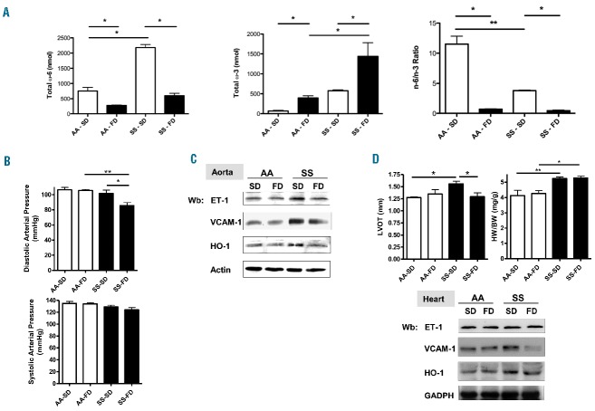
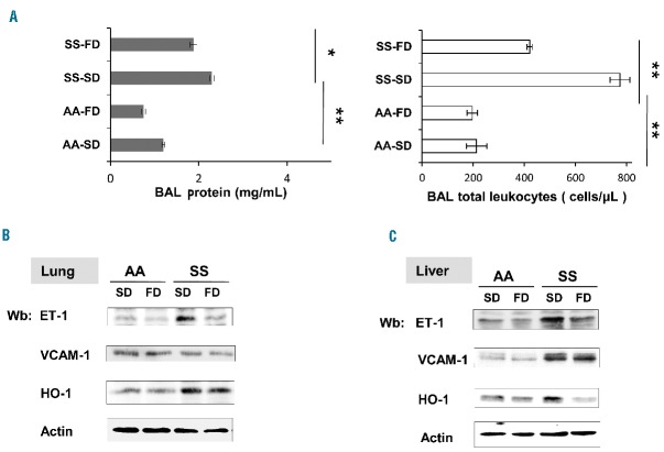
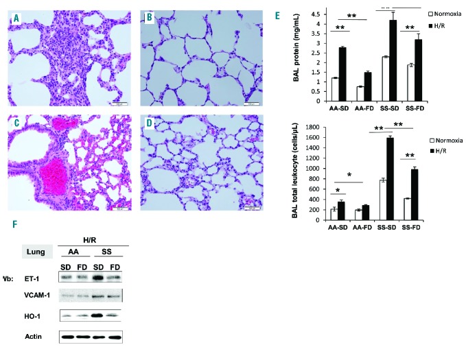
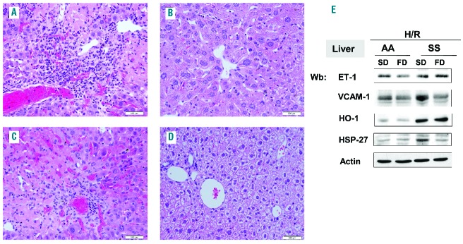
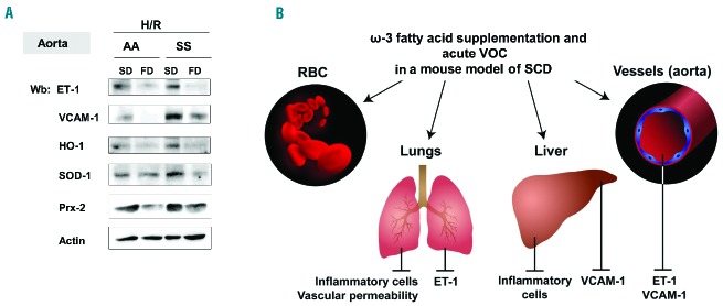
The anemia of sickle cell disease is associated with a severe inflammatory vasculopathy and endothelial dysfunction, which leads to painful and life-threatening clinical complications. Growing evidence supports the anti-inflammatory properties of ω-3 fatty acids in clinical models of endothelial dysfunction. Promising but limited studies show potential therapeutic effects of ω-3 fatty acid supplementation in sickle cell disease. Here, we treated humanized healthy and sickle cell mice for 6 weeks with ω-3 fatty acid diet (fish-oil diet). We found that a ω-3 fatty acid diet: (i) normalizes red cell membrane ω-6/ω-3 ratio; (ii) reduces neutrophil count; (iii) decreases endothelial activation by targeting endothelin-1 and (iv) improves left ventricular outflow tract dimensions. In a hypoxia-reoxygenation model of acute vaso-occlusive crisis, a ω-3 fatty acid diet reduced systemic and local inflammation and protected against sickle cell-related end-organ injury. Using isolated aortas from sickle cell mice exposed to hypoxia-reoxygenation, we demonstrated a direct impact of a ω-3 fatty acid diet on vascular activation, inflammation, and anti-oxidant systems. Our data provide the rationale for ω-3 dietary supplementation as a therapeutic intervention to reduce vascular dysfunction in sickle cell disease.


Figures

Figure 1.
Figure 1.
FD modified red cell membrane fatty acid composition and beneficially affects cardiovascular system in sickle cell mice. (A) Total ω-6 fatty acids (n-6) and ω-3 fatty acid/ω-6 fatty acid ratio (n3/n6 ratio) red cell membrane content as determined by mass spectroscopy in AA and SS mice under a soy-diet (SD) or fish-oil diet (FD). (B) Measurements of diastolic and systolic blood pressure (upper panel) in AA and SS mice under a SD or FD. (C) Immunoblot analysis with specific antibodies against endothelin-1 (ET-1), vascular adhesion molecule-1 (VCAM-1) and heme oxygenase-1 (HO-1) of isolated aortas from AA and SS mice under a SD or FD. One representative gel from six with similar results is presented. Densitometric analysis of anti- ET-1, VCAM-1 and HO-1 immunoblots is shown in Online Supplementary Figure S1A. (D) Upper panel. Left ventricular outflow tract (LVOT) diameter and left ventricular hypertrophy expressed as heart weight/body weight (HW/BW) in AA and SS mice under a SD or FD. Lower panel. Immunoblot analysis with specific antibodies against ET-1, VCAM-1, and HO-1 of hearts from AA and SS mice under a SD or FD. One representative gel from six with similar results is presented. Densitometric analysis of immunoblots is shown in Online Supplementary Figure S1B.
Figure 2.
Figure 2.
FD ameliorates vascular dysfunction in lung and liver from SS mice. (A) Left panel. Bronchoalveolar (BAL) fluid protein content from AA and SS mice under a soy-diet (SD) or fish-oil diet (FD). Right panel. BAL fluid leukocyte content from AA and SS mice under a SD or FD. (B) Immunoblot analysis with specific antibodies against endothelin-1 (ET-1); vascular adhesion molecule-1 (VCAM-1) and heme-oxygenase-1 (HO-1) of lungs from AA and SS mice under a SD or FD. One representative gel from six with similar results is presented. Densitometric analysis of ET-1, VCAM-1 and HO-1 immunoblots is shown in Online Supplementary Figure S2A. (C) Immunoblot analysis with specific antibodies against ET-1, VCAM-1, and HO-1 of livers from AA and SS mice under a SD or FD. One representative gel from six with similar results is presented. Densitometric analysis of ET-1, VCAM-1 and HO-1 immunoblots is shown in Online Supplementary Figure S2B.
Figure 3.
Figure 3.
FD prevents H/R-induced dense cell formation and increased neutrophil counts in SS mice. (A) Hematocrit (Hct), hemoglobin (Hb), and red cell distribution width (HDW) of AA and SS mice under a soy-diet (SD) or fish-oil diet (FD) under normoxia (white bars) and exposed to hypoxia (black bars) (8% oxygen; 10 h) followed by reoxygenation (21% oxygen; 3 h) (H/R). (B) Red cell distribution histograms generated for erythrocyte cell volume (RBC Volume) and cell hemoglobin concentration (RBC-HC). Red cell morphology is shown for each condition. (C) Neutrophil counts in AA and SS mice treated as in (A) under normoxia (white bars) and exposed to hypoxia (black bars) (8% oxygen; 10 h) followed by reoxygenation (21% oxygen; 3 h) (H/R). All statistical data are presented as means ± standard deviation (n=6; *P<0.005; **P<0.002).
Figure 4.
Figure 4.
FD reduces H/R-induced lung damage and prevents H/R-induced ET-1 up-regulation. (AD) Hematoxylin and eosin-stained sections of lung tissue at 400x magnification from SS mice under a SD (A and C) or supplemented with FD (B and D) exposed to hypoxia (8% oxygen; 10 h) followed by reoxygenation (21% oxygen; 3 h) (H/R). Lungs from SS mice given a FD have less inflammatory cellular infiltrate (B) and thrombi (D) than SS mice not fed FD (A and C). (E) Upper panel. BAL protein content from AA and SS mice treated as in (A–E). Lower panel. BAL leukocyte content from AA and SS mice treated as in (A–E). White bars show data from mice under normoxia and black bars show data from mice under H/R. (F) Immunoblot analysis with specific antibodies against ET-1, VCAM-1, and HO-1 of lung from AA and SS mice treated as in (A–E). One representative gel from six with similar results is presented. Densitometric analysis of ET-1, VCAM-1 and HO-1 immunoblots is shown in Online Supplementary Figure S4B.
Figure 5.
Figure 5.
FD reduces H/R-induced liver damage and vascular activation. (A–D) Hematoxylin and eosin-stained sections of liver tissue at 400x magnification from SS mice under a SD (A and C) or FD supplementation (B and D) exposed to H/R. Livers from mice given FD have less inflammatory cellular infiltrate (B) and thrombi (D) than livers from SS mice fed a SD. The infiltrate shown best in (B) is composed mostly of lymphocytes. Scattered hemosiderin deposits and areas of necrosis are also present. (E) Immunoblot analysis with specific antibodies against ET-1, VCAM-1, HO-1, and heat shock protein-27 (HSP27) of liver from AA and SS mice treated as in (A–D). One representative gel from six with similar results is presented. Densitometric analysis of ET-1, VCAM-1, HO-1 and HSP27 immunoblots is shown in Online Supplementary Figure S5.
Figure 6.
Figure 6.
FD prevents H/R-induced ET-1 expression, vascular activation and modulates anti-oxidant systems. (A) Immunoblot analysis with specific antibodies against ET-1, VCAM-1, HO-1, SOD-1, and peroxiredoxin-2 (Prx-2) of isolated aortas from AA and SS under a SD or FD and exposed to H/R. One representative gel from six with similar results is presented. Densitometric analysis of ET-1, VCAM-1 and HO-1 immunoblots is shown in Online Supplementary Figure S7. (B) Schematic diagram of effects of ω-3 fatty supplementation on acute vaso-occlusive crises (VOC) in a mouse model of SCD. ω-3 fatty supplementation (i) beneficially affects sickle red cell membrane composition, with a shift towards an anti-inflammatory substrate (ii) has potent anti-inflammatory action in target organs for SCD such as lung and liver; (iii) improves vascular dysfunction through either a reduction of ET-1 (lung) or VCAM-1 (liver) or the synergizing decrease of both molecules in aorta.

Similar articles

Cited by

References

    1. Bunn HF. Pathogenesis and treatment of sickle cell disease. N Engl J Med. 1997;337(11):762–769. - PubMed
    1. Sparkenbaugh E, Pawlinski R. Interplay between coagulation and vascular inflammation in sickle cell disease. Br J Haematol. 2013;162(1):3–14. - PMC - PubMed
    1. De Franceschi L, Cappellini MD, Olivieri O. Thrombosis and sickle cell disease. Semin Thromb Hemost. 2011;37(3):226–236. - PubMed
    1. Hebbel RP, Vercellotti G, Nath KA. A systems biology consideration of the vasculopathy of sickle cell anemia: the need for multi-modality chemo-prophylaxsis. Cardiovasc Hematol Disord Drug Targets. 2009;9(4):271–292. - PMC - PubMed
    1. Massaro M, Scoditti E, Carluccio MA, De Caterina R. Basic mechanisms behind the effects of n-3 fatty acids on cardiovascular disease. Prostaglandins Leukot Essent Fatty Acids. 2008;79(3–5):109–115. - PubMed

Publication types

MeSH terms