

Evaluation of CTA, time-resolved 4D CE-MRA and DSA in the follow-up of an intracranial aneurysm treated with a flow diverter stent: Experience from a single case
- PMID: 25934778
- PMCID: PMC4757195
- DOI: 10.15274/inr-2014-10092
Evaluation of CTA, time-resolved 4D CE-MRA and DSA in the follow-up of an intracranial aneurysm treated with a flow diverter stent: Experience from a single case
Abstract
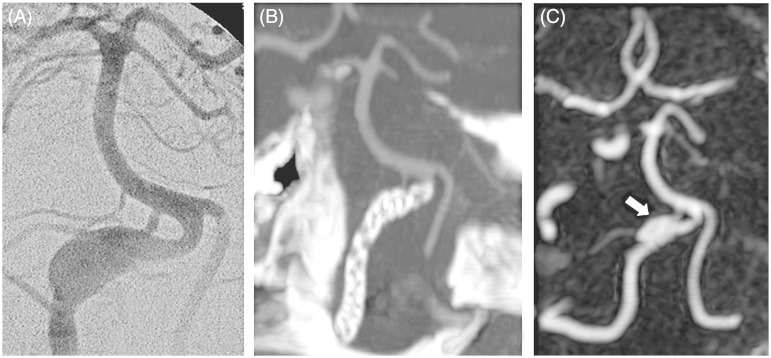
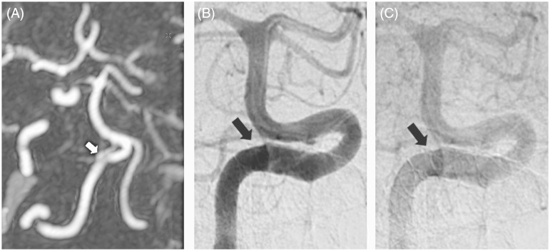
Endovascular treatment of giant, fusiform and dissecting aneurysms with flow diverter stents is becoming more and more popular. However, very few studies on the follow-up have been published. We describe a patient with a dissecting aneurysm of the right vertebral artery treated with flow diverter stent placement. The patient was followed up with CT angiography (CTA), time-resolved contrast-enhanced MR angiography (CE-MRA) and digital subtraction angiography (DSA). CTA had false negative results in two instances, whereas time-resolved CE-MRA and DSA were the most accurate in depicting the residual flow in the aneurysmal sac. However, in the case of DSA the demonstration of residual flow proved quite difficult and required a very thorough examination with oblique projections. Our 2.5-year experience with this patient led us to believe that time-resolved CE-MRA is a valuable tool in the follow-up of flow diverter-treated stents.
Keywords: CT angiography; Intracranial aneurysm; contrast-enhanced MR angiography; digital subtraction angiography; flow diverter.
© The Author(s) 2015 Reprints and permissions:]br]sagepub.co.uk/journalsPermissions.nav.
Figures
Similar articles
-
Follow-up of intracranial aneurysms treated with stent-assisted coiling: Comparison of contrast-enhanced MRA, time-of-flight MRA, and digital subtraction angiography.J Neuroradiol. 2017 Feb;44(1):44-51. doi: 10.1016/j.neurad.2016.10.004. Epub 2016 Nov 9. J Neuroradiol. 2017. PMID: 27836654
-
Follow-up of intracranial aneurysms treated by flow diverter: comparison of three-dimensional time-of-flight MR angiography (3D-TOF-MRA) and contrast-enhanced MR angiography (CE-MRA) sequences with digital subtraction angiography as the gold standard.J Neurointerv Surg. 2016 Jan;8(1):81-6. doi: 10.1136/neurintsurg-2014-011449. Epub 2014 Oct 28. J Neurointerv Surg. 2016. PMID: 25352582
-
Usefulness of pointwise encoding time reduction with radial acquisition sequence in subtraction-based magnetic resonance angiography for follow-up of the Neuroform Atlas stent-assisted coil embolization for cerebral aneurysms.Acta Radiol. 2021 Sep;62(9):1193-1199. doi: 10.1177/0284185120952784. Epub 2020 Aug 31. Acta Radiol. 2021. PMID: 32867507
-
MRA versus DSA for the follow-up imaging of intracranial aneurysms treated using endovascular techniques: a meta-analysis.J Neurointerv Surg. 2019 Oct;11(10):1009-1014. doi: 10.1136/neurintsurg-2019-014936. Epub 2019 May 2. J Neurointerv Surg. 2019. PMID: 31048457 Review.
-
Flow diverter treatment of intracranial vertebral artery dissecting pseudoaneurysms.J Neurointerv Surg. 2017 Nov;9(11):1064-1068. doi: 10.1136/neurintsurg-2017-013020. Epub 2017 Apr 24. J Neurointerv Surg. 2017. PMID: 28438894 Review.
Cited by
-
[Automatic detection method of intracranial aneurysms on maximum intensity projection images based on SE-CaraNet].Sheng Wu Yi Xue Gong Cheng Xue Za Zhi. 2024 Apr 25;41(2):228-236. doi: 10.7507/1001-5515.202301008. Sheng Wu Yi Xue Gong Cheng Xue Za Zhi. 2024. PMID: 38686402 Free PMC article. Chinese.
-
Diagnostic Value of Low-Dose 256-Slice Spiral CT Angiography, MR Angiography, and 3D-DSA in Cerebral Aneurysms.Dis Markers. 2020 Jan 13;2020:8536471. doi: 10.1155/2020/8536471. eCollection 2020. Dis Markers. 2020. PMID: 32399089 Free PMC article.
-
Comparison of postsurgical clinical sequences between completely embolized and incompletely embolized patients with wide nicked intracranial aneurysms treated with stent assisted coil embolization technique: A STROBE-compliant study.Medicine (Baltimore). 2018 Jun;97(23):e10987. doi: 10.1097/MD.0000000000010987. Medicine (Baltimore). 2018. PMID: 29879055 Free PMC article.
-
Aneurysmal and Perianeurysmal Changes After Endovascular Treatment: from Inflammation to Microbleed. A Case Report.Clin Neuroradiol. 2016 Jun;26(2):239-42. doi: 10.1007/s00062-015-0442-7. Epub 2015 Jul 31. Clin Neuroradiol. 2016. PMID: 26227620 No abstract available.
-
Application of computed tomography angiography for evaluating clinical morphology in intracranial aneurysms - monocentric study.J Int Med Res. 2020 Apr;48(4):300060519894790. doi: 10.1177/0300060519894790. Epub 2019 Dec 29. J Int Med Res. 2020. PMID: 31884845 Free PMC article.
References
-
- Cirillo L, Dall'olio M, Princiotta C, et al. The use of flow-diverting stents in the treatment of giant cerebral aneurysms: preliminary results. Neuroradiol J 2010; 23(2): 220–224. - PubMed
-
- Toni F, Marliani AF, Cirillo L, et al. 3T MRI in the evaluation of brain aneurysms treated with flow-diverting stents: preliminary experience. Neuroradiol J 2009; 22(5): 588–599. - PubMed
Publication types
MeSH terms
Substances
LinkOut - more resources
Full Text Sources
Medical
