. 2015 Jul 7;132(1):47-58.
doi: 10.1161/CIRCULATIONAHA.115.015656. Epub 2015 May 1.

Platelet Extracellular Regulated Protein Kinase 5 Is a Redox Switch and Triggers Maladaptive Platelet Responses and Myocardial Infarct Expansion

Affiliations

Platelet Extracellular Regulated Protein Kinase 5 Is a Redox Switch and Triggers Maladaptive Platelet Responses and Myocardial Infarct Expansion

Scott J Cameron et al. Circulation. .

Abstract

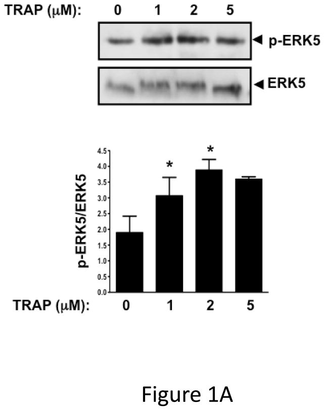
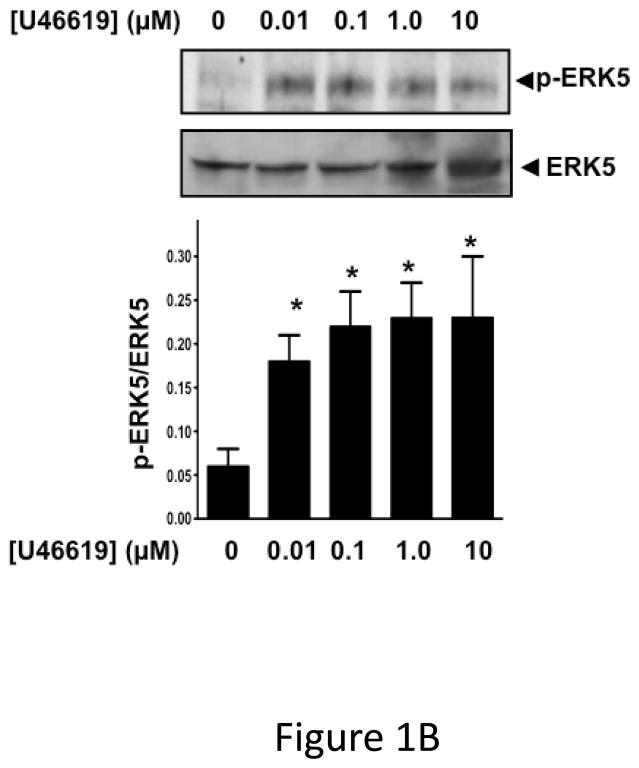
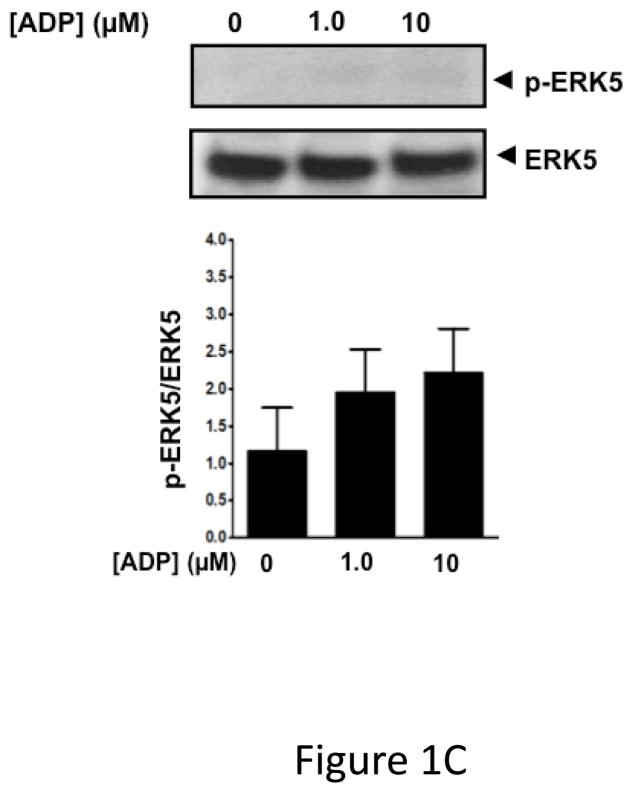
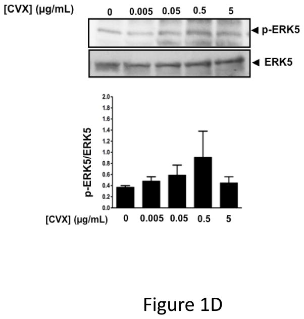
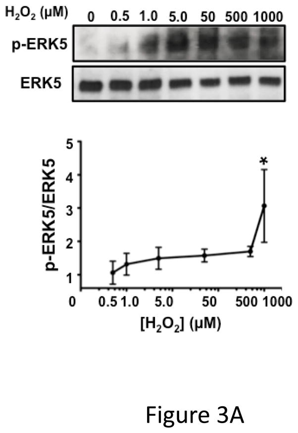
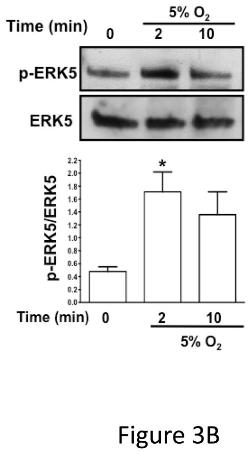
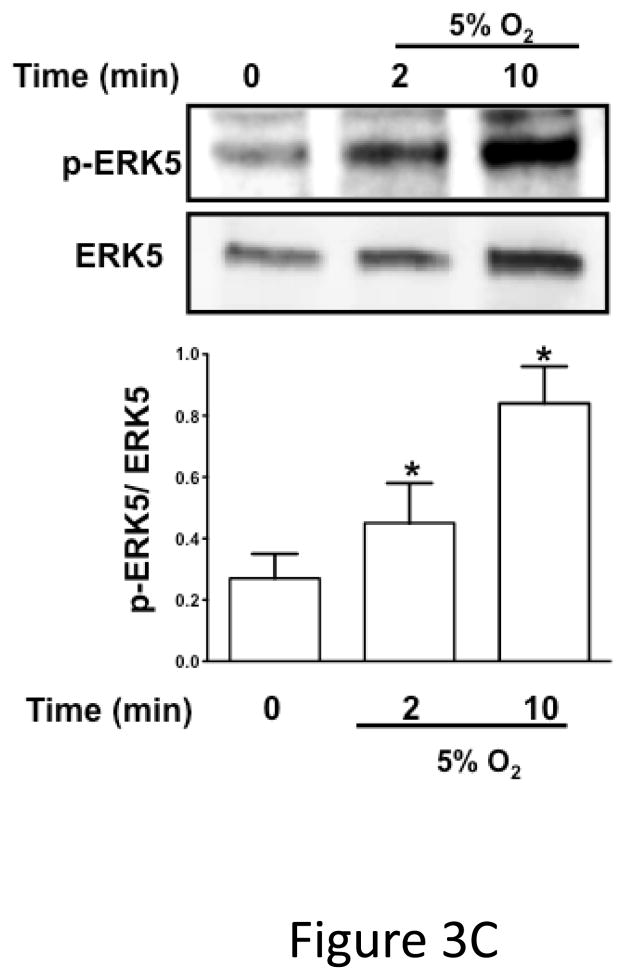
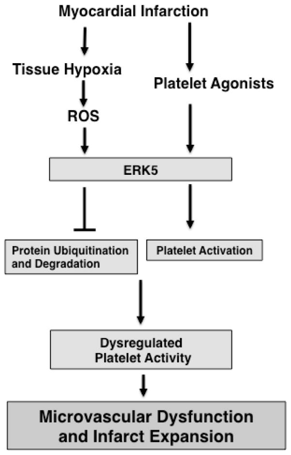
Background: Platelets have a pathophysiologic role in the ischemic microvascular environment of acute coronary syndromes. In comparison with platelet activation in normal healthy conditions, less attention is given to mechanisms of platelet activation in diseased states. Platelet function and mechanisms of activation in ischemic and reactive oxygen species-rich environments may not be the same as in normal healthy conditions. Extracellular regulated protein kinase 5 (ERK5) is a mitogen-activated protein kinase family member activated in hypoxic, reactive oxygen species-rich environments and in response to receptor-signaling mechanisms. Prior studies suggest a protective effect of ERK5 in endothelial and myocardial cells after ischemia. We present evidence that platelets express ERK5 and that platelet ERK5 has an adverse effect on platelet activation via selective receptor-dependent and receptor-independent reactive oxygen species-mediated mechanisms in ischemic myocardium.

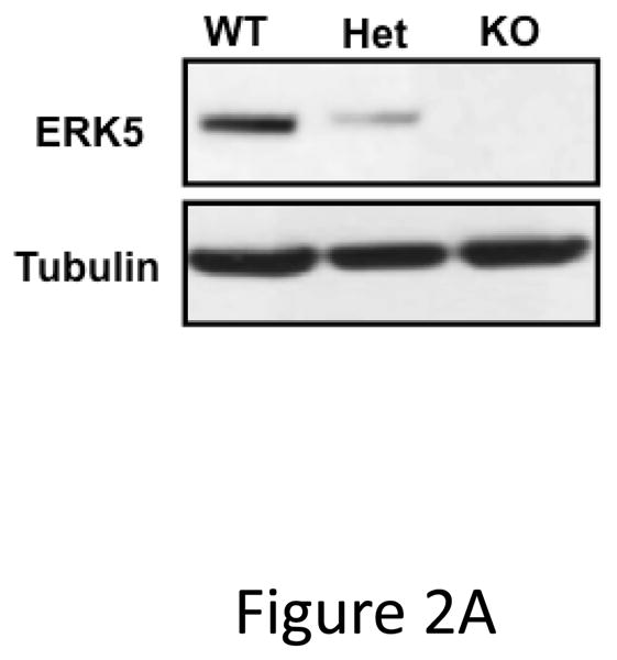
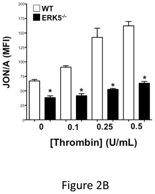
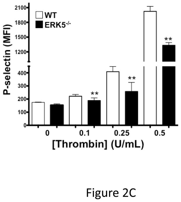
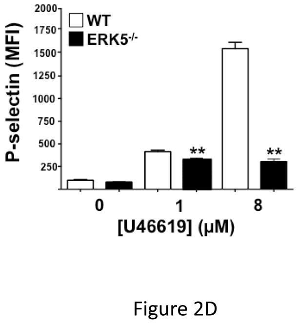
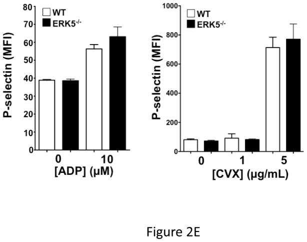
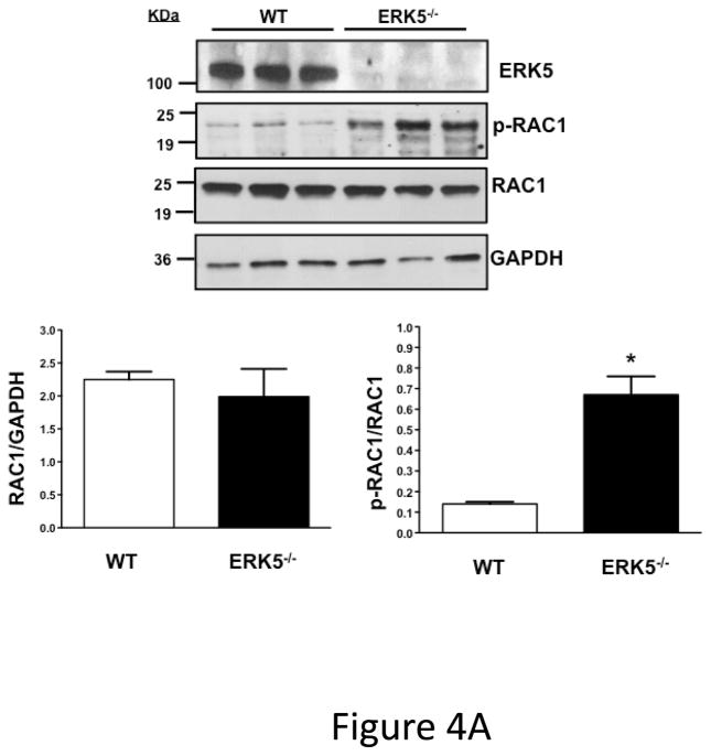
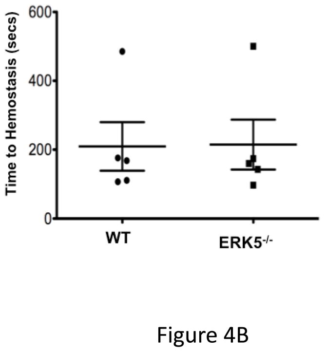
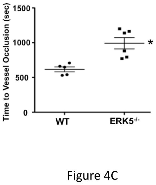
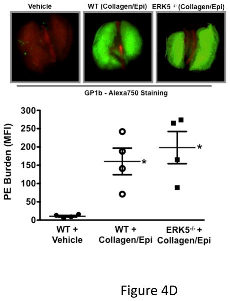
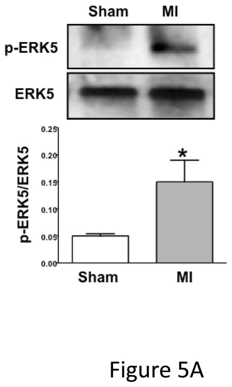
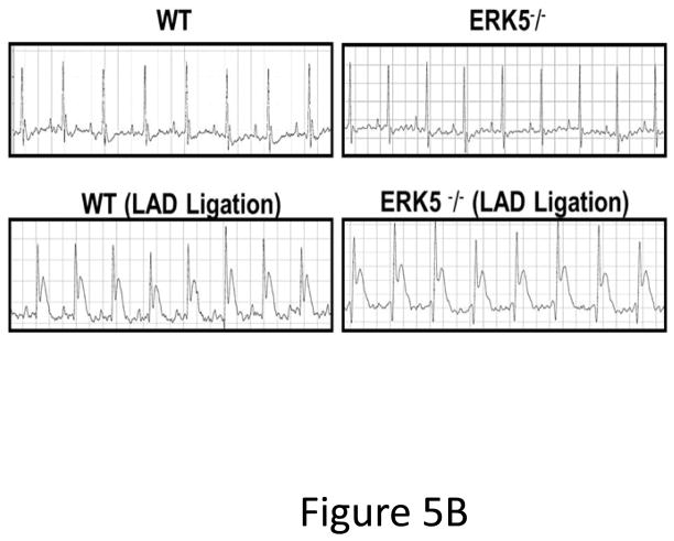
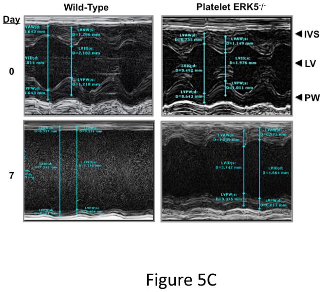
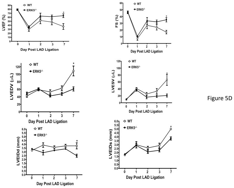
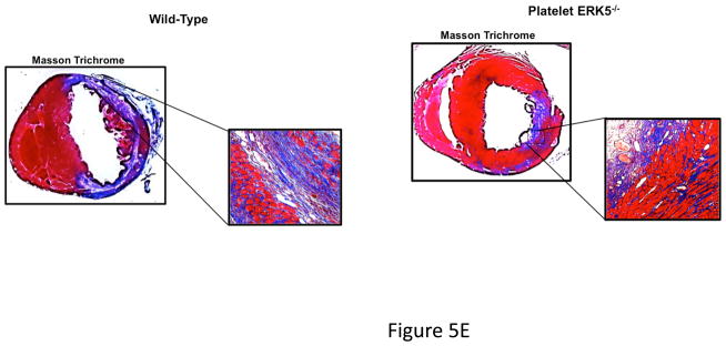
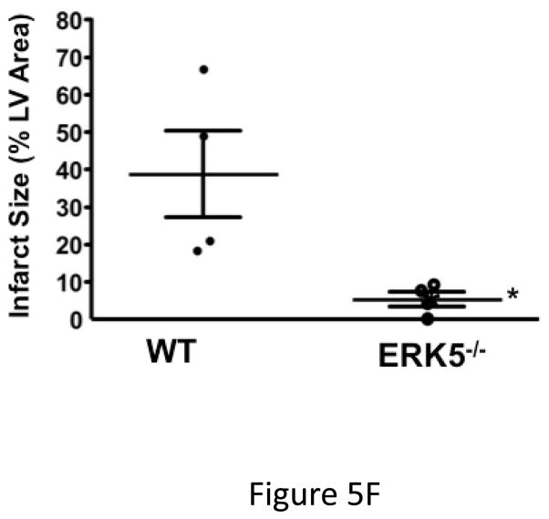
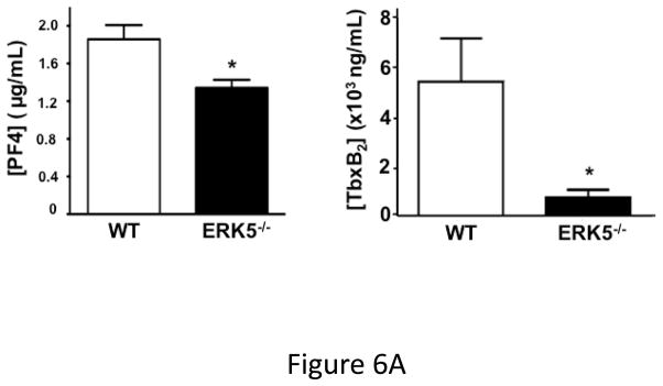
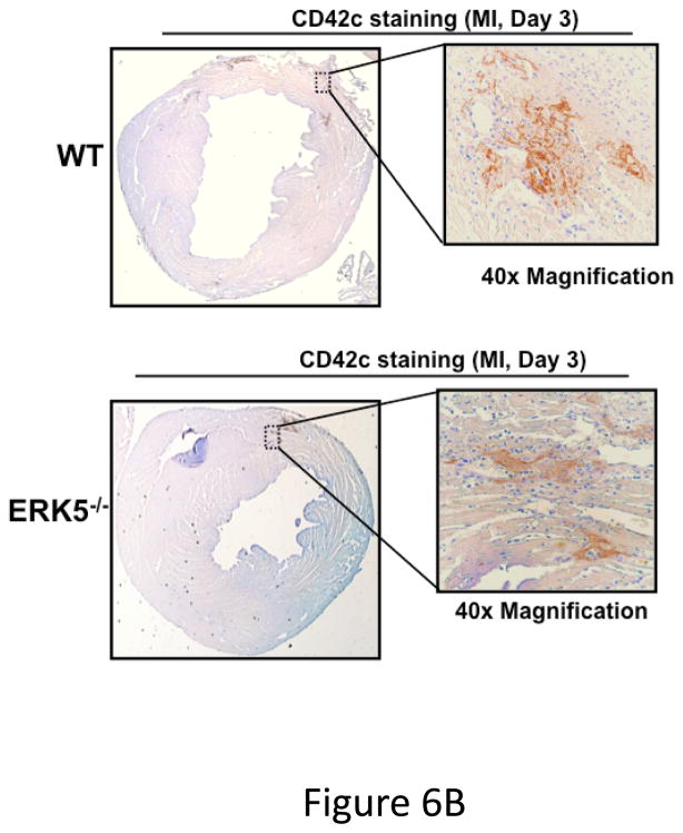
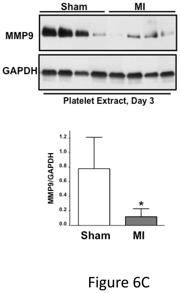
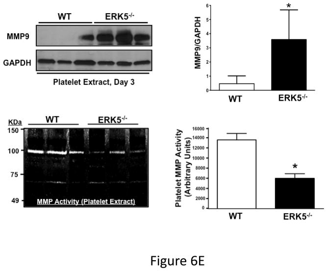
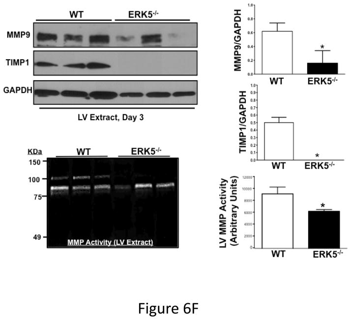
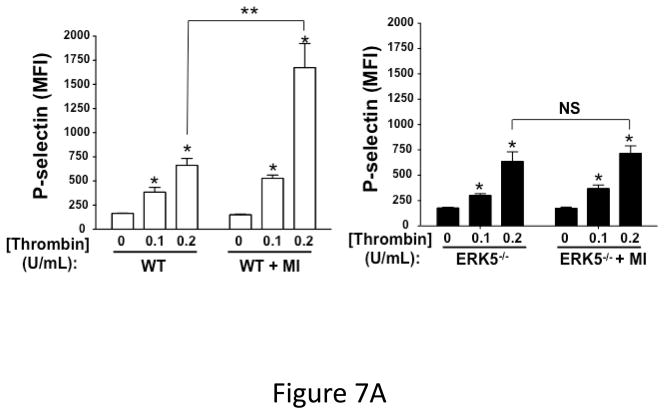
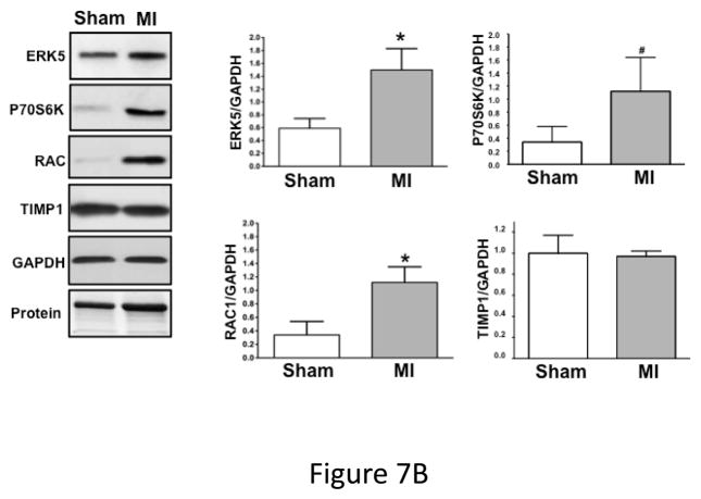
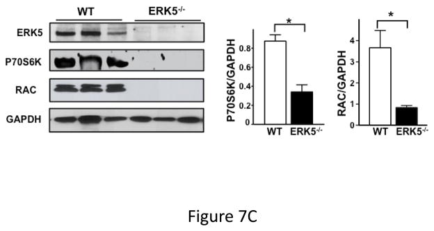
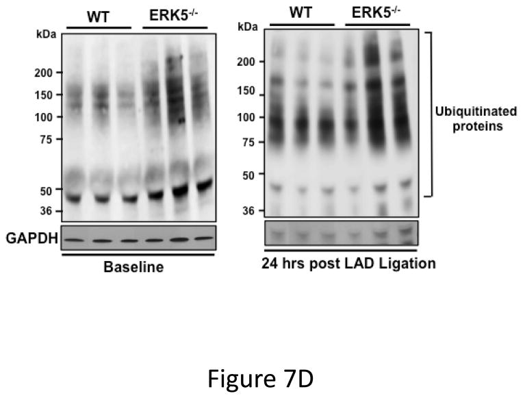
Methods and results: Using isolated human platelets and a mouse model of myocardial infarction (MI), we found that platelet ERK5 is activated post-MI and that platelet-specific ERK5(-/-) mice have less platelet activation, reduced MI size, and improved post-MI heart function. Furthermore, the expression of downstream ERK5-regulated proteins is reduced in ERK5(-/-) platelets post-MI.

Conclusions: ERK5 functions as a platelet activator in ischemic conditions, and platelet ERK5 maintains the expression of some platelet proteins after MI, leading to infarct expansion. This demonstrates that platelet function in normal healthy conditions is different from platelet function in chronic ischemic and inflammatory conditions. Platelet ERK5 may be a target for acute therapeutic intervention in the thrombotic and inflammatory post-MI environment.

Keywords: blood platelets; echocardiography; infarction; microcirculation.


Figures

Figure 1
Figure 1
ERK5 is expressed in human platelets and activated by thrombin and thromboxane. Washed human platelets were stimulated with A) 1–5 μM TRAP, B) 0.01–10 μM U46619, C) 1–10 μM 2-methyl adenosine diphosphate (ADP), or D) 5–5000 ng/mL convulxin, each for 10 mins. Western blotting was performed for p-ERK5 (blots over-exposed for emphasis in C and D, and total ERK5 as loading control). ERK5 activation reported as mean p-ERK5/ERK5 (± SEM, N=4–7, * P <0.05 vs 0, paired t-test). P=0.08 for A), P=0.06 for B), P=0.35 for C), P =0.85 for D) by Friedman’s test.
Figure 1
Figure 1
ERK5 is expressed in human platelets and activated by thrombin and thromboxane. Washed human platelets were stimulated with A) 1–5 μM TRAP, B) 0.01–10 μM U46619, C) 1–10 μM 2-methyl adenosine diphosphate (ADP), or D) 5–5000 ng/mL convulxin, each for 10 mins. Western blotting was performed for p-ERK5 (blots over-exposed for emphasis in C and D, and total ERK5 as loading control). ERK5 activation reported as mean p-ERK5/ERK5 (± SEM, N=4–7, * P <0.05 vs 0, paired t-test). P=0.08 for A), P=0.06 for B), P=0.35 for C), P =0.85 for D) by Friedman’s test.
Figure 2
Figure 2
ERK5−/− platelets have decreased activation in response to PAR and thromboxane receptor stimulation. A) Washed platelets were isolated from WT, ERK5+/− (Het) or ERK5−/− mice and immunoblotted for ERK5. ERK5−/− platelets do not express ERK5 (representative blot). B–D) Washed platelets from WT and ERK5−/− mice were stimulated with B–C) thrombin, D) U46619 which is a thromboxane receptor agonist, E) ADP or convulxin. Platelet activation was determined by JON/A antibody staining or surface P-selectin expression via FACS (MFI ± SD, N=4, * P < 0.05. vs. WT at same agonist concentration and **P < 0.01. vs WT at same agonist concentration, t-test for equal variances).
Figure 2
Figure 2
ERK5−/− platelets have decreased activation in response to PAR and thromboxane receptor stimulation. A) Washed platelets were isolated from WT, ERK5+/− (Het) or ERK5−/− mice and immunoblotted for ERK5. ERK5−/− platelets do not express ERK5 (representative blot). B–D) Washed platelets from WT and ERK5−/− mice were stimulated with B–C) thrombin, D) U46619 which is a thromboxane receptor agonist, E) ADP or convulxin. Platelet activation was determined by JON/A antibody staining or surface P-selectin expression via FACS (MFI ± SD, N=4, * P < 0.05. vs. WT at same agonist concentration and **P < 0.01. vs WT at same agonist concentration, t-test for equal variances).
Figure 3
Figure 3
Platelet ERK5 is activated by ROS. A) Dose-dependent platelet ERK5 ROS activation. Washed human platelets were stimulated with H2O2 (0.5–1000 μM) for 5 mins and p-ERK5 determined at multiple time points by p-ERK5 immunoblotting normalized to total ERK5 (* P<0.05 vs 0, paired t-test; P =0.12 by Friedman’s test). B) Washed human platelets were incubated under normoxic or reduced oxygen (5% O2) conditions for 2–10 mins (P <0.05 vs 0, paired t-test and P =0.02, Friedman’s test). C) Washed mouse platelets were incubated under normoxic or reduced oxygen (5% O2) conditions for 2–10 mins. (P <0.05 vs 0, paired t-test; P=0.02, Friedman’s test). ERK5 activation reported by Western blotting as mean p-ERK5/ERK5 (± SEM, N=4–6).
Figure 3
Figure 3
Platelet ERK5 is activated by ROS. A) Dose-dependent platelet ERK5 ROS activation. Washed human platelets were stimulated with H2O2 (0.5–1000 μM) for 5 mins and p-ERK5 determined at multiple time points by p-ERK5 immunoblotting normalized to total ERK5 (* P<0.05 vs 0, paired t-test; P =0.12 by Friedman’s test). B) Washed human platelets were incubated under normoxic or reduced oxygen (5% O2) conditions for 2–10 mins (P <0.05 vs 0, paired t-test and P =0.02, Friedman’s test). C) Washed mouse platelets were incubated under normoxic or reduced oxygen (5% O2) conditions for 2–10 mins. (P <0.05 vs 0, paired t-test; P=0.02, Friedman’s test). ERK5 activation reported by Western blotting as mean p-ERK5/ERK5 (± SEM, N=4–6).
Figure 4
Figure 4
ERK5 mediates in vivo platelet activation. A) RAC1 basal activation is reduced in ERK5−/− platelets. Washed WT and ERK5−/− platelet lysates were prepared and immunoblotted for total RAC or RAC1 phosphorylated on serine 71 (p-RAC S71 is inactive RAC1). Representative blots (each lane is an individual mouse platelet extract) run on separate protein gels (top), and densitometry (bottom). GAPDH was determined by re-probing as an additional loading control (mean ± SEM, N=4–7, *P < 0.05 vs WT, t-test for unequal variances). B) Platelet ERK5−/− mice have normal bleeding times. Mouse tail bleeding times were determined by time to visual cessation of bleeding (mean ± SEM, N=5–6). C) Platelet ERK5−/− mice have prolonged time to in vivo thrombus formation. Mesenteric ferric chloride injury model with time to vessel occlusion recorded (mean ± SEM, N=5, *p = 0.004 vs. WT, t-test for unequal variances). D) WT and platelet ERK5−/− mice have similar thrombus burden in a PE model. Mice were injected with Alexa750 labeled anti-GP1b antibody and then treated with collagen/epinephrine or vehicle control via the jugular vein to induce PE. Perfused lungs were isolated and imaged to quantify lung thrombus burden (MFI ± SEM, N=4, *p =0.02 vs. vehicle=PBS, t-test for unequal variances. Representative image from are from individual mice).
Figure 4
Figure 4
ERK5 mediates in vivo platelet activation. A) RAC1 basal activation is reduced in ERK5−/− platelets. Washed WT and ERK5−/− platelet lysates were prepared and immunoblotted for total RAC or RAC1 phosphorylated on serine 71 (p-RAC S71 is inactive RAC1). Representative blots (each lane is an individual mouse platelet extract) run on separate protein gels (top), and densitometry (bottom). GAPDH was determined by re-probing as an additional loading control (mean ± SEM, N=4–7, *P < 0.05 vs WT, t-test for unequal variances). B) Platelet ERK5−/− mice have normal bleeding times. Mouse tail bleeding times were determined by time to visual cessation of bleeding (mean ± SEM, N=5–6). C) Platelet ERK5−/− mice have prolonged time to in vivo thrombus formation. Mesenteric ferric chloride injury model with time to vessel occlusion recorded (mean ± SEM, N=5, *p = 0.004 vs. WT, t-test for unequal variances). D) WT and platelet ERK5−/− mice have similar thrombus burden in a PE model. Mice were injected with Alexa750 labeled anti-GP1b antibody and then treated with collagen/epinephrine or vehicle control via the jugular vein to induce PE. Perfused lungs were isolated and imaged to quantify lung thrombus burden (MFI ± SEM, N=4, *p =0.02 vs. vehicle=PBS, t-test for unequal variances. Representative image from are from individual mice).
Figure 5
Figure 5
Platelet ERK5−/− mice have improved post-MI heart function. A) Washed mouse platelets isolated on day 3 following MI were assessed for activated ERK5 (p-ERK5) (mean p-ERK5/ERK5 ± SEM, N=5, *P = 0.03 vs. sham operated, t-test for unequal variances). B) ECG before and 30 seconds after LAD coronary artery ligation indicates transmural myocardial ischemia (ST-segment elevation). C) Representative M-mode echocardiograms for WT and ERK5−/− mice pre-LAD coronary ligation (day 0) and day 7 post-LAD coronary ligation. D) Echocardiography quantification of LVEF, FS, LVEDV, LVESV, LVIDd, LVIDs (mean ± SEM, N=4–6, *P < 0.05 vs. WT, t-test for unequal variances). E) Platelet ERK5−/− mice have smaller infarcts (reduced Masson Trichrome stain of LV, insert 40x magnification). F) Quantification of Masson Trichome positive staining (mean % of LV area, N=4 ± SEM,*P = 0.02 vs WT, t-test for equal variances).
Figure 5
Figure 5
Platelet ERK5−/− mice have improved post-MI heart function. A) Washed mouse platelets isolated on day 3 following MI were assessed for activated ERK5 (p-ERK5) (mean p-ERK5/ERK5 ± SEM, N=5, *P = 0.03 vs. sham operated, t-test for unequal variances). B) ECG before and 30 seconds after LAD coronary artery ligation indicates transmural myocardial ischemia (ST-segment elevation). C) Representative M-mode echocardiograms for WT and ERK5−/− mice pre-LAD coronary ligation (day 0) and day 7 post-LAD coronary ligation. D) Echocardiography quantification of LVEF, FS, LVEDV, LVESV, LVIDd, LVIDs (mean ± SEM, N=4–6, *P < 0.05 vs. WT, t-test for unequal variances). E) Platelet ERK5−/− mice have smaller infarcts (reduced Masson Trichrome stain of LV, insert 40x magnification). F) Quantification of Masson Trichome positive staining (mean % of LV area, N=4 ± SEM,*P = 0.02 vs WT, t-test for equal variances).
Figure 6
Figure 6
Post-MI platelet activation is reduced in platelet ERK5−/− mice. A) Plasma PF4 or TxB2 were measured by ELISA as markers of ongoing platelet activation 3 days following LAD coronary artery ligation (mean ± S.D, N=6. *P <0.05 vs. WT, t-test for unequal variances). B) Representative parasternal short axis sections of the heart on day 3 following LAD coronary ligation. CD42c (platelet) immunostaining is less in platelet ERK5−/− mouse hearts compared to WT hearts at the infarct border (additional images are shown in S11). C) Platelet MMP content by Western blotting on day 3 following LAD coronary ligation is reduced compared to sham-operated mice (mean MMP9/GAPDH ± SEM, N=4–5. *p = 0.02 vs. sham-operated, t-test for unequal variances). D) LV MMP9 and TIMP1 content is reduced in platelet ERK5−/− mice after MI (mean intensity ± SEM, N=5 each group, *P =0.02 vs. WT, t-test for unequal variances). LV extracts on day 3 following LAD coronary artery ligation were protein-normalized and run in duplicate on non-reducing gels with a gelatin matrix. Gelatinase activity is shown as light bands on the final zymogram. Total LV MMP activity (all isoforms) was calculated for each lane (mean intensity ± SEM, N=6–10, *P < 0.001 vs. WT, t-test for unequal variances). Washed platelet MMP9 content is increased in platelet ERK5−/− mice compared to WT mice on day 3 following LAD coronary ligation (mean MMP9/GAPDH ± SEM, N=5. *p < 0.05 vs. WT mouse platelets). E) Platelet MMP9 content and MMP activity are reduced in platelet ERK5−/− mice. Washed platelets were isolated on day 3 following LAD coronary artery ligation, homogenized, protein-normalized and immunoblotted for MMP9 content (mean intensity ± SEM, N=3–5, *P < 0.01 vs. WT, t-test for unequal variances). Washed platelets were isolated on day 3 following LAD coronary artery ligation and were also run on non-reducing gels with a gelatin matrix. Gelatinase activity is shown as light bands on the final zymogram. Total MMP activity (all isoforms) was calculated for each lane. (Protein expression normalized to GAPDH and gelatinase activity is demonstrated by light bands on the final zymogram (total platelet MMP activity, mean ± SEM, N=8–10, *P=0.04 vs. WT, t-test for unequal variances).
Figure 6
Figure 6
Post-MI platelet activation is reduced in platelet ERK5−/− mice. A) Plasma PF4 or TxB2 were measured by ELISA as markers of ongoing platelet activation 3 days following LAD coronary artery ligation (mean ± S.D, N=6. *P <0.05 vs. WT, t-test for unequal variances). B) Representative parasternal short axis sections of the heart on day 3 following LAD coronary ligation. CD42c (platelet) immunostaining is less in platelet ERK5−/− mouse hearts compared to WT hearts at the infarct border (additional images are shown in S11). C) Platelet MMP content by Western blotting on day 3 following LAD coronary ligation is reduced compared to sham-operated mice (mean MMP9/GAPDH ± SEM, N=4–5. *p = 0.02 vs. sham-operated, t-test for unequal variances). D) LV MMP9 and TIMP1 content is reduced in platelet ERK5−/− mice after MI (mean intensity ± SEM, N=5 each group, *P =0.02 vs. WT, t-test for unequal variances). LV extracts on day 3 following LAD coronary artery ligation were protein-normalized and run in duplicate on non-reducing gels with a gelatin matrix. Gelatinase activity is shown as light bands on the final zymogram. Total LV MMP activity (all isoforms) was calculated for each lane (mean intensity ± SEM, N=6–10, *P < 0.001 vs. WT, t-test for unequal variances). Washed platelet MMP9 content is increased in platelet ERK5−/− mice compared to WT mice on day 3 following LAD coronary ligation (mean MMP9/GAPDH ± SEM, N=5. *p < 0.05 vs. WT mouse platelets). E) Platelet MMP9 content and MMP activity are reduced in platelet ERK5−/− mice. Washed platelets were isolated on day 3 following LAD coronary artery ligation, homogenized, protein-normalized and immunoblotted for MMP9 content (mean intensity ± SEM, N=3–5, *P < 0.01 vs. WT, t-test for unequal variances). Washed platelets were isolated on day 3 following LAD coronary artery ligation and were also run on non-reducing gels with a gelatin matrix. Gelatinase activity is shown as light bands on the final zymogram. Total MMP activity (all isoforms) was calculated for each lane. (Protein expression normalized to GAPDH and gelatinase activity is demonstrated by light bands on the final zymogram (total platelet MMP activity, mean ± SEM, N=8–10, *P=0.04 vs. WT, t-test for unequal variances).
Figure 7
Figure 7
Dysregulated platelet activity following MI. A) Washed platelets from WT and platelet ERK5−/− mice on day 6 following LAD coronary ligation were examined for thrombin-induced activation. Platelet activation was determined by surface P-selectin expression via FACS (MFI ± SD, N=3–4. *P =0.03, Friedman’s test for WT + thrombin; P =0.04, Friedman’s test for WT/MI + thrombin; P =0.04, Friedman’s test for ERK5−/− + thrombin; P =0.03, Friedman’s test for ERK5−/−/MI + thrombin; ** P=0.04, t-test for unequal variance). B) Washed platelets on day 3 following LAD coronary ligation were examined for the expression of ERK5, P70S6K, RAC, TIMP1 and GAPDH by Western blotting. Protein gels were also stained by Coomassie Blue for total protein. Representative data (mean protein/GAPDH ± SEM, N=5. * P < 0.05 vs. platelets from sham-operated mice; # P =0.053 vs. platelets from sham-operated mice, t-test for unequal variances). C) Washed platelets from WT and platelet ERK5−/− mice on day 7 following LAD coronary ligation were examined for the expression of P70S6K and RAC by Western blotting (representative data are expressed as mean protein/GAPDH ± SEM, N=4–5. * P < 0.05 vs. WT, t-test for equal variances). D) Washed platelets at baseline and after 24hrs of LAD coronary ligation were examined for ubiquitinated proteins by Western blotting in WT and platelet ERK5−/− mice. Proteins ubiquitination was increased in ERK5−/− platelets, representative immunoblots.
Figure 7
Figure 7
Dysregulated platelet activity following MI. A) Washed platelets from WT and platelet ERK5−/− mice on day 6 following LAD coronary ligation were examined for thrombin-induced activation. Platelet activation was determined by surface P-selectin expression via FACS (MFI ± SD, N=3–4. *P =0.03, Friedman’s test for WT + thrombin; P =0.04, Friedman’s test for WT/MI + thrombin; P =0.04, Friedman’s test for ERK5−/− + thrombin; P =0.03, Friedman’s test for ERK5−/−/MI + thrombin; ** P=0.04, t-test for unequal variance). B) Washed platelets on day 3 following LAD coronary ligation were examined for the expression of ERK5, P70S6K, RAC, TIMP1 and GAPDH by Western blotting. Protein gels were also stained by Coomassie Blue for total protein. Representative data (mean protein/GAPDH ± SEM, N=5. * P < 0.05 vs. platelets from sham-operated mice; # P =0.053 vs. platelets from sham-operated mice, t-test for unequal variances). C) Washed platelets from WT and platelet ERK5−/− mice on day 7 following LAD coronary ligation were examined for the expression of P70S6K and RAC by Western blotting (representative data are expressed as mean protein/GAPDH ± SEM, N=4–5. * P < 0.05 vs. WT, t-test for equal variances). D) Washed platelets at baseline and after 24hrs of LAD coronary ligation were examined for ubiquitinated proteins by Western blotting in WT and platelet ERK5−/− mice. Proteins ubiquitination was increased in ERK5−/− platelets, representative immunoblots.
Figure 8
Figure 8
Proposed model. Following MI, platelet ERK5 is activated by both agonists and ROS. ERK5 activation leads to both platelet activation and increased expression of proteins important for platelet activation, in part via decreased protein ubiquitination.

References

    1. Smyth SS, McEver RP, Weyrich AS, Morrell CN, Hoffman MR, Arepally GM, French PA, Dauerman HL, Becker RC, Platelet Colloquium P. Platelet functions beyond hemostasis. J Thromb Haemost. 2009;7:1759–1766. - PubMed
    1. Jia ZB, Tian H, Kang K, Miao HZ, Liu KY, Jiang SL, Wang LP. Expression of the tissue inhibitor of metalloproteinase-3 by transplanted vsmcs modifies heart structure and function after myocardial infarction. Transpl Immunol. 2014;30:149–158. - PubMed
    1. Kandalam V, Basu R, Abraham T, Wang X, Awad A, Wang W, Lopaschuk GD, Maeda N, Oudit GY, Kassiri Z. Early activation of matrix metalloproteinases underlies the exacerbated systolic and diastolic dysfunction in mice lacking timp3 following myocardial infarction. Am J Physiol Heart Circ Physiol. 2010;299:H1012–1023. - PMC - PubMed
    1. Matsumura S, Iwanaga S, Mochizuki S, Okamoto H, Ogawa S, Okada Y. Targeted deletion or pharmacological inhibition of mmp-2 prevents cardiac rupture after myocardial infarction in mice. J Clin Invest. 2005;115:599–609. - PMC - PubMed
    1. Heymans S, Luttun A, Nuyens D, Theilmeier G, Creemers E, Moons L, Dyspersin GD, Cleutjens JP, Shipley M, Angellilo A, Levi M, Nube O, Baker A, Keshet E, Lupu F, Herbert JM, Smits JF, Shapiro SD, Baes M, Borgers M, Collen D, Daemen MJ, Carmeliet P. Inhibition of plasminogen activators or matrix metalloproteinases prevents cardiac rupture but impairs therapeutic angiogenesis and causes cardiac failure. Nat Med. 1999;5:1135–1142. - PubMed

Publication types

Substances