. 2015 May 1;21(9):2167-76.
doi: 10.1158/1078-0432.CCR-14-1826.

IL2 Inducible T-cell Kinase, a Novel Therapeutic Target in Melanoma

Affiliations

IL2 Inducible T-cell Kinase, a Novel Therapeutic Target in Melanoma

Craig C Carson et al. Clin Cancer Res. .

Abstract

Purpose: IL2 inducible T-cell kinase (ITK) promoter CpG sites are hypomethylated in melanomas compared with nevi. The expression of ITK in melanomas, however, has not been established and requires elucidation.

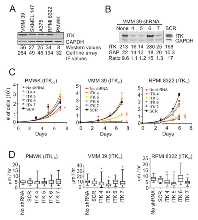
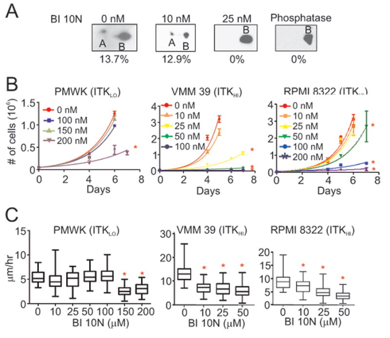
Experimental design: An ITK-specific monoclonal antibody was used to probe sections from deidentified, formalin-fixed paraffin-embedded tumor blocks or cell line arrays and ITK was visualized by IHC. Levels of ITK protein differed among melanoma cell lines and representative lines were transduced with four different lentiviral constructs that each contained an shRNA designed to knockdown ITK mRNA levels. The effects of the selective ITK inhibitor BI 10N on cell lines and mouse models were also determined.

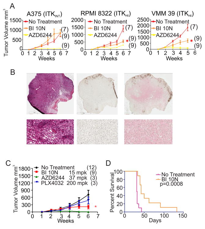
Results: ITK protein expression increased with nevus to metastatic melanoma progression. In melanoma cell lines, genetic or pharmacologic inhibition of ITK decreased proliferation and migration and increased the percentage of cells in the G0-G1 phase. Treatment of melanoma-bearing mice with BI 10N reduced growth of ITK-expressing xenografts or established autochthonous (Tyr-Cre/Pten(null)/Braf(V600E)) melanomas.

Conclusions: We conclude that ITK, formerly considered an immune cell-specific protein, is aberrantly expressed in melanoma and promotes tumor development and progression. Our finding that ITK is aberrantly expressed in most metastatic melanomas suggests that inhibitors of ITK may be efficacious for melanoma treatment. The efficacy of a small-molecule ITK inhibitor in the Tyr-Cre/Pten(null)/Braf(V600E) mouse melanoma model supports this possibility.


Conflict of interest statement

Disclosure of Potential Conflicts of Interest. The authors have no conflict of interest to disclose.

Figures

Figure 1
Figure 1
ITK expression in melanocytic tissues. (A) Representative tissue sections from normal skin (a), benign nevus (b), primary melanoma (c), and metastatic melanoma (d) were stained with H&E (top row) or probed with ITK-Cy5 (red) and S100-Alexa 555 (green) primary and secondary antibodies and counterstained with DAPI (blue). Orange indicates colocation of ITK with S100, a melanocytic lineage marker. The clinical and histologic characteristics of the melanocytic tissues stained and their ITK levels are in Supplementary Table S1. Staining protocols are in Supplementary Table S2. (B) Box-and-Whisker plots (median with the 25-75th percentiles and outliers) of ITK expression in S100+ cells from nevi (n = 30), primary melanomas (n = 20), and metastatic melanomas (n = 70). Of the metastatic melanomas, 91% (64/70) had ITK expression above that of the range of ITK expression found in nevi.
Figure 2
Figure 2
ITK Expression in melanoma cell lines and effects of ITK depletion on their proliferation and motility. (A) Western blot analysis for total ITK in whole cell protein lysates obtained from five melanoma cell lines. Signal intensities of the protein bands were normalized to GAPDH. Cell array IF values from Supplementary Table S3 are also shown for comparison. (B) Western blot analysis for total ITK in protein lysates obtained from VMM 39 melanoma cells that were transduced with shRNA sequences designed to target ITK mRNA (ITK 4, 5, 6, and 7) and with scrambled (SCR) shRNA. The integrated values of ITK containing bands shown in the figure were normalized against the GAPDH values to produce the ratio shown in the figure. (C) Effects of the five shRNAs on the proliferation rates of PMWK, VMM 39 and RPMI 8322 melanoma cells. Asterisks denote melanoma cell lines whose proliferation was significantly reduced compared to SCR shRNA (P < 0.05, Bonferroni corrected; Supplementary Table S4A). (D) Single-cell motility analysis of PMWK, VMM 39 and RPMI 8322 cells transduced with various shRNAs. Asterisks denote significant changes in motility compared to SCR shRNA (P < 0.05, Bonferroni corrected; Supplementary Table S4B).
Figure 3
Figure 3
Effects of BI 10N on the phosphorylation of ITK and on the proliferation and motility of melanoma cells. (A) Western blot analysis on whole cell lysates obtained from RPMI 8322 cells. Cells were treated with BI 10N for 3 days at the indicated concentrations prior to the analysis. Extracts of cells that did not receive BI 10N were treated with phosphatase prior to analysis. Proteins from lysates were separated by two-dimensional polyacrylamide gel electrophoresis prior to Western blot analysis using an antibody against total ITK. The percentages indicate the area of the smaller band as a percent of the whole signal. (B) Effects of increasing BI 10N concentrations on the proliferation rates of PMWK, VMM 39 and RPMI 8322 melanoma cells. Asterisks denote significant decreases in proliferation compared to untreated cells (P < 0.05, Bonferroni corrected; Supplementary Table S4C). (C) Effects of increasing BI 10N concentrations on the motility of PMWK, VMM 39 and RPMI 8322 melanoma cells treated for 24 hours prior to the assay. Asterisks denote significant decreases in motility in BI 10N-treated compared to untreated melanoma cells (P < 0.05, Bonferroni corrected; Supplementary Table S4D).
Figure 4
Figure 4
Effects of BI 10N on tumor growth in vivo. (A) Effects of BI 10N (15mpk) on the growth of human melanoma xenografts (mean and standard error of mean). Asterisks indicate significant tumor growth inhibition by BI 10N-treatment compared to untreated mice (P < 0.05, Bonferroni corrected). AZD6244 (37 mpk), an active agent against melanoma, is shown for comparison. Samples sizes are indicated in parentheses. (B) Representative sections from an untreated melanoma tumor tissue that was induced within the CRE-ERT2 B-RafL/+PtenL/L genetically engineered mouse model (GEMM). The left panel is stained with H&E, The center panel is stained with the biotin linked secondary antibody, while the right panel is stained both with antibodies against total ITK (red) and biotin linked secondary antibody then counterstained with hematoxylin. The lower panels are magnified areas of each of these images. (C) Effect of BI 10N on tumor growth (mean and standard error of mean) in the PTEN/BRAF GEMM. Asterisks indicate significant tumor growth inhibition of BI 10N-treated compared to untreated mice (P < 0.05, Bonferonni corrected). The effects of AZD6244 and PLX4032 on tumor growth are also shown. The activity of PLX4032 shown in C is typical for the GEMM model. Samples sizes are indicated in parentheses. (D) Melanoma bearing mice survive longer when orally dosed with 15 milligrams/kilogram BI 10N. According to the protocol governing the use of mice, the animals have to be sacrificed when the tumors reach a specific size. Of the 9 BI 10N treated animals, 7 were culled due to a multiple masses developing at the end of therapy while the primaries were still responding or had not reached a terminal burden. 1 animal had a primary that achieved a complete response and was culled at 133 days due to age and IACUC time limits. 1 animal was culled due to a progressing primary with no secondary mass.

References

    1. Chapman PB, Hauschild A, Robert C, Haanen JB, Ascierto P, Larkin J, et al. Improved survival with vemurafenib in melanoma with BRAF V600E mutation. N Engl J Med. 2011;364:2507–16. - PMC - PubMed
    1. Hauschild A, Grob JJ, Demidov LV, Jouary T, Gutzmer R, Millward M, et al. Dabrafenib in BRAF-mutated metastatic melanoma: a multicentre, open-label, phase 3 randomised controlled trial. Lancet. 2012;380:358–65. - PubMed
    1. Flaherty KT, Robert C, Hersey P, Nathan P, Garbe C, Milhem M, et al. Improved survival with MEK inhibition in BRAF-mutated melanoma. N Engl J Med. 2012;367:107–14. - PubMed
    1. Flaherty KT, Infante JR, Daud A, Gonzalez R, Kefford RF, Sosman J, et al. Combined BRAF and MEK inhibition in melanoma with BRAF V600 mutations. N Engl J Med. 2012;367:1694–703. - PMC - PubMed
    1. Hodis E, Watson IR, Kryukov GV, Arold ST, Imielinski M, Theurillat JP, et al. A landscape of driver mutations in melanoma. Cell. 2012;150:251–63. - PMC - PubMed

Publication types

MeSH terms

Substances