. 2015 Aug 1;24(15):4212-24.
doi: 10.1093/hmg/ddv154. Epub 2015 May 1.

Second-generation compound for the modulation of utrophin in the therapy of DMD

Affiliations

Second-generation compound for the modulation of utrophin in the therapy of DMD

Simon Guiraud et al. Hum Mol Genet. .

Abstract

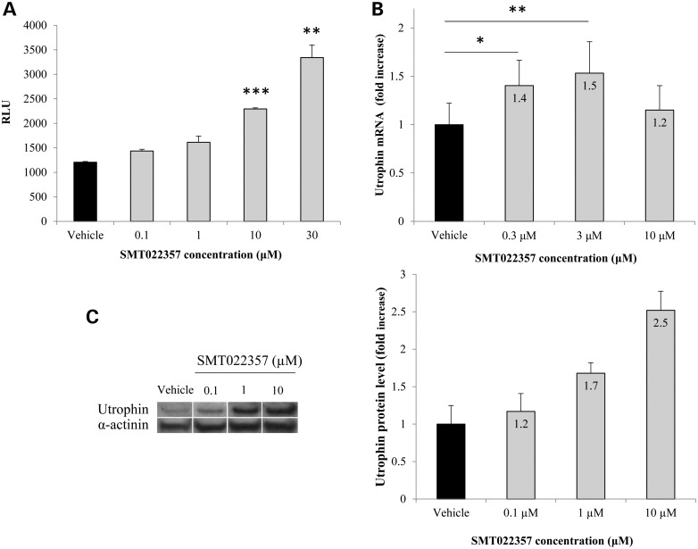
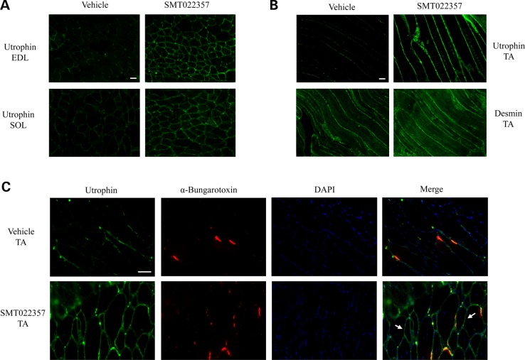
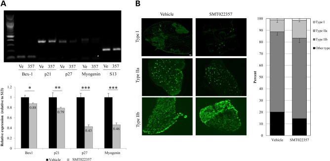
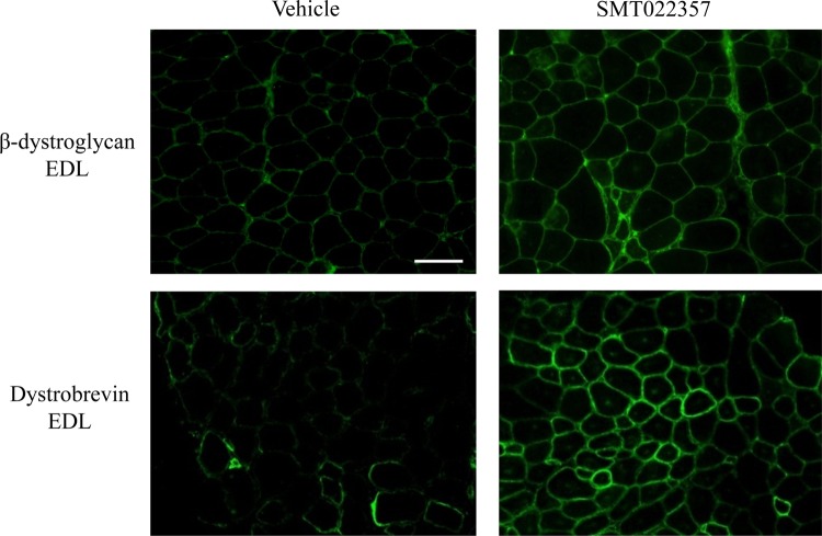
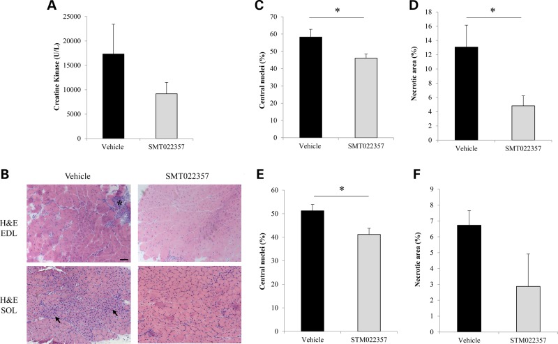
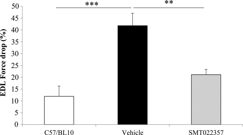
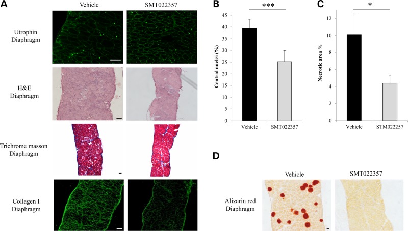
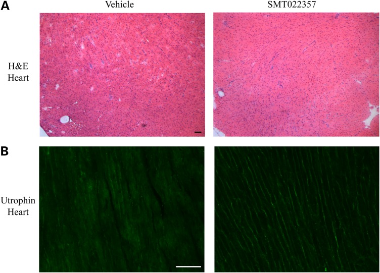
Duchenne muscular dystrophy (DMD) is a lethal, X-linked muscle-wasting disease caused by lack of the cytoskeletal protein dystrophin. There is currently no cure for DMD although various promising approaches are progressing through human clinical trials. By pharmacologically modulating the expression of the dystrophin-related protein utrophin, we have previously demonstrated in dystrophin-deficient mdx studies, daily SMT C1100 treatment significantly reduced muscle degeneration leading to improved muscle function. This manuscript describes the significant disease modifying benefits associated with daily dosing of SMT022357, a second-generation compound in this drug series with improved physicochemical properties and a more robust metabolism profile. These studies in the mdx mouse demonstrate that oral administration of SMT022357 leads to increased utrophin expression in skeletal, respiratory and cardiac muscles. Significantly, utrophin expression is localized along the length of the muscle fibre, not just at the synapse, and is fibre-type independent, suggesting that drug treatment is modulating utrophin transcription in extra-synaptic myonuclei. This results in improved sarcolemmal stability and prevents dystrophic pathology through a significant reduction of regeneration, necrosis and fibrosis. All these improvements combine to protect the mdx muscle from contraction induced damage and enhance physiological function. This detailed evaluation of the SMT C1100 drug series strongly endorses the therapeutic potential of utrophin modulation as a disease modifying therapeutic strategy for all DMD patients irrespective of their dystrophin mutation.


Figures

Figure 1.
Figure 1.
In vitro activity of SMT022357. (A) SMT022357 in vitro dose response in murine H2k-mdx utrnA-luc cells expressing the human utrophin promoter linked to a firefly luciferase reporter gene. Cells were treated with compound for 24 h in standard growth medium containing 1% DMSO. The chart shows relative luminescence (RLU) in relation to five different doses (0.1, 0.3, 1, 3 and 10 µM) of SMT022357. (B) SMT022357 significantly increased mRNA copy number of the utrophin transcript in murine myoblast cells. Cells were exposed to different dose of SMT022357 in standard media with 0.1% DMSO (vehicle) for 24 hours with six biological replicates. Utrophin transcripts were normalised with 18s and b2m. Values are mean ± SEM of n = 6 per condition; *P < 0.05, **P < 0.01, ***P < 0.001. (C) Relative utrophin protein expression in murine cells treated with SMT022357 was determined by western blot and standardized for α-actinin loading. Western blots showing 2.5-fold increase of utrophin protein expression with 10 µM of SMT022357 when compared with vehicle. Relative utrophin expression is shown as mean ± SEM of n = 3 per condition.
Figure 2.
Figure 2.
SMT022357 increases utrophin expression in skeletal muscles. (A) Immunofluoresence staining for utrophin in EDL and SOL muscles of 7 weeks old mdx mice treated for 5 weeks with 30 mg/kg/day SMT022357 or vehicle. Transverse sections were stained with anti-utrophin polyclonal antibody URD40 and anti-rabbit secondary antibody. Scale bar: 100 µm. (B) Longitudinal cryosections of TA muscle following treatment of mdx mice homogenously increased the utrophin expression along the myofiber after treatment with SMT022357 compared to vehicle group. Scale bar: 100 µm. (C) Co-immunohistochemical staining of TA muscle with utrophin, α-Bungarotoxin and DAPI prepared from mdx mice treated with SMT022357. White arrows indicate utrophin expression in extra-synaptic regions of the sarcolemma. Scale bar: 50 µm.
Figure 3.
Figure 3.
SMT022357 treatment results in a decrease in regeneration with no change in fibre type composition in the skeletal muscle. (A) Semi-quantitative RT-PCR demonstrates a decrease of skeletal muscle regeneration/myogenic differentiation markers Bex1, myogenin and cell cycle dependent-kinase inhibitors (CDKI) p21 and p27 from EDL of mdx mice dosed with SMT022357 (357, n = 8) compared to vehicle (Ve, n = 8). S13 ribosomal protein was used as an internal control for the RT-PCR. A 12% decrease in Bex1 (P = 0.034), 21% decrease in p21 (P = 0.002), 67% decrease in p27 (P < 0.001) and 64% decrease in Myogenin (P < 0.001) transcript levels were quantified by ImageJ software in SMT022357 group compared to vehicle. (B) Immunofluoresence staining for type 1a, 2a and 2b fibre types in the EDL of 7 weeks old mdx mice treated for 5 weeks with 30 mg/kg/day SMT022357 or vehicle (n = 6 per group). Sections were stained with anti-MYHC1, anti-MYHC2A anti-MYHC2B and anti-mouse secondary antibody. Percentage of type 1a, 2a and 2b fibre types was quantified by ImageJ software in the whole EDL muscle (n = 6 per group). No change was observed in fibre type composition of whole EDL muscle treated with SMT022357 compared to treatment with vehicle only. Scale bar: 100 µm.
Figure 4.
Figure 4.
SMT022357 treatment improves fibre membrane stability. Immunofluoresence staining for β-dystroglycan, and dystrobrevin in EDL muscle of 7 weeks old mdx mice treated for 5 weeks with 30 mg/kg/day SMT022357 or vehicle shows that key components of the DAPC complex are properly localised to the sarcolemma after SMT022357 treatment. n = 3 per group; Scale bar: 100 µm.
Figure 5.
Figure 5.
SMT022357 prevents muscular dystrophy in skeletal muscle. (A) Creatine kinase (CK) levels in serum following daily oral gavage of mdx mice with 30 mg/kg of SMT022357 or vehicle from two weeks of age for five weeks. A 47% decrease in serum CK was observed after treatment with SMT022357 compared to vehicle-treated animals (n = 7-9 per group). (B) H&E-stained transverse muscle sections of EDL muscle (7 weeks of age) in untreated vs. treated mdx mice (n = 10 per group) showing necrotic areas (black stars) and regenerating fibres (black arrows). Scale bar: 100 µm. (C) Muscle from mice dosed with SMT022357 showed a significant 20.1% (P = 0.03) decrease in centrally nucleated fibres compared to the vehicle group in EDL muscles. Values are mean ± SEM of n = 9 per groups; *P < 0.05. (D) The necrotic muscle area in EDL of mice treated with SMT022357 significantly decreased by 63.1% (P = 0.02) compared to the vehicle group. Values are mean ± SEM of n = 9 per groups; *P < 0.05. (E) Muscle from mice dosed with SMT022357 showed a significant 19.6% (P = 0.02) decrease in centrally nucleated fibres compared to the vehicle group in SOL muscles. Values are mean ± SEM of n = 7 per groups; *P < 0.05. (F) The necrotic muscle area in SOL of mice treated with SMT022357 decreased by 57.3% (P = 0.06) compared to the vehicle group. Values are mean ± SEM of n = 7 per groups.
Figure 6.
Figure 6.
SMT022357 protects the muscle against damage and improves the muscle function. The difference in force produced between the first and fifth stretch is represented as a sensitive indicator of the resistance of the muscle to stretch-induced damage. EDL muscles were stretched at 15% of their fibre length whilst contracting tetanically. Values are mean ± SEM of n = 10 per group; **P < 0.01, ***P < 0.001.
Figure 7.
Figure 7.
SMT022357 improves the pathology in the diaphragm. (A) Immunofluoresence staining for utrophin in the diaphragm of 7 weeks old mdx mice treated for 5 weeks with 30 mg/kg/day SMT022357 or vehicle (n = 10 per group). Sections were stained with anti-utrophin polyclonal antibody URD40 and anti-rabbit secondary antibody. H&E-stained transverse muscle sections of diaphragm muscle (7 weeks of age) showed improved morphology in treated vs. vehicle mdx mice (n = 10 per group). Masson's trichrome staining of diaphragm in untreated vs. treated mdx mice (n = 8) indicated that SMT022357 treatment reduced the amount of collagen infiltration (stained in blue); immunofluorescence using anti-collagen type I antibody confirmed a decrease of collagen in the muscular endomysium of SMT022357 treated diaphragm. Scale bar 100 µm. (B) Diaphragms of mice dosed with SMT022357 showed a significant 35.9% decrease in centrally nucleated fibres compared to the vehicle group. Values are mean ± SEM of n = 8 per groups; ***P < 0.001. (C) The necrotic diaphragm area of mice treated with SMT022357 significantly decreased by 56.6% compared to the vehicle group. Values are mean ± SEM of n = 8 per group; *P < 0.05. (D) The vehicle mdx diaphragm exhibits positive staining with Alizarin Red indicating the presence of calcium deposits. SMT022357 treatment completely prevented the calcification. (n = 10 per group); Scale bar: 100 µm.
Figure 8.
Figure 8.
SMT022357 increases utrophin expression in the mdx heart. (A) H&E staining of transverse heart sections in 7-week-old, vehicle- and SMT022357-treated mdx mice. n = 10 per group. Scale bar: 100 µm. (B) Immunostaining showed an increase of utrophin in the heart of 7-week-old mdx mice after SMT022357 (30 mg/kg/day) treatment compared to the vehicle group. (n = 10 per group); Scale bar: 100 µm.

Similar articles

Cited by

References

    1. Cohn R.D., Campbell K.P. (2000) Molecular basis of muscular dystrophies. Muscle Nerve, 23, 1456–1471. - PubMed
    1. Dickson G., Love D.R., Davies K.E., Wells K.E., Piper T.A., Walsh F.S. (1991) Human dystrophin gene transfer: production and expression of a functional recombinant DNA-based gene. Hum. Genet., 88, 53–58. - PubMed
    1. Emery A.E. (1993) Duchenne muscular dystrophy--Meryon's disease. Neuromusc. Disord., 3, 263–266. - PubMed
    1. Bogdanovich S., Perkins K.J., Krag T.O., Khurana T.S. (2004) Therapeutics for Duchenne muscular dystrophy: current approaches and future directions. J. Mol. Med., 82, 102–115. - PubMed
    1. Bach J.R., O'Brien J., Krotenberg R., Alba A.S. (1987) Management of end stage respiratory failure in Duchenne muscular dystrophy. Muscle Nerve, 10, 177–182. - PubMed

Publication types

MeSH terms