. 2015 Sep;44(9):1188-96.
doi: 10.1016/j.ijom.2015.04.001. Epub 2015 Apr 29.

Fast three-dimensional superimposition of cone beam computed tomography for orthopaedics and orthognathic surgery evaluation

Affiliations

Fast three-dimensional superimposition of cone beam computed tomography for orthopaedics and orthognathic surgery evaluation

A Weissheimer et al. Int J Oral Maxillofac Surg. 2015 Sep.

Abstract

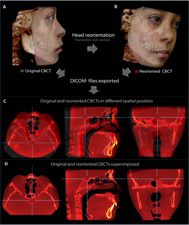
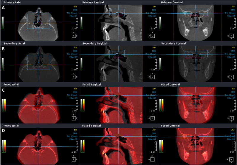
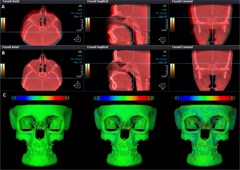
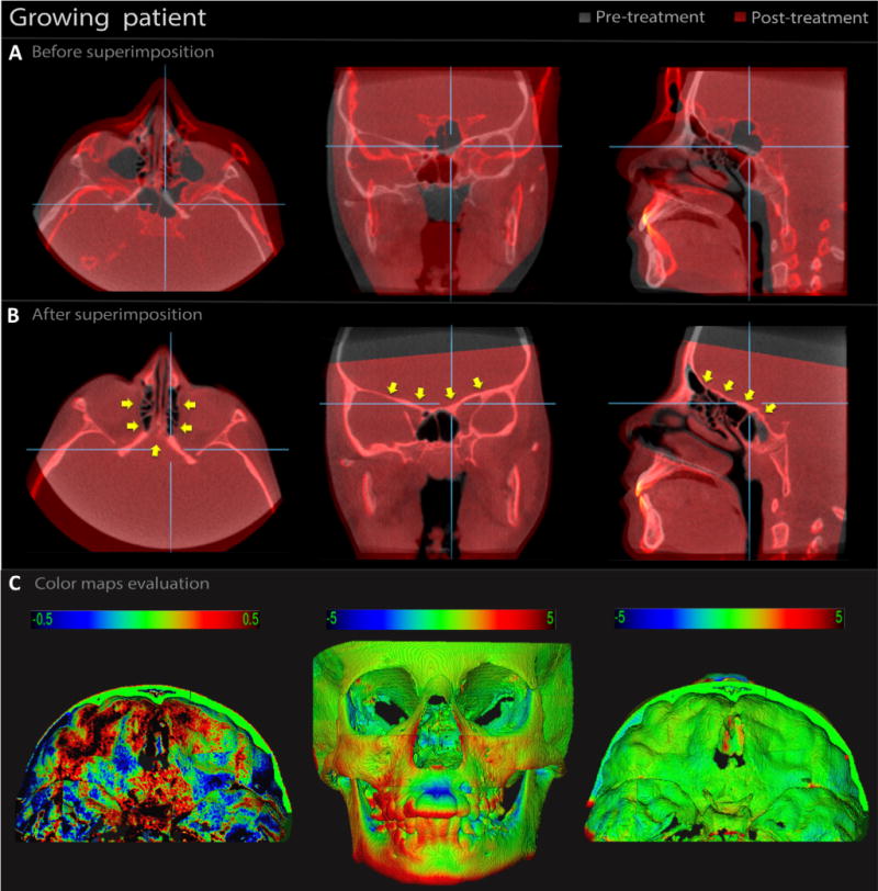
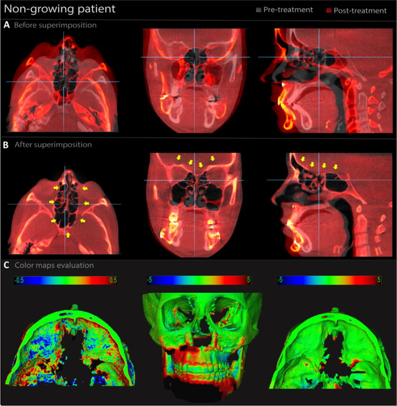
The aim of this study was to validate a method for fast three-dimensional (3D) superimposition of cone beam computed tomography (CBCT) in growing patients and adults (surgical cases). The sample consisted of CBCT scans of 18 patients. For 10 patients, as the gold standard, the spatial position of the pretreatment CBCT was reoriented, saved as a reoriented volume, and then superimposed on the original image. For eight patients, four non-growing and four growing, the pre- and post-treatment scans were superimposed. Fast voxel-based superimposition was performed, with registration at the anterior cranial base. This superimposition process took 10-15s. The fit of the cranial base superimposition was verified by qualitative visualization of the semi-transparent axial, sagittal, and coronal cross-sectional slices of all corresponding anatomical structures. Virtual 3D surface models of the skull were generated via threshold segmentation, and superimposition errors in the reoriented models and the results of treatment for the treated cases were evaluated by 3D surface distances on colour-coded maps. The superimposition error of the spatial reorientation and for growing and non-growing patients was <0.5mm, which is acceptable and clinically insignificant. The voxel-based superimposition method evaluated was reproducible in different clinical conditions, rapid, and applicable for research and clinical practice.

Keywords: 3D image registration; cone-beam computed tomography; orthodontic and orthopaedic treatment; orthognathic surgery; voxel-based superimposition.


Conflict of interest statement

Competing interests

None declared.

Figures

Fig. 1
Fig. 1
Example of a case used as the gold standard. (A) Original CBCT. (B) Same patient CBCT reoriented to a different spatial position. (C) Slices of multiplanar reconstruction showing original (grey) and reoriented CBCT (red) before superimposition. (D) Slices showing both CBCTs after superimposition at the anterior cranial base.
Fig. 2
Fig. 2
OnDemand3D voxel-based superimposition process. (A) Anterior cranial base selection (blue rectangle) in multiplanar slices of pretreatment CBCT and (B) post-treatment CBCT. (C) Multiplanar slices view of pre-treatment (grey) and post-treatment (red) CBCT files before superimposition and (D) after superimposition at the anterior cranial base. (For interpretation of the references to colour in figure legend, the reader is referred to the web version of the article.)
Fig. 3
Fig. 3
CBCT superimposition of a gold standard patient. (A) Multiplanar slices of original CBCT (grey) and reoriented CBCT (red) files before superimposition and (B) after superimposition at the anterior cranial base, in OnDemand3D. Note the complete correspondence of registration in all areas of the skull. (C) Skull models after superimposition showing 3D displacements (registration error) via colour maps with scales of 0.5 mm, 0.25 mm, and 0.10 mm. Positive and negative values indicate outward (red) and inward (blue) changes, respectively. Note the error was less than 0.25 mm (CMF application software). (For interpretation of the references to colour in figure legend, the reader is referred to the web version of the article.)
Fig. 4
Fig. 4
CBCT superimposition of a growing patient subjected to rapid palatal expansion (RPE) with 1-year follow-up. (A) Multiplanar slices of pre-treatment CBCT (grey) and post-treatment CBCT (red) before superimposition and (B) after superimposition, in OnDemand3D. Note the complete correspondence of registration at the anterior cranial base (yellow arrows). (C) 3D models after superimposition showing 3D displacements via colour maps. In the 0.5-mm colour map, the black areas represent changes of 0.5 mm, blue/red areas represent changes less than 0.5 mm, and green areas represent no changes. In the 5-mm colour map, the areas in black, red, and blue are due to the RPE and normal growth. The changes at the cranial base were minimal (green) (CMF application software). (For interpretation of the references to colour in figure legend, the reader is referred to the web version of the article.)
Fig. 5
Fig. 5
CBCT superimposition of an adult patient subjected to orthognathic surgery with 1-year follow-up. (A) Multiplanar slices of pre-treatment CBCT (grey) and post-treatment CBCT (red) before superimposition and (B) after superimposition, in OnDemand3D. Note the complete correspondence of registration at the anterior cranial base (yellow arrows). (C) 3D models after superimposition showing 3D displacements via colour maps. In the 0.5-mm colour map, the black areas represent changes of 0.5 mm, blue/red areas represent changes less than 0.5 mm, and green areas represents no changes. In the 5-mm colour map, the areas in black, red, and blue are changes due to the surgical treatment. The changes at the cranial base were minimal (green) (CMF application software). (For interpretation of the references to colour in figure legend, the reader is referred to the web version of the article.)

References

    1. Kapila S, Conley RS, Harrell WE., Jr The current status of cone beam computed tomography imaging in orthodontics. Dentomaxillofac Radiol. 2011;40:24–34. - PMC - PubMed
    1. Weissheimer A, Menezes LM, Sameshima GT, Enciso R, Pham J, Grauer D. Imaging software accuracy for 3-dimensional analysis of the upper airway. Am J Orthod Dentofacial Orthop. 2012;142:801–13. - PubMed
    1. Grauer D, Cevidanes LS, Proffit WR. Working with DICOM craniofacial images. Am J Orthod Dentofacial Orthop. 2009;136:460–70. - PMC - PubMed
    1. Cevidanes LH, Heymann G, Cornelis MA, DeClerck HJ, Tulloch JF. Superimposition of 3-dimensional cone-beam computed tomography models of growing patients. Am J Orthod Dentofacial Orthop. 2009;136:94–9. - PMC - PubMed
    1. Cevidanes LH, Motta A, Proffit WR, Ackerman JL, Styner M. Cranial base superimposition for 3-dimensional evaluation of soft-tissue changes. Am J Orthod Dentofacial Orthop. 2010;137(4 Suppl. 1):S120–9. - PMC - PubMed

Publication types

LinkOut - more resources