. 2015 Aug;12(2):2417-24.
doi: 10.3892/mmr.2015.3694. Epub 2015 Apr 28.

Colorectal cancer stem cell and chemoresistant colorectal cancer cell phenotypes and increased sensitivity to Notch pathway inhibitor

Affiliations

Colorectal cancer stem cell and chemoresistant colorectal cancer cell phenotypes and increased sensitivity to Notch pathway inhibitor

Rui Huang et al. Mol Med Rep. 2015 Aug.

Abstract

Colorectal cancer stem cells (Co-CSCs) are a small subpopulation of tumor cells which have been proposed to be tumor-initiating cells in colorectal cancer (CRC) and to be implicated in resistance to standard chemotherapy. Chemoresistance is a common problem in the clinic. However, the interrelation between Co-CSCs and chemoresistant cells has yet to be elucidated. The present study investigated the Co-CSC phenotype in colonospheres and chemoresistant CRC cell lines and aimed to identify targets for therapy. Colonospheres and chemoresistant CRC cells were found to be enriched with the CSC markers CD133 and CD44, and exhibited similar phenotypes. Furthermore, it was found that Notch signaling may simultaneously regulate Co-CSCs and chemoresistant cells and may represent a novel strategy for targeting this pathway in CRC.


Figures

Figure 1
Figure 1
Colonospheres and chemoresistant cell lines are enriched with Co-CSC markers. (A) Western blot analysis revealed that expression of the Co-CSC markers CD133 and CD44 was higher in the colonospheres and HCT116/5FU-R and HCT116/OxR chemoresistant cells compared with the parental HCT116 human CRC cells. β-actin was used as a loading control. (B) Flow cytometric analysis revealed that the colonospheres and chemoresistant cell lines were enriched with cells expressing CD133 and CD44 compared with the parental cell line. A total of 33% of the HCT116/5FU-R cells, 47% of the HCT116/OxR cells and 65% of the HCT116/colonosphere cells expressed CD133 compared with 2% of the parental HCT116 cells. Similarly, 84% of the HCT116/5FU-R cells, 93% of the chemoresistant cells and 92% of the HCT116/colonosphere cells expressed CD44 compared with 48% of the parental cells. Cytometric analysis plots using isotype control antibodies were used as staining controls. (C) CD44 and CD133 labelling and flow cytometric analysis revealed a 4.8-, 22- and 24.7-fold enrichment of double-positive cells in the HCT116/5FU-R, HCT116/OxR and colonosphere cells compared with the parental HCT116 cell line. SCC, side scatter; Co-CSC, colorectal cancer stem cell; CD, cluster of differentiation; 5-FU, 5-fluorouracil; R, resistant; Ox, oxaliplatin.
Figure 2
Figure 2
Colonospheres and chemoresistant cell exhibit a cancer stem cell phenotype. (A) Colonospheres and chemoresistant cells proliferated at a significantly slower rate than parental cells, detected using the Cell Counting Kit-8 assay. (B) Parental cells were sensitive to 5FU and Ox following exposure for 72 h, with only 34 and 21% of cells remaining, respectively, compared with the untreated cells. By contrast, HCT116/5FU-R cells were resistant to 5FU; however, they also exhibited increased resistance to Ox compared with the parental cells. Similarly, HCT116/Ox-R cells were resistant to Ox, as well as 5FU. Colonosphere cells were also resistant to Ox and 5-FU. (C) Cells were plated in an ultralow-attachment 96-well plate in the absence of serum and after 14 days, the rate of viable sphere-forming cells was assessed. The colonosphere formation rate was significantly increased in the colonospheres and chemoresistant cells compared with the parental cells. (D) Clonogenic assay revealed that the number of colonies larger than 50 µm in diameter which were formed under standard growth conditions was significantly higher in the colonospheres and chemoresistant compared with the parental HCT116 cells. Data are presented as the mean ± standard error. *P<0.05 vs. HCT116 cells. OD, optical density, 5-FU, 5-fluorouracil; Ox, oxaliplatin; R, resistant.
Figure 3
Figure 3
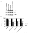
Effect of Notch pathway inhibition on colonospheres and chemoresistant cells. (A) Analysis of whole-cell lysates from parental HCT116 cells, chemoresistant HCT116/5FU-R and HCT116/Ox-R cells and colonosphere cells revealed an increase in Notch1 and Hes1 in the chemoresistant and colonosphere cells compared with the parental cells. (B) Cells were treated with DMSO (control) or a Notch pathway inhibitor DAPT and the cell number was assessed. Upon DAPT treatment, the cell number was significantly decreased in the chemoresistant and colonosphere cells (42–51%) compared with the HCT116 cells (12%). *P<0.05 vs. HCT116 cells. 5-FU, 5-fluorouracil; R, resistant; Ox, oxaliplatin; DAPT, N-[N-(3,5-difluorophenacetyl)-l-alanyl]-S-phen ylglycine t-butyl ester; DMSO, dimethyl sulfoxide; Hes, hairy and enhancer of split.
Figure 4
Figure 4
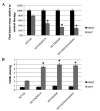
Effect of Notch pathway inhibition on in vivo tumor growth, proliferation and apoptosis. Mice were subcutaneously injected with 1×10⁶ HCT116, HCT116/5FU-R, HCT116/OxR or HCT116/colonosphere cells and treated with DMSO (control) or DAPT twice weekly. Final tumor masses were measured and compared between mice bearing tumors from each cell line. (A) In the DAPT-treated mice, the HCT116/5FU-R-, HCT116/OxR- and HCT116/colonosphere-derived tumors showed significantly greater growth inhibition than the HCT116-derived tumors. (B) In the DAPT-treated mice, TUNEL staining revealed significantly greater apoptosis in the HCT116/5FU-R-, HCT116/OxR- and HCT116/colonospheres-derived tumors than in tumors derived from the HCT116 cells. Data are presented as the mean ± standard error. *P<0.05 vs. HCT116 cells. 5-FU, 5-fluorouracil; R, resistant; Ox, oxaliplatin; DAPT, N-[N-(3,5-difluorophenacetyl)-l-alanyl]-S-phenylglycine t-butyl ester; DMSO, dimethyl sulfoxide; TUNEL, terminal deoxynucleotidyltransferase-mediated dUTP nick-end labeling.
Figure 5
Figure 5
Effect of Notch pathway inhibition on in vivo tumor characteristics. Immunohistochemical analysis of tumors was performed and multiple tumor fields were analyzed per group. Representative images of all groups and treatments are presented (magnification, x100). H&E staining revealed similar subcutaneous tumor morphology among all groups of tumors. Ki67 staining showed decreased cell proliferation in the tumors treated with DAPT; however, no significant differences were observed in the cell proliferation between the HCT116-, colonosphere-, HCT116/5FU-R- and HCT116/Ox-R-derived tumor sections. TUNEL staining revealed significantly increased apoptosis in response to DAPT in colonosphere-, HCT116/5FU-R- and HCT116/Ox-R-derived tumors compared with tumors derived from HCT116 cells (P<0.05). 5-FU, 5-fluorouracil; R, resistant; Ox, oxaliplatin; DAPT, N-[N-(3,5-difluorophenacetyl)-l-alanyl]-S-phenylglycine t-butyl ester; DMSO, dimethyl sulfoxide; TUNEL, terminal deoxynucleotidyltransferase-mediated dUTP nick-end labeling; H&E, hematoxylin and eosin.

References

    1. Jemal A, Bray F, Center MM, Ferlay J, Ward E, Forman D. Global cancer statistics. CA Cancer J Clin. 2011;61:69–90. doi: 10.3322/caac.20107. - DOI - PubMed
    1. Reya T, Morrison SJ, Clarke MF, Weissman IL. Stem cells, cancer, and cancer stem cells. Nature. 2001;414:105–111. doi: 10.1038/35102167. - DOI - PubMed
    1. O’Brien CA, Pollett A, Gallinger S, Dick JE. A human colon cancer cell capable of initiating tumour growth in immunodeficient mice. Nature. 2007;445:106–110. doi: 10.1038/nature05372. - DOI - PubMed
    1. Ricci-Vitiani L, Lombardi DG, Pilozzi E, et al. Identification and expansion of human colon-cancer-initiating cells. Nature. 2007;445:111–115. doi: 10.1038/nature05384. - DOI - PubMed
    1. Yeung TM, Mortensen NJ. Colorectal cancer stem cells. Dis Colon Rectum. 2009;52:1788–1796. doi: 10.1007/DCR.0b013e3181a8738c. - DOI - PubMed

MeSH terms