. 2015 Nov;40(12):2753-63.
doi: 10.1038/npp.2015.125. Epub 2015 May 4.

Increased GABAergic Efficacy of Central Amygdala Projections to Neuropeptide S Neurons in the Brainstem During Fear Memory Retrieval

Affiliations

Increased GABAergic Efficacy of Central Amygdala Projections to Neuropeptide S Neurons in the Brainstem During Fear Memory Retrieval

Kay Jüngling et al. Neuropsychopharmacology. 2015 Nov.

Abstract

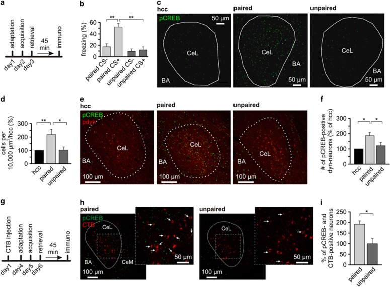
The canonical view on the central amygdala has evolved from a simple output station towards a highly organized microcircuitry, in which types of GABAergic neurons in centrolateral (CeL) and centromedial (CeM) subnuclei regulate fear expression and generalization. How these specific neuronal populations are connected to extra-amygdaloid target regions remains largely unknown. Here we show in mice that a subpopulation of GABAergic CeL and CeM neurons projects monosynaptically to brainstem neurons expressing neuropeptide S (NPS). The CeL neurons are PKCδ-negative and are activated during conditioned fear. During fear memory retrieval, the efficacy of this GABAergic influence on NPS neurons is enhanced. Moreover, a large proportion of these neurons (~50%) contain prodynorphin and somatostatin, two neuropeptides inhibiting NPS neurons. We conclude that CeL and CeM neurons inhibit NPS neurons in the brainstem by GABA release and that efficacy of this connection is strengthened upon fear memory retrieval. Thereby, this pathway provides a possible feedback mechanism between amygdala and brainstem routes involved in fear and stress coping.


Figures

Figure 1
Figure 1
CeL neurons project to NPS neurons in the periLC region. (a) Scheme of rAAV-hSynI-mCherry injection into the CeL for anterograde tracing (modified after: mbl.org) to detect axons in the periLC region originating from CeL neurons. (b) Example of an injection site in the CeL in a horizontal slice preparation. Infected neurons express mCherry (red; VL, lateral ventricle; c, caudal; r, rostral). (c) In transgene NPS-EGFP mice (n=4), mCherry-positive fibers are overlapping with the NPS neurons cluster of the periLC region (4V, fourth ventricle). The tracing indicates that among other neurons, NPS neurons might be targeted by CeL neurons.
Figure 2
Figure 2
Retrogradely labeled neurons in the CeL are PKCδ-negative. (a) Scheme depicting the injection of the retrograde tracer CTB-Alexa594 into the periLC region (left panel; modified after: mbl.org). An example of a cholera toxin B-Alexa594 (CTB; red, right panel) unilateral injection in the NPS-EGFP cluster at the LC (f4V: fourth ventricle). Solid circle marks the injection and the dashed circle outlines the location of the NPS-EGFP neurons within the slice. (b) Immunohistochemical stainings from CTB-injected animals against pdyn reveals co-localization of retrogradely transported CTB and pdyn within CeL neurons. (c) Immunohistochemical stainings from CTB-injected animals against PKCδ reveals no co-localization of retrogradely transported CTB and PKCδ within CeL neurons (d) Immunohistochemical staining against PKCδ (green) and pdyn (red) in coronal slices containing the CeL revealing that these peptides are expressed separately. (e) Immunostaining against SOM in the CeL of CTB-injected mouse. (f) Co-immunostaining against pdyn (red) and SOM (green) in the CeL. There was about two times more SOM- than pdyn-positive neurons detected. The vast majority (>80%) of pdyn-neurons was positive for SOM (n=3). (g) Percentage of CeL neurons that were CTB and pdyn-positive (n=4), CTB and SOM-positive (n=4), CTB and PKCδ-positive (n=2), pdyn- and PKCδ-positive (n=3), and pdyn- and SOM-positive (n=2).
Figure 3
Figure 3
Expression of pCREB in dynorphinergic CeL neurons. (a) Scheme of the experimental design. (b) Quantification of freezing responses during retrieval in paired and unpaired groups. (c) Examples of immunohistochemical staining against pCREB(S133; green) in the CeL of home-cage controls (HCC) and after fear retrieval of paired and unpaired groups. (d) Quantification of pCREB-positive nuclei of the CeL neurons in the HCC (n=9 animals), paired (n=8 animals), and unpaired group (n=7 animals). After counting pCREB-positive nuclei per 10 000 μm2 of the CeL, data were normalized to the HCC of each individual set. (e) Examples of co-staining for pdyn and pCREB(S133) in CeL of the HCC (n=9), paired (n=8), and unpaired (n=7) group. (f) Quantification of the normalized pCREB-positive nuclei in pdyn-positive neurons, normalized to the HCC of each individual set. (g) Scheme of experimental design. (h) Examples of immunohistochemical staining against pCREB (S133; green) in the CeL of CTB-injected mice after fear retrieval of the paired (n=6 animals) and unpaired (n=6 animals) groups. CTB was injected into the LC/periLC region and CTB-positive neurons (red) were detectable in the CeL. Note also the presence of some CTB-positive neurons in the CeM. (i) Quantification of the normalized percentage of pCREB-positive nuclei in CTB-positive neurons, normalized to the unpaired group of each individual set.
Figure 4
Figure 4
CeL neurons form GABAergic synapses on NPS neurons in the periLC region. (a) Scheme of the experimental design. rAAV solution was injected into the CeL and NPS-EGFP neurons in the periLC were recorded in horizontal slice preparations. GABA release was triggered by brief blue-light exposure (modified after: mbl.org). Example of an injection site (b) example of a horizontal slice with the injection site (ChR2-EYFP fluorescence) within the cluster of PKCδ-positive neurons of the CeL. (c) Quantification of freezing responses during retrieval in paired and unpaired groups of transgenic NPS-EGFP mice used for ex vivo recordings. (d) Examples of light-evoked GABAergic responses in the voltage-clamp mode in NPS neurons of paired and unpaired trained animals 1.5 h after fear retrieval. Light-evoked responses were sensitive to gabazine (GBZ; middle). (e) Quantification of the light-evoked IPSC failure rate recorded in paired (n=23 neurons/6 animals) and unpaired (n=16 neurons/4 animals) trained mice using maximal stimulation intensity. (f) Quantification of the failure rates at decreasing stimulation intensities recorded in the paired and unpaired mice. The data were fitted with an asymptotic function (solid lines) and the confidence intervals of the fit (95%) are depicted by the dashed lines. (g) Quantification of the mean success amplitude during maximal stimulation. (h) Input–output relationship (normalized IPSC amplitude vs relative light intensity) for paired (n=15 neurons/4 animals) and unpaired (n=13 neurons/4 animals) trained mice. The data were fitted with an asymptotic function (solid lines) and the confidence intervals of the fit (95%) are depicted by the dashed lines. (i) Quantification of the paired-pulse ratio (100 ms interval) of light-evoked IPSCs at maximal stimulation.
Figure 5
Figure 5
Dynorphin and somatostatin inhibit NPS neurons in the periLC region. (a) Immunostaining against pdyn and NPS-EGFP (top), and SOM and NPS-EGFP (bottom) in horizontal slice preparations. The stainings indicate the presence of both neuropeptides in the vicinity of NPS neurons in the periLC region. (b) Example of a current-clamp recording of the NPS neuron. Application of dynA hyperpolarizes the recorded neuron from a membrane potential of −65 mV. Hyperpolarizing current injections (−40 pA) were applied to analyze the input resistance. During maximal drug effect, the membrane potential was briefly manually clamped back to baseline values. A similar hyperpolarization was observed upon SOM-application at a membrane potential of −60 mV in a different NPS neuron (c and d) Quantification of the substance-induced changes of the membrane potential in NPS neurons. (e) Fear responsive (fearon) neurons of the CeL inhibit via GABAergic synapses fearoff, PKCδ-positive CeL neurons, which form GABAergic synapses on the CeM output neurons. Activity of fearon neurons would disinhibit CeM neurons and thus allow eg freezing behavior (Ciocchi et al, 2010; Haubensak et al, 2010; Li et al, 2013). In addition, the CeL fearon neurons and CeM neurons form inhibitory GABAergic synapses on NPS neurons in the periLC region. LC and/or LPB (lateral parabrachial nucleus) NPS neurons in turn project to LA/BA of the amygdala and reduce anxiety or facilitate fear extinction via NPS-release. Terminals from the CeL and CeM release GABA onto NPS neurons and thus reduce their activity. A subset of CeL GABAergic inputs might contain, eg dynorphin and/or SOM, which could inactivate NPS neurons in the periLC when released.

References

    1. Carey AN, Lyons AM, Shay CF, Dunton O, McLaughlin JP (2009). Endogenous kappa opioid activation mediates stress-induced deficits in learning and memory. J Neurosci 29: 4293–4300. - PMC - PubMed
    1. Charney DS (2003). Neuroanatomical circuits modulating fear and anxiety behaviors. Acta Psychiatr Scand Suppl 108: 38–50. - PubMed
    1. Chauveau F, Lange MD, Jungling K, Lesting J, Seidenbecher T, Pape HC (2012). Prevention of stress-impaired fear extinction through neuropeptide s action in the lateral amygdala. Neuropsychopharmacology 37: 1588–1599. - PMC - PubMed
    1. Chessell IP, Black MD, Feniuk W, Humphrey PP (1996). Operational characteristics of somatostatin receptors mediating inhibitory actions on rat locus coeruleus neurones. Br J Pharmacol 117: 1673–1678. - PMC - PubMed
    1. Ciocchi S, Herry C, Grenier F, Wolff SB, Letzkus JJ, Vlachos I et al (2010). Encoding of conditioned fear in central amygdala inhibitory circuits. Nature 468: 277–282. - PubMed

Publication types

MeSH terms

LinkOut - more resources