Review
. 2015 Sep 11;427(18):2919-30.
doi: 10.1016/j.jmb.2015.04.013. Epub 2015 Apr 30.

The Mechanism and Function of Group II Chaperonins

Affiliations
Review

The Mechanism and Function of Group II Chaperonins

Tom Lopez et al. J Mol Biol. .

Abstract

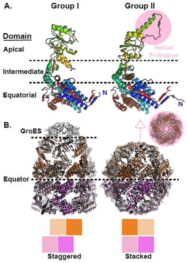
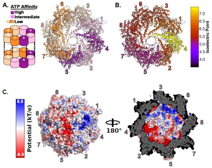
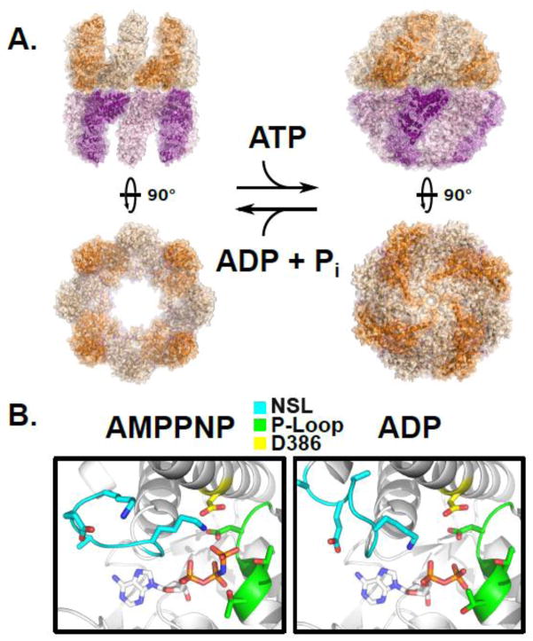
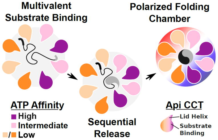
Protein folding in the cell requires the assistance of enzymes collectively called chaperones. Among these, the chaperonins are 1-MDa ring-shaped oligomeric complexes that bind unfolded polypeptides and promote their folding within an isolated chamber in an ATP-dependent manner. Group II chaperonins, found in archaea and eukaryotes, contain a built-in lid that opens and closes over the central chamber. In eukaryotes, the chaperonin TRiC/CCT is hetero-oligomeric, consisting of two stacked rings of eight paralogous subunits each. TRiC facilitates folding of approximately 10% of the eukaryotic proteome, including many cytoskeletal components and cell cycle regulators. Folding of many cellular substrates of TRiC cannot be assisted by any other chaperone. A complete structural and mechanistic understanding of this highly conserved and essential chaperonin remains elusive. However, recent work is beginning to shed light on key aspects of chaperonin function and how their unique properties underlie their contribution to maintaining cellular proteostasis.

Keywords: TRiC/CCT; chaperones; chaperonin; protein folding; proteostasis.


Figures

Figure 1
Figure 1
Structural comparison of group I and group II chaperonins. A) Domain architecture is conserved between group I and group II chaperonins. Left, chain A from the cis-cavity of a GroE crystal structure. Right, chain A from the MmCpn crystal structure. B) Comparison of the GroEL (left) and MmCpn (right) complex architectures. The GroE and MmCpn crystal structures used were PDBID: 1AON [16] and PDBID: 3RUW [40] respectively.
Figure 2
Figure 2
The subunit arrangement of the hetero-oligomeric eukaryotic chaperonin TRiC. A) Left, a schematic of the subunit arrangement of TRiC showing the inter-ring register. Right, the crystal structure of TRiC showing the subunit arrangement and the partitioning of ATP affinities. Subunits are colored by their ATP affinity. B) Influence of subunit arrangement on charge distribution. Subunits are colored by their isoelectric points. Isoelectric points were estimated for the Saccharomyces cerevisiae CCT subunits using the pepstats program from the EMBOSS suite [112]. C) The surface charge characteristics inside the closed TRiC cavity. Left, a view of the outside of the chaperonin folding chamber colored by surface electrostatic potential. Right, a view of the lid of the folding chamber from the inside illustrating the polarized nature of the TRiC cavity. Surface electrostatics were rendered in PyMOL [113] using APBS [114] Tools [115]. The TRiC structure is PDBID: 4V94 [57] prepared by adding missing heavy atoms in PDBFixer [116].
Figure 3
Figure 3
Structural basis for substrate release during lid closure. The apical domains of the group II chaperonin MmCpn in the closed state, highlighting the substrate binding surface at the interface between two adjacent apical subunits, from PDBID: 3RUW [40]. The substrate binding surface of the left subunit comprising Helix 11 and the Proximal Loop is indicated as well as the RLS Loop of the right hand subunit which is responsible for evicting bound substrate during closure.
Figure 4
Figure 4
The ATP driven conformational cycle of the group II chaperonin. A) Left, the open apo state of a group II chaperonin MmCpn determined by cryoelectron microscopy, PDBID: 3IYF. Right, the cryoelectron microscopy structure of wild type MmCpn in the ATP-induced closed state, PDBID: 3LOS [64]. B) The position of the nucleotide sensing loop (NSL) is dependent upon the presence of the γ-phosphate. Left, the crystal structure of the archaeal chaperonin MmCpn in a pre-hydrolysis state, complexed with the non-hydrolyzable ATP analog AMPPNP, PDBID: 3RUV. Right, the crystal structure of the same MmCpn complexed with ADP, PDBID: 3RUW [40]. The nucleotide sensing loop, P-loop, and catalytic aspartate, D386 are indicated in cyan, green, and yellow respectively. The conformation of the NSL is altered by the scission of the gamma phosphate between the left and right panels.
Figure 5
Figure 5
A schematic view of factors promoting substrate folding by TRiC and other group II chaperonins. The substrate folding process involves multivalent binding of distinct substrate epitopes by the different TRiC apical domains. Release of substrate may proceed in a sequential fashion owing to the asymmetric usage of ATP by the eukaryotic group II chaperonin and the nonconcerted nature of lid closure. Once encapsulated, substrates experience a polarized charge environment with one lobe of the complex demonstrating a positive electrostatic potential while the other is negative.

Similar articles

Cited by

References

    1. Anfinsen CB, Haber E, Sela M, White FH., Jr The kinetics of formation of native ribonuclease during oxidation of the reduced polypeptide chain. Proceedings of the National Academy of Sciences of the United States of America. 1961;47:1309–14. - PMC - PubMed
    1. Levinthal C. How To Fold Graciously. University of Illinois Bulletin. 1969;67:22–4.
    1. Baldwin RL. The search for folding intermediates and the mechanism of protein folding. Annual review of biophysics. 2008;37:1–21. - PubMed
    1. Sosnick TR, Barrick D. The folding of single domain proteins--have we reached a consensus? Current opinion in structural biology. 2011;21:12–24. - PMC - PubMed
    1. van den Berg B, Ellis RJ, Dobson CM. Effects of macromolecular crowding on protein folding and aggregation. The EMBO journal. 1999;18:6927–33. - PMC - PubMed

Publication types

MeSH terms