. 2015 May 12;11(6):851-858.
doi: 10.1016/j.celrep.2015.04.001. Epub 2015 Apr 30.

Somatosensory substrates of flight control in bats

Affiliations

Somatosensory substrates of flight control in bats

Kara L Marshall et al. Cell Rep. .

Abstract

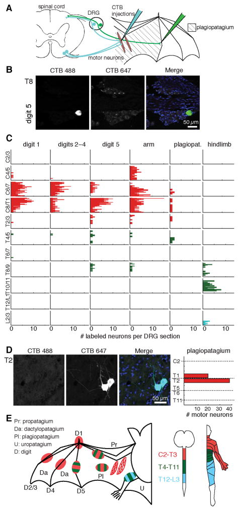
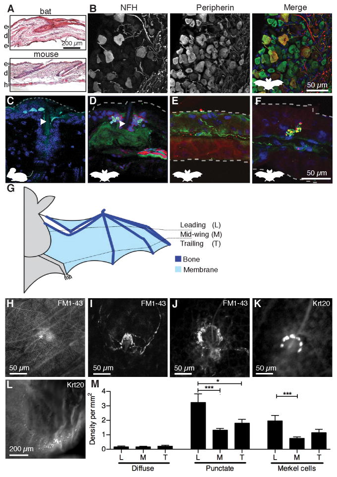
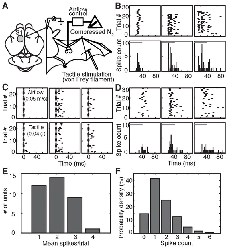
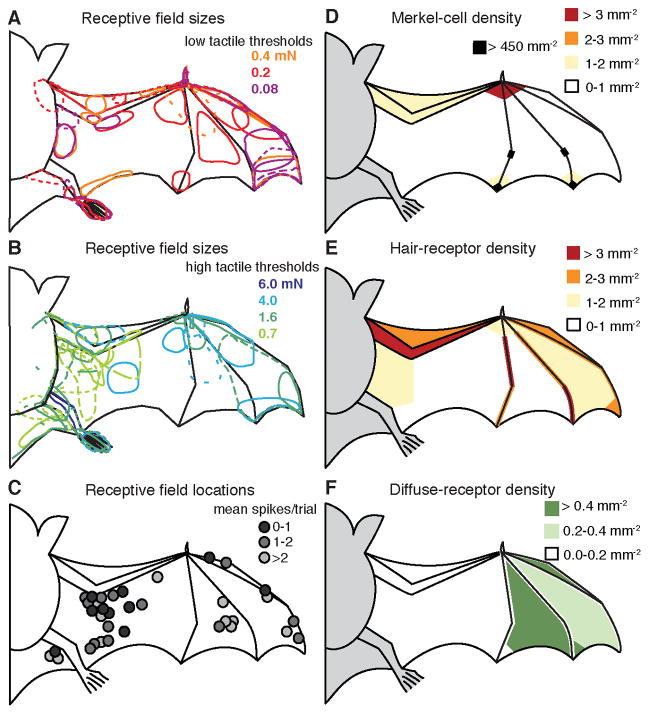
Flight maneuvers require rapid sensory integration to generate adaptive motor output. Bats achieve remarkable agility with modified forelimbs that serve as airfoils while retaining capacity for object manipulation. Wing sensory inputs provide behaviorally relevant information to guide flight; however, components of wing sensory-motor circuits have not been analyzed. Here, we elucidate the organization of wing innervation in an insectivore, the big brown bat, Eptesicus fuscus. We demonstrate that wing sensory innervation differs from other vertebrate forelimbs, revealing a peripheral basis for the atypical topographic organization reported for bat somatosensory nuclei. Furthermore, the wing is innervated by an unusual complement of sensory neurons poised to report airflow and touch. Finally, we report that cortical neurons encode tactile and airflow inputs with sparse activity patterns. Together, our findings identify neural substrates of somatosensation in the bat wing and imply that evolutionary pressures giving rise to mammalian flight led to unusual sensorimotor projections.


Conflict of interest statement

The authors declare no conflict of interest.

Figures

Figure 1
Figure 1. Bat wing neuronal tracing reveals atypical somatosensory-motor innervation
(A) Schematic of neuronal tracing approach. (B) T8 DRG section from bat wing injected at digit 5 with CTB Alexa-488 (green). Merged images shows DAPI-stained nuclei (blue). (C) Histograms show the number of neurons labeled at each spinal level from all injections (≤1.5 μl per injection). Each column shows labeling from a separate wing site (N=2–3 injections per site from 2–3 bats). See also Figure S1. Color key in panel E. (D) Motor neurons in upper thoracic spinal cord were labeled by injection of CTB Alexa-647 into plagiopatagial muscles. Merged image shows DAPI-stained nuclei (blue). Right, motor neuron quantification (N=6 injections in 2 bats). Dashed lines indicate transection levels of dissected spinal cords (see Supplemental Methods). (E) Dermatome and myotome maps. Left, injection sites colored according to spinal level of innervation. Motor pools are represented by hatched areas. Middle: spinal level color key. Right, map of corresponding human dermatomes.
Figure 2
Figure 2. An unusual repertoire of touch receptors innervates bat wings
(A) Skin histology of bat wing and mouse limb [epidermis (e), dermis (d), hypodermis (h)]. (B) Bat DRG labeled with antibodies against neurofilament H (NFH; red) and peripherin (green). DAPI (blue) labeled nuclei. Labeling and colors apply to B–F. See also Figure S2A. (C–F) Immunohistochemistry of mouse limb (C) and bat wing skin (D–F). Dashed lines denote skin surfaces. (C) Keratin 8 (Krt8) antibodies (cyan) labeled mouse Merkel cells adjacent to a guard hair (arrowhead). (D) Krt20 antibodies (cyan) labeled bat Merkel cells around a wing hair (arrowhead). (E) Free nerve ending. (F) Knob-like ending. Scale applies to C–F. (G) Schematic of wing areas. (H–J) In vivo FM1-43 injections labeled (H) diffuse endings (asterisk) (I) lanceolate endings and (j) sensory neurons similar to mouse Merkel-cell afferents (see also Figure S2B–D). (K–L) Merkel cells were surveyed using whole-mount Krt20 immunostaining of 12 wing areas (see Figure S2E). Merkel cells were found near hairs (K) and along fingertips (L). (M) Sensory ending density at wing areas defined in (G). [N=4 wings from four bats (diffuse and punctate), N=4 wings from three bats (Merkel cells)]. Punctate endings and Merkel cells were unevenly distributed across wing areas (One-way ANOVA; P=0.0004 and P=0.002, respectively). Asterisks denote significance between groups by Bonferroni’s multiple comparison test. ***P≤0.001, **P≤0.01, *P≤0.05. Bars: mean ± SEM. See also Figure S2E–H.
Figure 3
Figure 3. SI neuronal response to airflow is encoded by onset latency rather than spike times
(A) Schematic of in vivo neurophysiological recordings. (B) Raster plots (top) and post-stimulus time histograms (PSTH, bottom, 1-ms bins) of single-unit responses from three example neurons. Gray bars: stimulus duration. (C) Responses of three neurons to airflow (top) and tactile stimulation (bottom). Responses were aligned to the first post-stimulus spike. (D) Airflow responses of three representative neurons recorded under ketamine-xylazine anesthesia. (E) Distribution of mean spikes/trial across all neurons (N=35) and stimulus conditions. See also Figure S3. (F) Distribution of number of spikes elicited by air puffs for sampled neurons (N=35) pooled across all stimuli.
Figure 4
Figure 4. Response properties of SI cortical neuron receptive fields and peripheral receptor densities
(A–B) Receptive field sizes and response thresholds for multiunit SI neurons responding to tactile stimulation. Colors correspond to von Frey thresholds. (C) Receptive field locations for air-puff sensitive single units. Grayscale indicates mean spikes per trial. (D–F) Density maps of anatomical sensory endings.

References

    1. Abraira VE, Ginty DD. The sensory neurons of touch. Neuron. 2013;79:618–639. - PMC - PubMed
    1. Ackert JE. Innervation of the integument of chiroptera. Journal of Morphology. 1914;25:301–343.
    1. Angelica-Almeida M, Casal D, Mafra M, Mascarenhas-Lemos L, Martins-Ferreira J, Ferraz-Oliveira M, Amarante J, Goyri-O’Neill J. Brachial plexus morphology and vascular supply in the wistar rat. Acta medica portuguesa. 2013;26:243–250. - PubMed
    1. Augurelle AS, Smith AM, Lejeune T, Thonnard JL. Importance of cutaneous feedback in maintaining a secure grip during manipulation of hand-held objects. J Neurophysiol. 2003;89:665–671. - PubMed
    1. Azim E, Jiang J, Alstermark B, Jessell TM. Skilled reaching relies on a V2a propriospinal internal copy circuit. Nature. 2014;508:357–363. - PMC - PubMed

Publication types

MeSH terms

LinkOut - more resources