. 2015 May 12;11(6):902-909.
doi: 10.1016/j.celrep.2015.04.016. Epub 2015 Apr 30.

Glucose-based regulation of miR-451/AMPK signaling depends on the OCT1 transcription factor

Affiliations

Glucose-based regulation of miR-451/AMPK signaling depends on the OCT1 transcription factor

Khairul I Ansari et al. Cell Rep. .

Abstract

In aggressive, rapidly growing solid tumors such as glioblastoma multiforme (GBM), cancer cells face frequent dynamic changes in their microenvironment, including the availability of glucose and other nutrients. These challenges require that tumor cells have the ability to adapt in order to survive periods of nutrient/energy starvation. We have identified a reciprocal negative feedback loop mechanism in which the levels of microRNA-451 (miR-451) are negatively regulated through the phosphorylation and inactivation of its direct transcriptional activator OCT1 by 5' AMP-activated protein kinase (AMPK), which is activated by glucose depletion-induced metabolic stress. Conversely, in a glucose-rich environment, unrestrained expression of miR-451 suppresses AMPK pathway activity. These findings uncover miR-451 as a major effector of glucose-regulated AMPK signaling, allowing tumor cell adaptation to variations in nutrient availability in the tumor microenvironment.


Figures

Figure 1
Figure 1. Glucose regulates the levels of pri-miR-451
(A) qRT-PCR analysis of pri-miR-451 expression after 18h in low glucose. See also Figure S1A-B. *p<0.05, **p<0.01 (B) Glucose depletion by proliferating cells (left panel). qRT-PCR analysis of pri-miR-451 expression in glucose-depleted media (right panels). See also Figure S1C. *p<0.05, **p<0.01 (C) Glucose regimens used in the experiment (left panel). qRT-PCR analysis of pri-miR-451 expression in different glucose regimens (right panels). See also Figure S1D. *p<0.05, **p<0.01
Figure 2
Figure 2. OCT1 is a positive transcriptional modulator of miR-451
(A) A schematic representation of OCT1 binding sites within the putative promoter of miR-451 and cloned fragments (C1-C5). Numbering is relative to the transcription start site. See also Figure S2A. (B) The effect of OCT1 on the expression of miR-451. Luciferase assay in 3T3 Oct1−/− cells co-transfected with wild type and mutant (S335A and S335D) OCT1 constructs and luciferase construct containing different putative OCT1 binding sites. See also Figure S2B. (C–E) Glucose deprivation decreases the recruitment of OCT1 to the promoter of miR-451. ChIP analysis in low glucose conditions in miR-451 promoter regions (C) was validated by qRT-PCR of OCT1 (D) and RNA Pol II (E). *p<0.05, **p<0.01
Figure 3
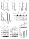
Figure 3. OCT1 plays critical roles in miR-451 expression
(A–B) qRT-PCR analysis of pri-miR-451 expression upon OCT1 knockdown (A) and in Oct1-deficient cells (B). See also Figure S3A–B. *p<0.05, **p<0.01 (C) qRT-PCR analysis of pri-miR-451 expression upon OCT1 overexpression. See also Figure S3C. *p<0.05, **p<0.01 (D) qRT-PCR analysis of pri-miR-451 expression in 3T3 Oct1−/− cells transfected with wild type and mutant OCT1 vectors and cultured in high and low glucose. See also Figure S3D–E. *p<0.05, **p<0.01 (E) The predominant nuclear localization of OCT1 in GBM cells is independent of glucose levels. Immunoblotting of cytoplasmic (c) and nuclear (n) cellular fractions of GBM cells cultured in high and low glucose. (F) OCT1 is phosphorylated at S335 in a glucose-dependent manner in GBM cells. Immunoblotting of GBM cells cultured in high and low glucose. (G) OCT1 is phosphorylated at S335 in glucose-dependent manner in mouse fibroblasts; phosphorylation of AMPK by low glucose conditions is not impaired in Oct1−/− cells. Immunoblotting of 3T3 cells cultured in high and low glucose. (H) Knockdown of AMPK a1, a2 and a1/a2 increases the expression of miR-451 in low glucose environment. Immunoblotting of GBM cells cultured in low glucose (upper panels). qRT-PCR analysis of pri-miR-451 expression upon AMPK knockdown (bottom). See also Figure S3G. *p<0.05, **p<0.01
Figure 4
Figure 4. Role of AMPK in phosphorylation of OCT1 and transcription of miR-451
(A) Phosphorylation of OCT1 at S335 is impaired in AMPKβ1/2-deficient GBM cells. Immunoblotting of AMPKβ1/2-deficient GBM cells cultured in high and low glucose. (B) qRT-PCR analysis of pri-miR-451 expression. See also Figure S4A. *p<0.05, **p<0.01 (C–D) AMPK directly phosphorylates OCT1 at S335. Kinase assays were performed with AMPK complex immuno-precipitated from U251 cells expressing Flag-AMPK in low glucose conditions (C), or recombinant, active AMPK complex containing AMPKa1, β1, and 1 (D), and GST-OCT1 peptide fragment containing S335 or S335A. See also Figure S4D-F. (E) A proposed miR-451/AMPK regulatory loop in a fluctuating glucose microenvironment.

References

    1. Bronisz A, Wang Y, Nowicki MO, Peruzzi P, Ansari KI, Ogawa D, Balaj L, De Rienzo G, Mineo M, Nakano I, et al. Extracellular vesicles modulate the glioblastoma microenvironment via a tumor suppression signaling network directed by miR-1. Cancer research. 2014;74:738–750. - PMC - PubMed
    1. Cheng C, Li W, Zhang Z, Yoshimura S, Hao Q, Zhang C, Wang Z. MicroRNA-144 is regulated by activator protein-1 (AP-1) and decreases expression of Alzheimer disease-related a disintegrin and metalloprotease 10 (ADAM10) J Biol Chem. 2013;288:13748–13761. - PMC - PubMed
    1. Dhruv HD, McDonough Winslow WS, Armstrong B, Tuncali S, Eschbacher J, Kislin K, Loftus JC, Tran NL, Berens ME. Reciprocal activation of transcription factors underlies the dichotomy between proliferation and invasion of glioma cells. PLoS One. 2013;8:e72134. - PMC - PubMed
    1. Dore LC, Amigo JD, Dos Santos CO, Zhang Z, Gai X, Tobias JW, Yu D, Klein AM, Dorman C, Wu W, et al. A GATA-1-regulated microRNA locus essential for erythropoiesis. Proc Natl Acad Sci U S A. 2008;105:3333–3338. - PMC - PubMed
    1. Farre D, Roset R, Huerta M, Adsuara JE, Rosello L, Alba MM, Messeguer X. Identification of patterns in biological sequences at the ALGGEN server: PROMO and MALGEN. Nucleic acids research. 2003;31:3651–3653. - PMC - PubMed

Publication types

LinkOut - more resources