Clinical Trial
. 2015 May 12;4(5):860-72.
doi: 10.1016/j.stemcr.2015.04.005. Epub 2015 Apr 30.

Treatment of macular degeneration using embryonic stem cell-derived retinal pigment epithelium: preliminary results in Asian patients

Affiliations
Clinical Trial

Treatment of macular degeneration using embryonic stem cell-derived retinal pigment epithelium: preliminary results in Asian patients

Won Kyung Song et al. Stem Cell Reports. .

Abstract

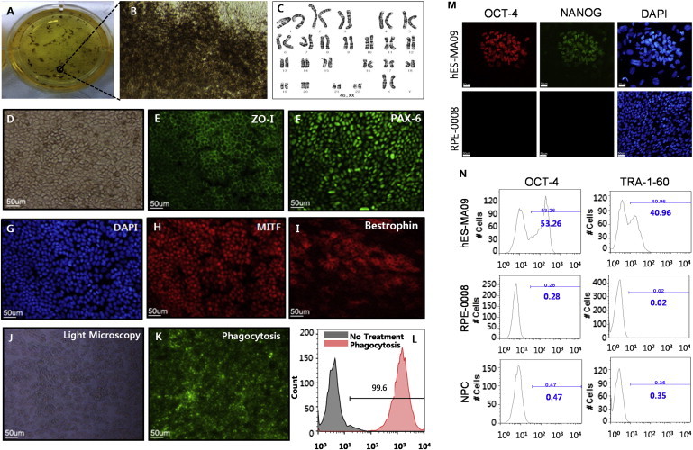
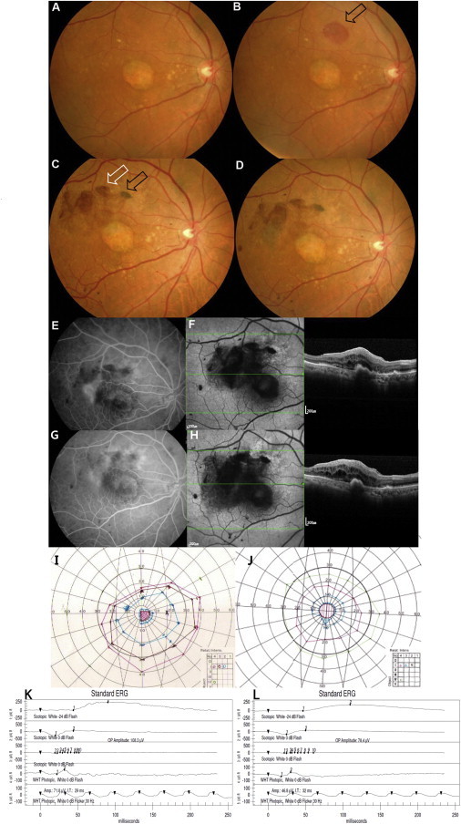
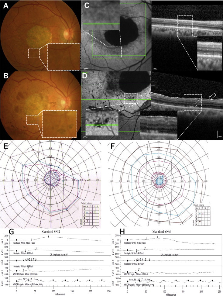
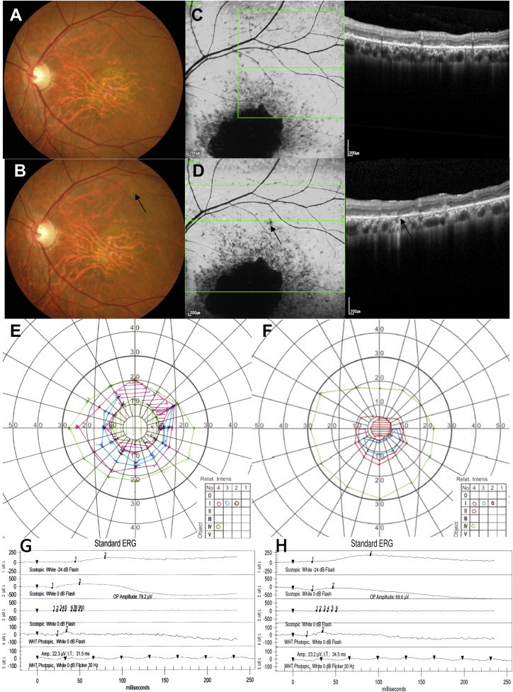
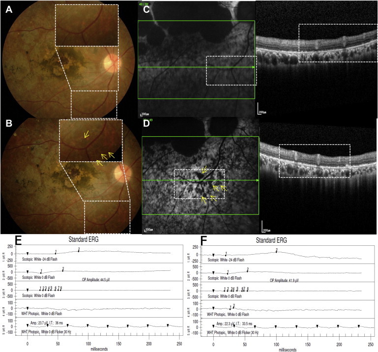
Embryonic stem cells hold great promise for various diseases because of their unlimited capacity for self-renewal and ability to differentiate into any cell type in the body. However, despite over 3 decades of research, there have been no reports on the safety and potential efficacy of pluripotent stem cell progeny in Asian patients with any disease. Here, we report the safety and tolerability of subretinal transplantation of human embryonic-stem-cell (hESC)-derived retinal pigment epithelium in four Asian patients: two with dry age-related macular degeneration and two with Stargardt macular dystrophy. They were followed for 1 year. There was no evidence of adverse proliferation, tumorigenicity, ectopic tissue formation, or other serious safety issues related to the transplanted cells. Visual acuity improved 9-19 letters in three patients and remained stable (+1 letter) in one patient. The results confirmed that hESC-derived cells could serve as a potentially safe new source for regenerative medicine.


Figures

Figure 1
Figure 1
Characterization of the Clinical Product: Identity, Potency, and Purity (A) RPE clusters were obtained by culturing an embryoid body attached to a six-well plate for about 8 weeks. (B) The cells at the edge of the pigmented cluster displayed typical morphology of hRPE with hypo-pigmentation of the leading edge. (C) A normal female karyotype (46XX) is shown. (D) A confluent cobblestone monolayer was observed via Hoffman modulation contrast microscopy. (E and F) Cells were positive for ZO-1 (E) and PAX-6 (F). (G and H) DAPI staining in (G) was used to identify the location of the nuclei corresponding to ZO-1 (in E) and MITF (H) at the same time. ZO-1 and MITF were double stained in one sample. (I) Mature RPE cells were recognized with anti-Bestrophin. (J–L) Phagocytosis assay results were shown. Fluorescence microscopy image and FACS analyses of the differentiated hESC-derived RPE cells demonstrate that most of the cells (99.6%) were phagocytized with the fluorescent-labeled particles. (M and N) Purity was assessed by the absence of hESCs of the final product by immunocytochemical staining for OCT-4 and NANOG (M) and FACS analysis demonstrating the absence of OCT-4 and TRA-1-60 (N). Scale bars, 50 μm.
Figure 2
Figure 2
Ophthalmologic Results of the First Dry AMD Patient (A) Baseline fundus photography with geographic atrophy and drusens. (B) Small subretinal hemorrhage at post-operative day 1 at the nasal injection site (arrow). (C) Fundus photography at post-operative 26 weeks showing absorption of hemorrhage. Subretinal pigment is present at the nasal injection site (black arrow) and preretinal pigmentation and epiretinal membrane are visible superotemporal to the fovea (white arrow). (D and E) Fundus photography (D) and fluorescence angiography (E) at post-operative 33 weeks reveal a neovascular membrane temporal to the fovea. (F) Autofluorescence imaging shows widespread hypo-autofluorescence (left); OCT demonstrates sub-RPE elevation and subretinal and intraretinal fluid (right). (G) The CNV is less active on fluorescein angiography at post-operative 52 weeks after three monthly intravitreal Lucentis treatments. (H) There is no significant change in autofluorescence and OCT. (I and J) There is minimal enlargement of central scotoma (J) compared with baseline (I) based on Goldmann visual field examination. (K and L) Electroretinography at baseline (K) and at the 1-year visit (L) showed no significant changes.
Figure 3
Figure 3
Ophthalmologic Results of the Second Dry AMD Patient (A and B) Baseline (A) and 52-week-postoperative (B) fundus photography of the second dry AMD patient, showing subretinal pigmentation (B, inset) after surgery. (C and D) Baseline (C) and 52-week-postoperative (D) autofluorescence imaging and SD-OCT. Note the stippled hypo-autofluorescence present after surgery at the border of the atrophic zone that may represent blockage from subretinal pigmentation and stippled hyper-autofluorescence at the same area underlying the bleb (D, left, inset). Subretinal deposits (D, right, dashed arrow on OCT) are seen in the cell-transplanted areas that were not present prior to surgery. There is an epiretinal membrane present postoperatively (D, right, solid arrow). Patchy hypo-autofluorescence present at preretinal pigmentation areas (D, left). (E and F) GVF examinations at baseline (E) and post-operative 52 weeks (F) show a central scotoma of diminished intensity. (G and H) ERG examinations at baseline (G) and at post-operative 52 weeks (H) show no significant changes.
Figure 4
Figure 4
Ophthalmologic Results of the First SMD Patient (A–D) Baseline (A) and 52-week-postoperative (B) fundus photography, baseline (C), and 52-week-postoperative (D) autofluorescence imaging and SD-OCT showing no signs of immune rejection, tumor formation, or adverse event related to the surgical procedure. No obvious pigmentation (arrow indicates the cell-injected retinotomy site) was noted after hESC-RPE transplantation. (E and F) GVF examinations at baseline (E) and post-operative 52 weeks (F) show a central scotoma of diminished size. (G and H) ERG examinations at baseline (G) and post-operative 52 weeks (H) show no change.
Figure 5
Figure 5
Ophthalmologic Results of the Second SMD Patient (A and B) Baseline (A) and 52-week postoperative (B) fundus photography show subretinal pigmentation (B, inset, yellow arrows) in the bleb area that may represent engraftment of injected hESC-RPE cells. (C and D) SD-OCT at baseline (C, right) and postoperative 52 weeks (D, right) at corresponding sites showing a monolayer of the RPE layer. Fundus autofluorescence images (left panels of C and D) show stippling of autofluorescence at 52 weeks after surgery (D, inset, yellow arrows). (E and F) There is no change in the electroretinogram from baseline (E) to postoperative 52 weeks (F).

References

    1. Algvere P.V., Berglin L., Gouras P., Sheng Y. Transplantation of fetal retinal pigment epithelium in age-related macular degeneration with subfoveal neovascularization. Graefes Arch. Clin. Exp. Ophthalmol. 1994;232:707–716. - PubMed
    1. Algvere P.V., Berglin L., Gouras P., Sheng Y., Kopp E.D. Transplantation of RPE in age-related macular degeneration: observations in disciform lesions and dry RPE atrophy. Graefes Arch. Clin. Exp. Ophthalmol. 1997;235:149–158. - PubMed
    1. Algvere P.V., Gouras P., Dafgård Kopp E. Long-term outcome of RPE allografts in non-immunosuppressed patients with AMD. Eur. J. Ophthalmol. 1999;9:217–230. - PubMed
    1. Anderson D.H., Stern W.H., Fisher S.K., Erickson P.A., Borgula G.A. The onset of pigment epithelial proliferation after retinal detachment. Invest. Ophthalmol. Vis. Sci. 1981;21:10–16. - PubMed
    1. Age-Related Eye Disease Study Research Group A randomized, placebo-controlled, clinical trial of high-dose supplementation with vitamins C and E, beta carotene, and zinc for age-related macular degeneration and vision loss: AREDS report no. 8. Arch. Ophthalmol. 2001;119:1417–1436. - PMC - PubMed

Publication types