. 2015 May 7;96(5):784-96.
doi: 10.1016/j.ajhg.2015.04.002. Epub 2015 Apr 30.

A Potential Contributory Role for Ciliary Dysfunction in the 16p11.2 600 kb BP4-BP5 Pathology

Collaborators, Affiliations

A Potential Contributory Role for Ciliary Dysfunction in the 16p11.2 600 kb BP4-BP5 Pathology

Eugenia Migliavacca et al. Am J Hum Genet. .

Abstract

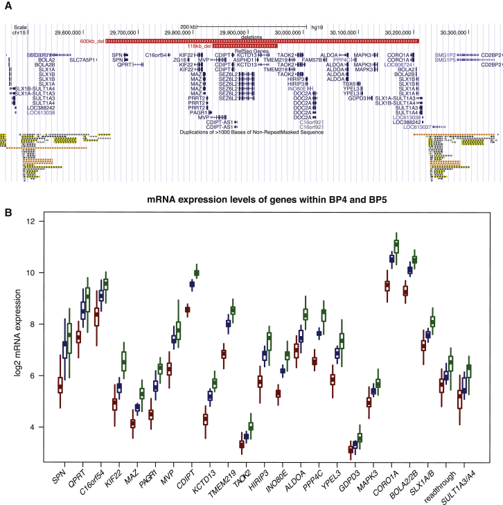
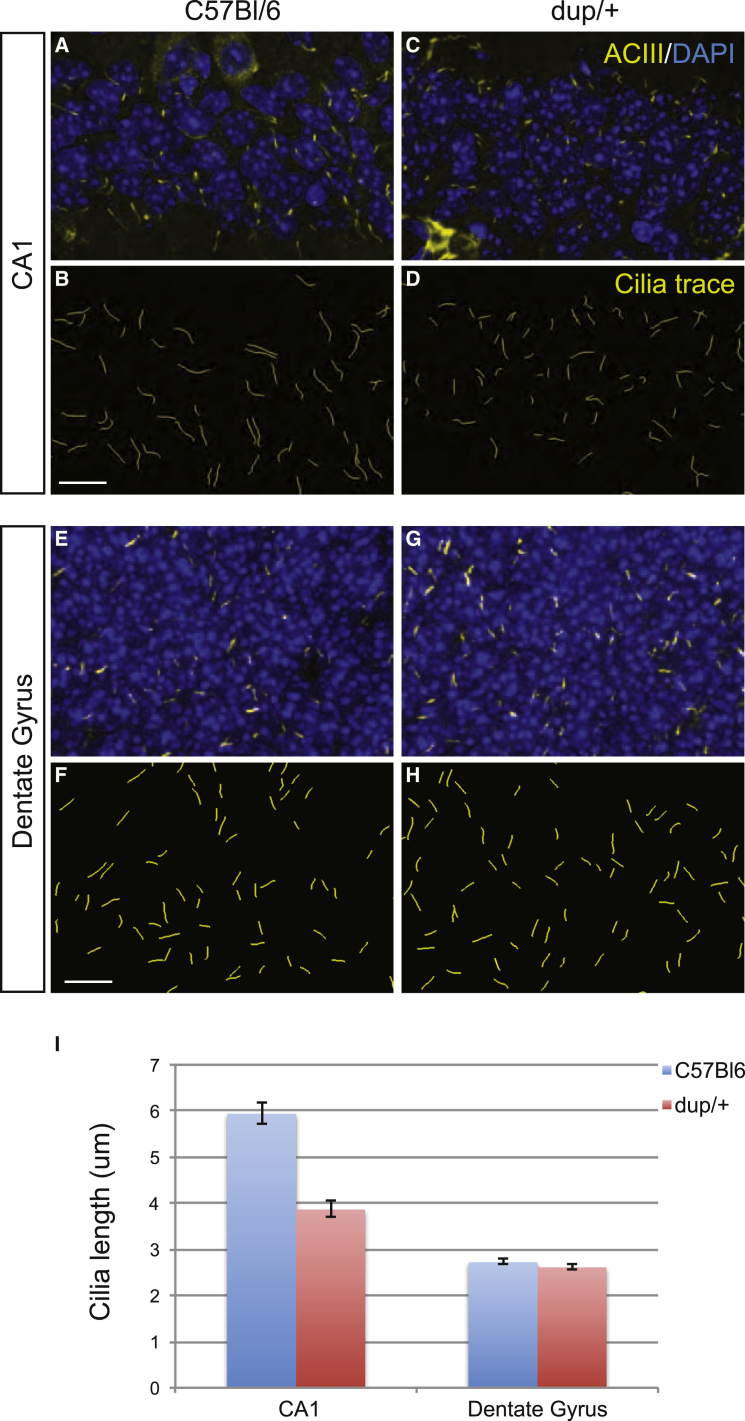
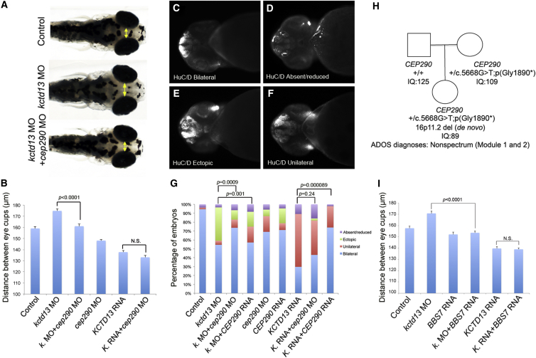
The 16p11.2 600 kb copy-number variants (CNVs) are associated with mirror phenotypes on BMI, head circumference, and brain volume and represent frequent genetic lesions in autism spectrum disorders (ASDs) and schizophrenia. Here we interrogated the transcriptome of individuals carrying reciprocal 16p11.2 CNVs. Transcript perturbations correlated with clinical endophenotypes and were enriched for genes associated with ASDs, abnormalities of head size, and ciliopathies. Ciliary gene expression was also perturbed in orthologous mouse models, raising the possibility that ciliary dysfunction contributes to 16p11.2 pathologies. In support of this hypothesis, we found structural ciliary defects in the CA1 hippocampal region of 16p11.2 duplication mice. Moreover, by using an established zebrafish model, we show genetic interaction between KCTD13, a key driver of the mirrored neuroanatomical phenotypes of the 16p11.2 CNV, and ciliopathy-associated genes. Overexpression of BBS7 rescues head size and neuroanatomical defects of kctd13 morphants, whereas suppression or overexpression of CEP290 rescues phenotypes induced by KCTD13 under- or overexpression, respectively. Our data suggest that dysregulation of ciliopathy genes contributes to the clinical phenotypes of these CNVs.


Figures

Figure 1
Figure 1
Expression Level Changes in 16p11.2 Deletion and Duplication LCLs (A) Comparison of the GRCh37/hg19 genomic boundaries of the recurrent 600 kb BP4-BP5 16p11.2 deletion with that of the 118 kb atypical deletion described in Crepel et al. The genes mapping within the interval are shown in blue. The low-copy repeats (LCRs) at the rearrangement breakpoints are indicated at the bottom. (B) Boxplot distribution of relative expression levels measured by microarrays in deletion (n = 50) and duplication (n = 31) carriers (red and green, respectively) and control LCLs (n = 17, blue) of the 19 “unique” and 3 multiple-copy genes (plus the read-through transcription between SLX1A/1B [MIM: 615822/615823] and SULT1A3/A4 [MIM: 600641/615819]) expressed in LCLs and mapping within the 16p11.2 BP4-BP5 interval and its flanking LCRs, respectively. Note that the expression level of these transcripts, de facto positive controls, is positively correlated with gene dosage.
Figure 2
Figure 2
Ciliary Length Analyses in Hippocampal Tissue from C57BL/6 and 16p11.2 dup/+ Mice (A–H) Staining of cilia in the CA1 (A, C) and dentate gyrus (E, G) regions of the hippocampus via ACIII (yellow) and counterstained with DAPI (blue) in C57BL/6 controls (A, E) and 16p11.2 duplication models (C, G). CA1 (B, D) and dentate gyrus (F, H) ciliary length analyses were performed by plotting pixel information from ACIII staining in C57BL/6 controls (A, E) and 16p11.2 duplication models (C, G). (I) Total ciliary length comparisons were measured and plotted for both the CA1 and dentate gyrus (SEM was plotted; ∗∗∗p < 0.001).
Figure 3
Figure 3
Ciliopathy Gene Suppression or Overexpression Rescues the Neuroanatomical Defects Associated with KCTD13 Imbalance (A) From top to bottom, dorsal views of representative 4.5 dpf controls, embryos injected with kctd13 MO, and co-injected with kctd13 and cep290 MOs. The yellow double-head arrows mark the distance between the convex tip of the eyecups, a proxy for head size. (B) Bar graph represents the distance between the eyecups for controls and injected embryos. SEM was plotted. Student’s t test was performed and the corresponding p value is denoted on the bar graph. (C–F) Representative photographs show ventral views of zebrafish embryos at 2 dpf stained with HuC/D. Embryos were binned into four classes: normal bilateral expression (C), absence or reduced expression (D), ectopic expression (E), and unilateral expression (F). (G) Percentage of embryos with normal bilateral HuC/D protein levels (blue) or unilateral HuC/D (red), ectopic (green), and absent/reduced protein levels (purple) in the anterior forebrain in embryo batches injected with kctd13, cep290 MOs, and/or KCTD13, CEP290 mRNAs. HuC/D levels in the anterior forebrain of the embryo injected with the kctd13 MO are considerably increased compared to those of the control embryo. This defect was rescued significantly by co-injection of cep290 MO. Likewise, HuC/D levels in the anterior forebrain of the embryo injected with the KCTD13 mRNA are considerably lower than those of the control embryo. This defect was rescued significantly by co-injection of full-length human CEP290 mRNA. Although significant differences were observed in unilateral and ectopic classes for embryos injected with CEP290 RNA and kctd13 MO compared to kctd13 morphants, the total percentage of abnormal embryos remained the same. Chi-square test was performed and the corresponding p values are denoted on the bar graph. (H) Two-generation pedigree shows mother-daughter transmission of a CEP290 nonsense variant and a de novo 16p11.2 deletion identified in the daughter. The genotypes, IQ, and ADOS diagnoses are indicated (see also Table S13). (I) Bar graph represents the distance between the eyecups for controls and embryos injected with kctd13 morpholino (MO), BBS7 RNA, and co-injected with kctd13 MO and BBS7 RNA. SEM was plotted. Student’s t test was performed and the corresponding p value is denoted on the bar graph.

References

    1. Weiss L.A., Shen Y., Korn J.M., Arking D.E., Miller D.T., Fossdal R., Saemundsen E., Stefansson H., Ferreira M.A., Green T., Autism Consortium Association between microdeletion and microduplication at 16p11.2 and autism. N. Engl. J. Med. 2008;358:667–675. - PubMed
    1. Glessner J.T., Wang K., Cai G., Korvatska O., Kim C.E., Wood S., Zhang H., Estes A., Brune C.W., Bradfield J.P. Autism genome-wide copy number variation reveals ubiquitin and neuronal genes. Nature. 2009;459:569–573. - PMC - PubMed
    1. Marshall C.R., Noor A., Vincent J.B., Lionel A.C., Feuk L., Skaug J., Shago M., Moessner R., Pinto D., Ren Y. Structural variation of chromosomes in autism spectrum disorder. Am. J. Hum. Genet. 2008;82:477–488. - PMC - PubMed
    1. Bochukova E.G., Huang N., Keogh J., Henning E., Purmann C., Blaszczyk K., Saeed S., Hamilton-Shield J., Clayton-Smith J., O’Rahilly S. Large, rare chromosomal deletions associated with severe early-onset obesity. Nature. 2010;463:666–670. - PMC - PubMed
    1. Walters R.G., Jacquemont S., Valsesia A., de Smith A.J., Martinet D., Andersson J., Falchi M., Chen F., Andrieux J., Lobbens S. A new highly penetrant form of obesity due to deletions on chromosome 16p11.2. Nature. 2010;463:671–675. - PMC - PubMed

Publication types

Substances