. 2015 Jul;27(1):38-45.
doi: 10.1016/j.intimp.2015.04.035. Epub 2015 Apr 29.

Blockage of P2X7 attenuates acute lung injury in mice by inhibiting NLRP3 inflammasome

Affiliations

Blockage of P2X7 attenuates acute lung injury in mice by inhibiting NLRP3 inflammasome

Shuang Wang et al. Int Immunopharmacol. 2015 Jul.

Abstract

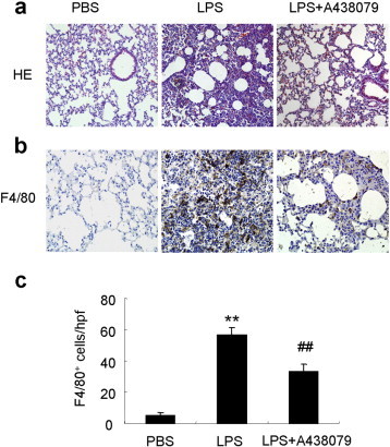
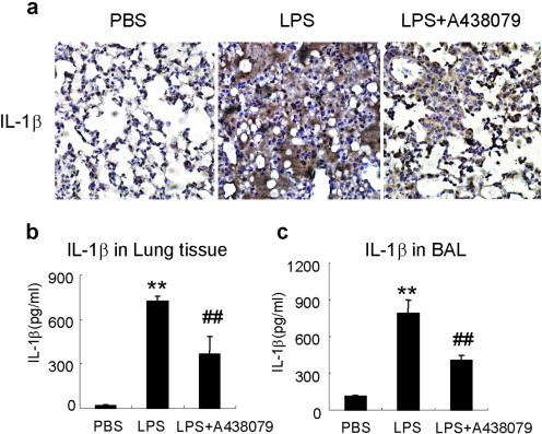
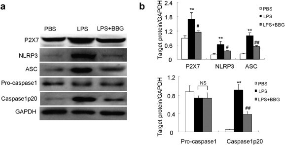
NLRP3 inflammasome is engaged in the inflammatory response during acute lung injury (ALI). Purinergic receptor P2X7 has been reported to be upstream of NLRP3 activation. However, the therapeutic implication of P2X7 in ALI remains to be explored. The present study used lipopolysaccharide (LPS)-induced mouse model to investigate the therapeutic potential of P2X7 blockage in ALI. Our results showed that P2X7/NLRP3 inflammasome pathway was significantly upregulated in the lungs of ALI mice as compared with control mice. P2X7 antagonist A438079 suppressed NLRP3/ASC/caspase 1 activation, production of IL-1β, IL-17A and IFN-γ and neutrophil infiltration but not the production of IL-10, resulting in a significant amelioration of lung injury. Moreover, blockage of P2X7 significantly inhibited NLRP3 inflammasome activation and IL-1β production in bone marrow derived macrophages. Similar results were obtained using another P2X7 inhibitor brilliant blue G (BBG) in vivo. Thus, pharmacological blockage of P2X7/NLRP3 pathway can be considered as a potential therapeutic strategy in patients with ALI.

Keywords: Acute lung injury; IL-1β; Inflammasome; Neutrophils; P2X7.


Figures

Fig. 1
Fig. 1
A438079 suppresses activation of P2X7/NLRP3 pathway in LPS-induced lung injury mice. a, representative Western blot bands showed the protein expression of P2X7, NLRP3, ASC, pro-caspase 1, caspase 1p20 and GAPDH in the lung tissue at day 2 after LPS induction. b, Quantitative data showed respective protein expressions normalized to the values for GAPDH. Each bar represents mean ± SEM (n = 6). **P < 0.01 versus normal control (PBS) mice; #P < 0.05, ##P < 0.01 versus LPS-induced lung injury mice. NS = not significant.
Fig. 2
Fig. 2
A438079 ameliorates lung injury and inflammatory cell infiltration in LPS-induced lung injury mice. a, HE staining of lung sections. Original magnification × 200. b, Immunohistochemistry staining of F4/80+ macrophages in lung tissues. c, Semiquantification of F4/80+ macrophages infiltration in the lungs. Results show that A438079 treatment attenuates pulmonary injury at day 2 after LPS induction. Each bar represents mean ± SEM (n = 6). **P < 0.01 versus normal control (PBS) mice; ##P < 0.01 versus LPS-induced lung injury mice.
Fig. 3
Fig. 3
A438079 decreases cell counts and protein level in BAL fluid. a, Wright–Giemsa staining of cytospun cells from BAL fluids. b, Total cell counts of BAL fluids in PBS (normal control), LPS (LPS-induced lung injury) and LPS + A438079 (treatment) groups. c, Neutrophil counts in BAL fluid. d, Total protein level in BAL fluids. Each bar represents mean ± SEM (n = 6). **P < 0.01 versus normal control (PBS) mice; ##P < 0.01 versus LPS-induced lung injury mice.
Fig. 4
Fig. 4
A438079 inhibits IL-1β production in lung tissues and BAL fluids. IL-1β in tissue homogenate or BAL fluid was measured by ELISA. a, Immunohistochemistry staining of IL-1β in lung tissues. b, IL-1β levels in lung tissues. c, IL-1β levels in BAL fluid. Each bar represents mean ± SEM (n = 6). **P < 0.01 versus normal control (PBS) mice; ##P < 0.01 versus control LPS-induced lung injury mice.
Fig. 5
Fig. 5
A438079 inhibits IFN-γ and IL-17A but not IL-10 production in lung tissues and BAL fluids. Cytokine level in tissue homogenate or BAL fluid was measured by ELISA. a and d, IFN-γ levels in lung tissues (a) and BAL fluid (d). b and e, IL-17A levels in lung tissues (b) and BAL fluid (e). c and f, IL-10 levels in lung tissues (c) and BAL fluid (f). Each bar represents mean ± SEM (n = 6). **P < 0.01 versus normal control (PBS) mice; #P < 0.05, ##P < 0.01 versus LPS-induced lung injury mice. NS = not significant.
Fig. 6
Fig. 6
A438079 inhibits NLRP3 inflammasome activation in mouse bone marrow derived macrophages (BMDMs). BMDMs were primed with 1 μg/ml LPS in the presence or absence of A438079 (5 mM) for 4 h followed by stimulation with ATP (5 mM) for 1 h. The NLRP3 inflammasome activation was evaluated by expression of NLRP3, ASC, pro-caspase 1 and caspase1p20. a, representative Western blot bands showed the protein expression of NLRP3, ASC, pro-caspase 1, caspase1p20 and GAPDH. b, Quantitative data showing respective protein expressions normalized to the values for GAPDH. c, IL-1β levels in cell supernatants from BMDMs determined by ELISA. Each bar represents mean ± SEM (n = 3). **P < 0.01 versus LPS only group; #P < 0.05, ##P < 0.01 versus LPS + ATP group. NS = not significant.
Fig. 7
Fig. 7
BBG suppresses activation of P2X7/NLRP3 pathway in LPS-induced lung injury mice. a, representative Western blot bands showed the protein expression of P2X7, NLRP3, ASC, pro-caspase 1, caspase 1p20 and GAPDH in the lung tissue at day 2 after LPS induction. b, Quantitative data showing respective protein expressions normalized to the values for GAPDH. Each bar represents mean ± SEM (n = 6). **P < 0.01 versus normal control (PBS) mice; #P < 0.05, ##P < 0.01 versus LPS-induced lung injury mice. NS = not significant.
Fig. 8
Fig. 8
BBG ameliorates lung injury and inflammatory cell infiltration in LPS-induced lung injury mice. a, HE staining of lung sections. Original magnification × 200. b, Semiquantification of F4/80+ macrophage infiltration in the lung tissues. c, Total cell counts of BAL fluids. d, Total protein level in BAL fluids. Each bar represents mean ± SEM (n = 6). **P < 0.01 versus normal control (PBS) mice; #P < 0.05, ##P < 0.01 versus LPS-induced lung injury mice.

References

    1. Rubenfeld G.D., Caldwell E., Peabody E., Weaver J., Martin D.P., Neff M. Incidence and outcomes of acute lung injury. N. Engl. J. Med. 2005;353:1685–1693. - PubMed
    1. Gullo A. Spectrum of acute lung injury, ARDS, sepsis. Minerva Anestesiol. 2002;68:45–53. - PubMed
    1. Schenten D., Medzhitov R. The control of adaptive immune responses by the innate immune system. Adv. Immunol. 2011;109:87–124. - PubMed
    1. Gross O., Thomas C.J., Guarda G., Tschopp J. The inflammasome: an integrated view. Immunol. Rev. 2011;243:136–151. - PubMed
    1. Saleh M. The machinery of Nod-like receptors: refining the paths to immunity and cell death. Immunol. Rev. 2011;243:235–246. - PubMed

Publication types

MeSH terms