

Energy dispersive CdTe and CdZnTe detectors for spectral clinical CT and NDT applications
- PMID: 25937684
- PMCID: PMC4415629
- DOI: 10.1016/j.nima.2014.10.079
Energy dispersive CdTe and CdZnTe detectors for spectral clinical CT and NDT applications
Abstract
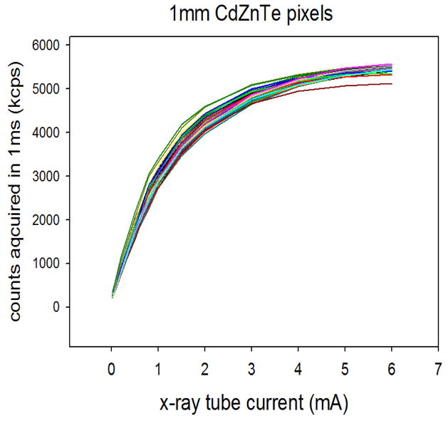
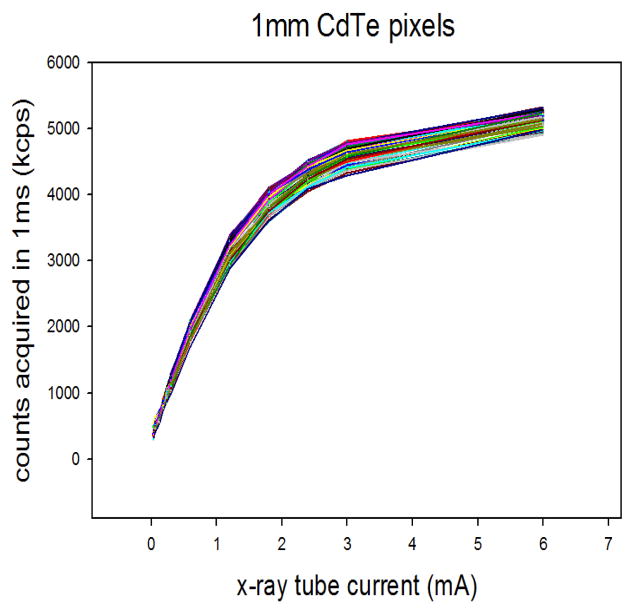
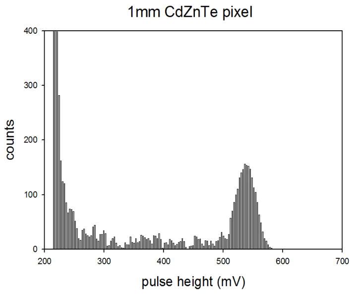
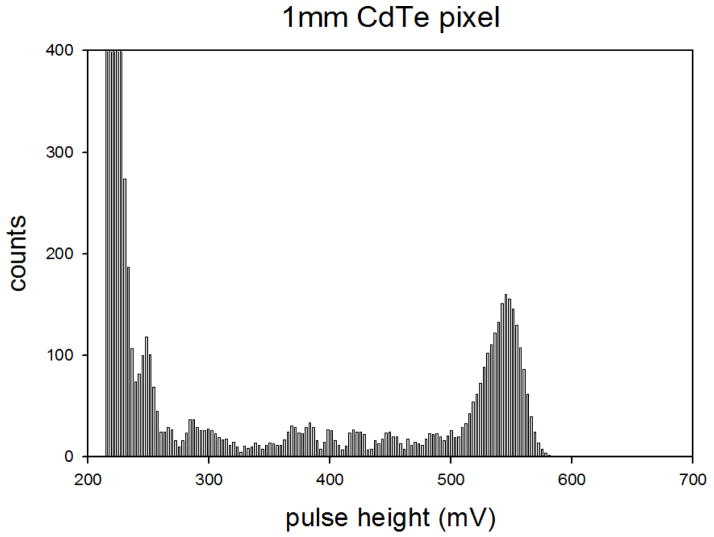
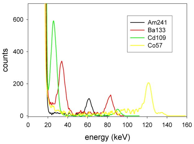
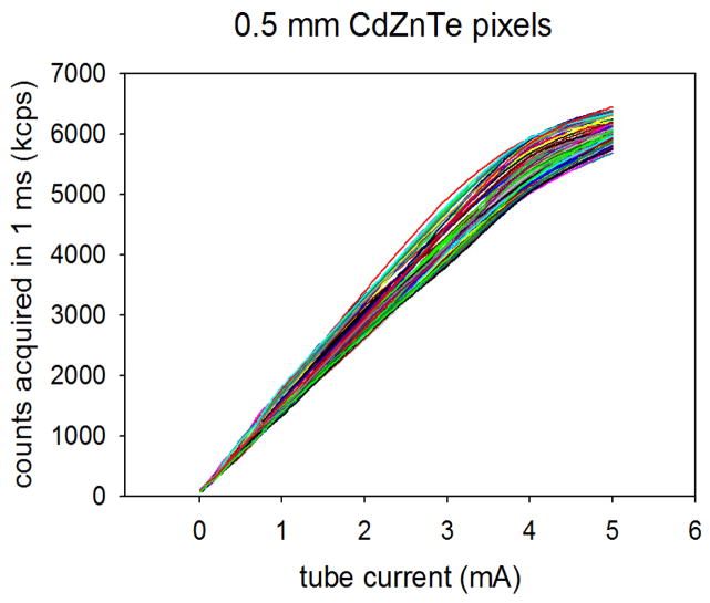
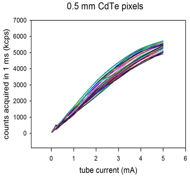
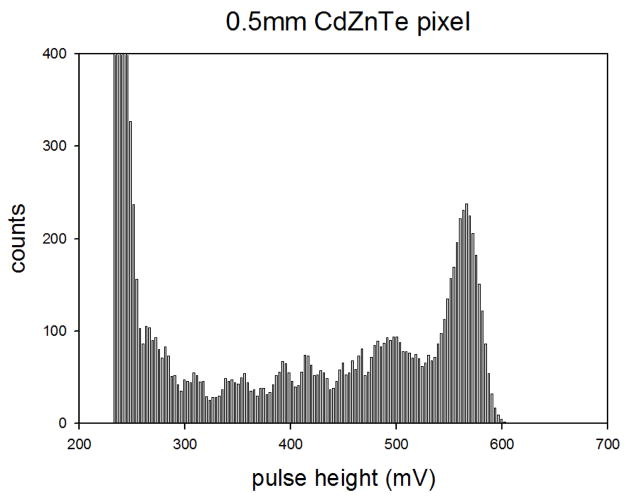
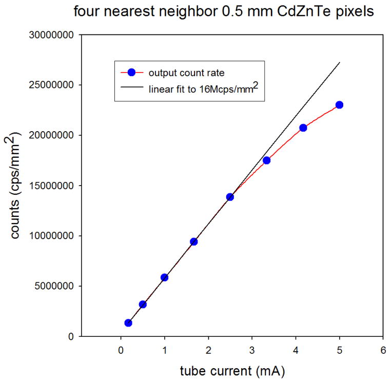
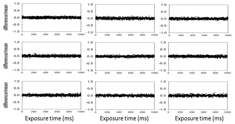
We are developing room temperature compound semiconductor detectors for applications in energy-resolved high-flux single x-ray photon-counting spectral computed tomography (CT), including functional imaging with nanoparticle contrast agents for medical applications and non destructive testing (NDT) for security applications. Energy-resolved photon-counting can provide reduced patient dose through optimal energy weighting for a particular imaging task in CT, functional contrast enhancement through spectroscopic imaging of metal nanoparticles in CT, and compositional analysis through multiple basis function material decomposition in CT and NDT. These applications produce high input count rates from an x-ray generator delivered to the detector. Therefore, in order to achieve energy-resolved single photon counting in these applications, a high output count rate (OCR) for an energy-dispersive detector must be achieved at the required spatial resolution and across the required dynamic range for the application. The required performance in terms of the OCR, spatial resolution, and dynamic range must be obtained with sufficient field of view (FOV) for the application thus requiring the tiling of pixel arrays and scanning techniques. Room temperature cadmium telluride (CdTe) and cadmium zinc telluride (CdZnTe) compound semiconductors, operating as direct conversion x-ray sensors, can provide the required speed when connected to application specific integrated circuits (ASICs) operating at fast peaking times with multiple fixed thresholds per pixel provided the sensors are designed for rapid signal formation across the x-ray energy ranges of the application at the required energy and spatial resolutions, and at a sufficiently high detective quantum efficiency (DQE). We have developed high-flux energy-resolved photon-counting x-ray imaging array sensors using pixellated CdTe and CdZnTe semiconductors optimized for clinical CT and security NDT. We have also fabricated high-flux ASICs with a two dimensional (2D) array of inputs for readout from the sensors. The sensors are guard ring free and have a 2D array of pixels and can be tiled in 2D while preserving pixel pitch. The 2D ASICs have four energy bins with a linear energy response across sufficient dynamic range for clinical CT and some NDT applications. The ASICs can also be tiled in 2D and are designed to fit within the active area of the sensors. We have measured several important performance parameters including; the output count rate (OCR) in excess of 20 million counts per second per square mm with a minimum loss of counts due to pulse pile-up, an energy resolution of 7 keV full width at half maximum (FWHM) across the entire dynamic range, and a noise floor about 20keV. This is achieved by directly interconnecting the ASIC inputs to the pixels of the CdZnTe sensors incurring very little input capacitance to the ASICs. We present measurements of the performance of the CdTe and CdZnTe sensors including the OCR, FWHM energy resolution, noise floor, as well as the temporal stability and uniformity under the rapidly varying high flux expected in CT and NDT applications.
Keywords: ASIC; CT; CZT; CdTe; X-ray; semiconductor.
Figures
References
-
- Cammin J, Srivastava S, Barber WC, Iwanczyk JS, Hartsough NE, Nygard E, Wessel JC, Malakhov N, Taguchi K. A tabletop clinical x-ray CT scanner with energy-resolving photon counting detectors. presented at the Medical Imaging 2011: Physics of Medical Imaging; Lake Buena Vista, Florida, USA. 2011. pp. 79611S–79611S–7.
-
- Tomita Y, Shirayanagi Y, Matsui S, Misawa M, Takahashi H, Aoki T, Hatanaka Y. X-ray color scanner with multiple energy differentiate capability. IEEE Nucl Sci Symp Conf Rec. 2004;6:3733–3737.
-
- Llopart X, Campbell M, Dinapoli R, San Segundo D, Pernigotti E. Medipix2: A 64-k pixel readout chip with 55-μm square elements working in single photon counting mode. IEEE Trans Nucl Sci. 2002;49(5):2279–2283.
Grants and funding
LinkOut - more resources
Full Text Sources
Other Literature Sources