A radiological evaluation of marginal bone around dental implants: An in-vivo study
- PMID: 25937721
- PMCID: PMC4405952
- DOI: 10.4103/0975-5950.154813
A radiological evaluation of marginal bone around dental implants: An in-vivo study
Abstract
Context: This article presents an original research conducted at Government Dental College, PGIDS, Rohtak.
Aims: (1) To evaluate the marginal bone level changes around dental implants based on the radiological examination. (2) To evaluate the relationship of various parameters, i.e., gender, implant length, implant diameter and location of implants on the amount of bone loss around dental implants.
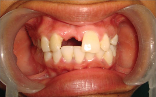
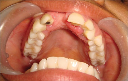
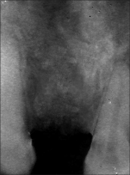
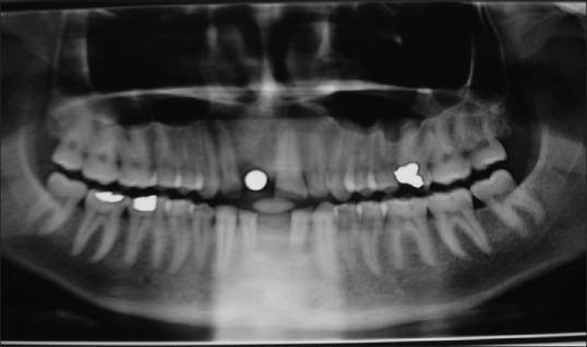
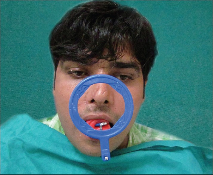
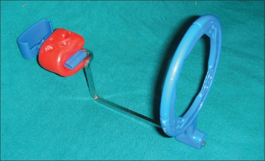
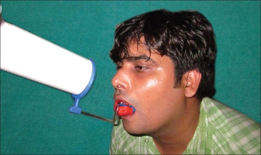
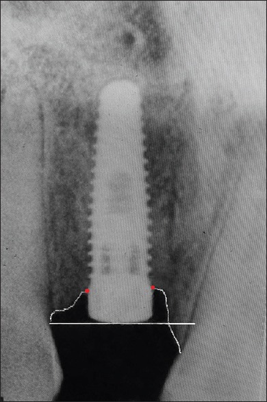
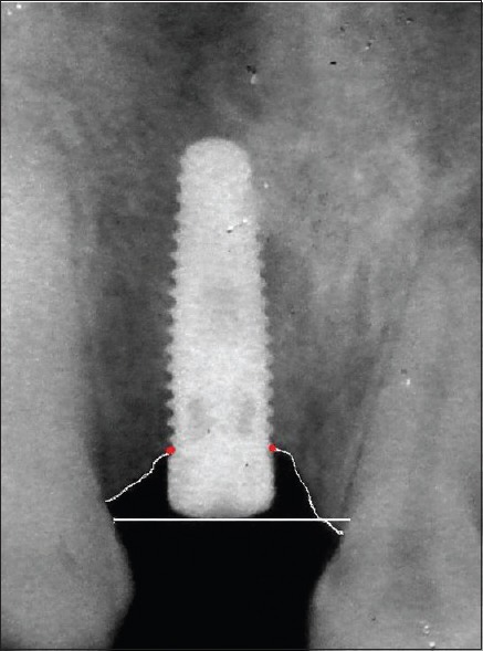
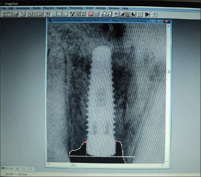
Materials and methods: An in-vivo study was undertaken to evaluate the crestal bone loss on mesial and distal aspect of implants, using standardized intra-oral periapical at the end of 6 months after placing the implants, but before prosthetically loading it.
Statistical analysis used: Student's unpaired t-test.
Results: Bone loss was measured and values were recorded immediately after implant placement and after 6 months.
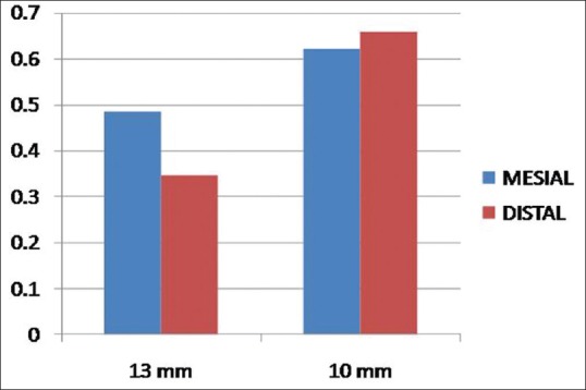
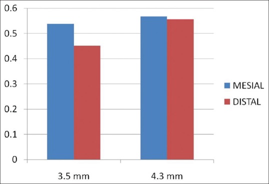
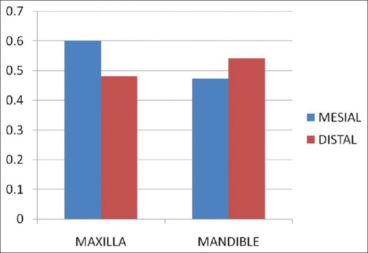
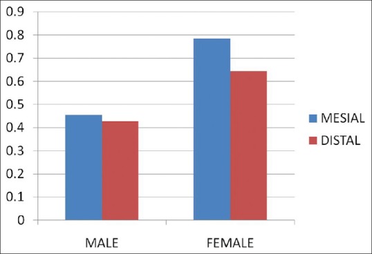
Conclusions: (1) Bone loss on mesial and distal aspects of implants was found to be same after period of 6 months. (2) Bone loss was found to be same in both 13 mm and 10 mm implants on mesial aspect, whereas on distal aspect, it was more in 10 mm implants. (3) Bone loss was found to be same in both 3.5 mm and 4.3 mm diameter implants on both mesial and distal aspects of implants. (4) Bone loss was found to be same in both maxilla and mandible on both mesial and distal aspects of implants. (5) Bone loss was found to be more in females on both mesial as well as distal aspects of implants.
Keywords: Bone loss; distal; implant; mesial.
Conflict of interest statement
Figures

References
-
- Branemark PI. Introduction to osseointegration. In: Branemark PI, Zarb GA, Albrektsson T, editors. Tissue-Integrated Prostheses: Osseointegration in Clinical Dentistry. Chicago: Quintessence Publ; 1985. pp. 11–76.
-
- Ashley ET, Covington LL, Bishop BG, Breault LG. Ailing and failing endosseous dental implants: A literature review. J Contemp Dent Pract. 2003;4:35–50. - PubMed
-
- Lavstedt S, Bolin A, Henrikson CO. Proximal alveolar bone loss in a longitudinal radiographic investigation. II. A 10-year follow-up study of an epidemiologic material. Acta Odontol Scand. 1986;44:199–205. - PubMed
-
- Bergman B. Evaluation of the results of treatment with osseointegrated implants by the Swedish National Board of Health and Welfare. J Prosthet Dent. 1983;50:114–5. - PubMed
-
- Akdeniz BG, Oksan T, Kovanlikaya I, Genç I. Evaluation of bone height and bone density by computed tomography and panoramic radiography for implant recipient sites. J Oral Implantol. 2000;26:114–9. - PubMed
LinkOut - more resources
Full Text Sources
Other Literature Sources