Accessory parotid gland with ectopic fistulous duct - Surgical view: A case report and review of current literature
- PMID: 25937745
- PMCID: PMC4405976
- DOI: 10.4103/0975-5950.154846
Accessory parotid gland with ectopic fistulous duct - Surgical view: A case report and review of current literature
Abstract
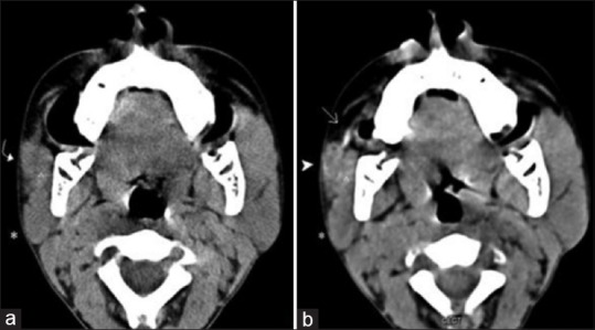
Accessory parotid glands are a common clinical occurrence and usually drain into the main Stenson's duct by small ductules and thereby, into the buccal cavity. Presence of an accessory parotid gland with an ectopic fistulous duct is a rare occurrence. Clinical findings, imaging studies, biochemical tests, histopathological examination are needed for appropriate surgical management. It is extremely rare case with ectopic fistulous duct in an accessory parotid gland managed surgically by internalization of the duct to open into the buccal mucosa and excision of pre-aural appendages. Further to this, we give a comprehensive review of literature on accessory parotid gland and duct anomalies.
Keywords: Accessory parotid gland; congenital parotid fistula; ectopic fistulous duct; pre-aural appendage.
Conflict of interest statement
Figures


References
-
- Frommer J. The human accessory parotid gland: Its incidence, nature, and significance. Oral Surg Oral Med Oral Pathol. 1977;43:671–6. - PubMed
-
- Toh H, Kodama J, Fukuda J, Rittman B, Mackenzie I. Incidence and histology of human accessory parotid glands. Anat Rec. 1993;236:586–90. - PubMed
-
- Naguru H, Miyazawa M. A case of congenital salivary fistula associated with aural appendix. Jpn J Oral Surg. 1972;18:165–8.
-
- Yamasaki H, Tashiro H, Watanabe T. Congenital parotid gland fistula. Int J Oral Maxillofac Surg. 1986;15:492–4. - PubMed
Publication types
LinkOut - more resources
Full Text Sources
Other Literature Sources