

Agreement of angle closure assessments between gonioscopy, anterior segment optical coherence tomography and spectral domain optical coherence tomography
- PMID: 25938053
- PMCID: PMC4413570
- DOI: 10.3980/j.issn.2222-3959.2015.02.23
Agreement of angle closure assessments between gonioscopy, anterior segment optical coherence tomography and spectral domain optical coherence tomography
Abstract
Aim: To determine angle closure agreements between gonioscopy and anterior segment optical coherence tomography (AS-OCT), as well as gonioscopy and spectral domain OCT (SD-OCT). A secondary objective was to quantify inter-observer agreements of AS-OCT and SD-OCT assessments.
Methods: Seventeen consecutive subjects (33 eyes) were recruited from the study hospital's Glaucoma clinic. Gonioscopy was performed by a glaucomatologist masked to OCT results. OCT images were read independently by 2 other glaucomatologists masked to gonioscopy findings as well as each other's analyses of OCT images.
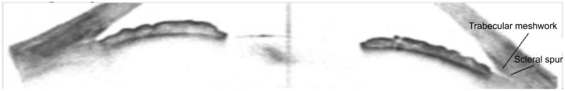
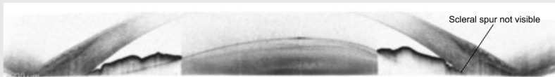
Results: Totally 84.8% and 45.5% of scleral spurs were visualized in AS-OCT and SD-OCT images respectively (P<0.01). The agreement for angle closure between AS-OCT and gonioscopy was fair at k=0.31 (95% confidence interval, CI: 0.03-0.59) and k=0.35 (95% CI: 0.07-0.63) for reader 1 and 2 respectively. The agreement for angle closure between SD-OCT and gonioscopy was fair at k=0.21 (95% CI: 0.07-0.49) and slight at k=0.17 (95% CI: 0.08-0.42) for reader 1 and 2 respectively. The inter-reader agreement for angle closure in AS-OCT images was moderate at 0.51 (95% CI: 0.13-0.88). The inter-reader agreement for angle closure in SD-OCT images was slight at 0.18 (95% CI: 0.08-0.45).
Conclusion: Significant proportion of scleral spurs were not visualised with SD-OCT imaging resulting in weaker inter-reader agreements. Identifying other angle landmarks in SD-OCT images will allow more consistent angle closure assessments. Gonioscopy and OCT imaging do not always agree in angle closure assessments but have their own advantages, and should be used together and not exclusively.
Keywords: angle closure assessment; anterior segment imaging; spectral domain imaging.
Figures
References
-
- Foster PJ. The epidemiology of primary angle closure and associated glaucomatous optic neuropathy. Semin Ophthalmol. 2002;17(2):50–58. - PubMed
-
- Pavlin CJ, Harasiewicz K, Sherar MD, Foster FS. Clinical use of ultrasound biomicroscopy. Ophthalmology. 1991;98(3):287–295. - PubMed
-
- Izatt JA, Hee MR, Swanson EA, Lin CP, Huang D, Schuman JS, Puliafito CA, Fujimoto JG. Micrometer-scale resolution imaging of the anterior eye in vivo with optical coherence tomography. Arch Ophthalmol. 1994;112(12):1584–1589. - PubMed
-
- Radhakrishnan S, Rollins AM, Roth JE, Yazdanfar S, Westphal V, Bardenstein DS, Izatt JA. Real-time optical coherence tomography of the anterior segment at 1310nm. Arch Ophthalmol. 2001;119(8):1179–1185. - PubMed
-
- Radhakrishnan S, Huang D, Smith SD. Optical coherence tomography imaging of the anterior chamber. Ophthalmol Clin North Am. 2005;18(3):375–381. - PubMed
LinkOut - more resources
Full Text Sources
Research Materials