. 2015 Apr 29;16(5):9654-92.
doi: 10.3390/ijms16059654.

Structural basis for carbapenem-hydrolyzing mechanisms of carbapenemases conferring antibiotic resistance

Affiliations

Structural basis for carbapenem-hydrolyzing mechanisms of carbapenemases conferring antibiotic resistance

Jeong Ho Jeon et al. Int J Mol Sci. .

Abstract

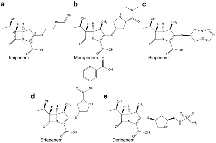
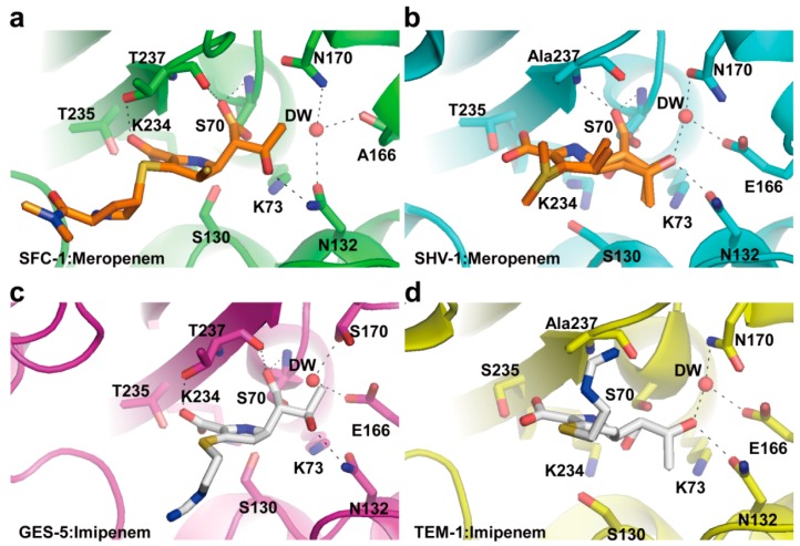
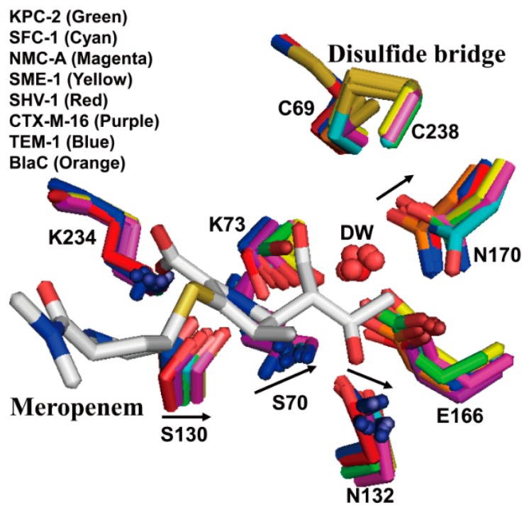
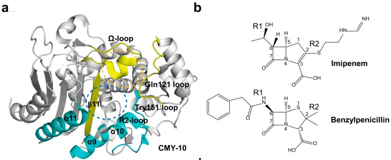
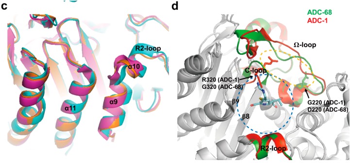
Carbapenems (imipenem, meropenem, biapenem, ertapenem, and doripenem) are β-lactam antimicrobial agents. Because carbapenems have the broadest spectra among all β-lactams and are primarily used to treat infections by multi-resistant Gram-negative bacteria, the emergence and spread of carbapenemases became a major public health concern. Carbapenemases are the most versatile family of β-lactamases that are able to hydrolyze carbapenems and many other β-lactams. According to the dependency of divalent cations for enzyme activation, carbapenemases can be divided into metallo-carbapenemases (zinc-dependent class B) and non-metallo-carbapenemases (zinc-independent classes A, C, and D). Many studies have provided various carbapenemase structures. Here we present a comprehensive and systematic review of three-dimensional structures of carbapenemase-carbapenem complexes as well as those of carbapenemases. We update recent studies in understanding the enzymatic mechanism of each class of carbapenemase, and summarize structural insights about regions and residues that are important in acquiring the carbapenemase activity.
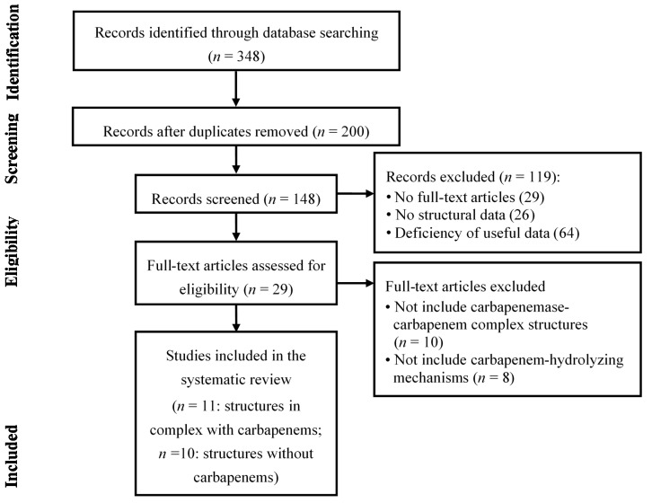

Figures

Figure 1
Figure 1
Chemical structures of (a) imipenem; (b) meropenem; (c) biapenem; (d) ertapenem; and (e) doripenem. The β-lactam nucleus is numbered.
Figure 2
Figure 2
(a) Superposition of active sites of KPC-2 (PDB entry 2OV5, green) and GES-5 (PDB entry 4GNU, yellow) is shown. The disulfide bridge between C69 and C238 (arrow) is shown in KPC-2 and GES-5; (b) Superposition of active sites of class A carbapenemases (KPC-2 (PDB entry 2OV5, green) and GES-5 (PDB entry 4GNU, yellow)) and class A non-carbapenemases (TEM-1 (PDB entry 1ZG4, red) and SHV-1 (PDB entry 1SHV, blue)). The residues (C69/M69, S70, K73, S130, N132, E166, N170/S170, T237/A237, and C238/G238) in the active-site cleft are shown as sticks. Superpositions were performed using SSM Superpose [53] to align the complete chains. This figure was prepared using PyMOL [54].
Figure 3
Figure 3
Imipenem acyl-enzyme intermediate complexes. (a) The active site of GES-1 (PDB entry 4GOG, green) with the bound imipenem (cyan) is shown; (b) The active site of GES-5 (PDB entry 4H8R, orange) with the bound imipenem (yellow) is shown; (c) Superposition of active sites of GES-1:imipenem and GES-5:imipenem is shown; (d) The active site of GES-2 (PDB entry 4QU3, light pink) with the bound ertapenem (magenta) is shown; (e) Superposition of active sites of GES-2:ertapenem and GES-5:imipenem is shown. The residues (S70, K73, S130, N132, E166, G170/S170/N170, T237, and R244) in the active-site cleft are shown as sticks. The hydrogen bond interactions are shown as dashed black lines. The partially occupied water molecules are shown as red and blue spheres. Superpositions were performed using SSM Superpose [53] to align the complete chains. These figures were prepared using PyMOL [54] and data adapted from Smith et al. [36] and Stewart et al. [57].
Figure 3
Figure 3
Imipenem acyl-enzyme intermediate complexes. (a) The active site of GES-1 (PDB entry 4GOG, green) with the bound imipenem (cyan) is shown; (b) The active site of GES-5 (PDB entry 4H8R, orange) with the bound imipenem (yellow) is shown; (c) Superposition of active sites of GES-1:imipenem and GES-5:imipenem is shown; (d) The active site of GES-2 (PDB entry 4QU3, light pink) with the bound ertapenem (magenta) is shown; (e) Superposition of active sites of GES-2:ertapenem and GES-5:imipenem is shown. The residues (S70, K73, S130, N132, E166, G170/S170/N170, T237, and R244) in the active-site cleft are shown as sticks. The hydrogen bond interactions are shown as dashed black lines. The partially occupied water molecules are shown as red and blue spheres. Superpositions were performed using SSM Superpose [53] to align the complete chains. These figures were prepared using PyMOL [54] and data adapted from Smith et al. [36] and Stewart et al. [57].
Figure 4
Figure 4
Comparison between carbapenem acyl-enzymes of class A carbapenemases (SFC-1 and GES-5) and non-carbapenemases (SHV-1 and TEM-1). (a) SFC-1 (E166A mutant):meropenem (PDB entry 4EV4, green); (b) SHV-1:meropenem (PDB entry 2ZD8, cyan); (c) GES-5:imipenem (PDB entry 4H8R, magenta); and (d) TEM-1:imipenem (PDB entry 1BT525, yellow). The residues (S70, K73, S130, N132, E166/A166, N170/S170, T235, and T237) in the active-site cleft are shown as sticks. Meropenem and imipenem carbon atoms are rendered in orange and white, respectively. Hydrogen bonds involving the deacylating water molecule (DW, red sphere), the acyl-enzyme carbonyl group, the carbapenem C3 carboxylate, the 6α-1R-hydroxyethyl group of carbapenem, and Asn132 NH2 are indicated by dashed black lines. These figures were prepared using PyMOL [54] and data adapted from Fonseca et al. [37].
Figure 5
Figure 5
Superposition of active sites of class A carbapenemases (KPC-2 (PDB entry 2OV5, green), SFC-1 (E166A mutant; PDB entry 4EQI, cyan), NMC-A (PDB entry 1BUE, magenta), and SME-1 (PDB entry 1DY6, yellow)) and non-carbapenemases (SHV-1 (PDB entry 1SHV, red), CTX-M-16 (PDB entry 1YLW, purple), TEM-1 (PDB entry 1ZG4, blue), and BlaC (PDB entry 2GDN, orange)). Meropenem, as bound in the SFC-1 (E166A mutant) acyl-enzyme complex structure, is shown in white. The residues (C69, S70, K73, S130, N132, E166, N170, K234, and C238) in the active-site cleft are shown as sticks. Arrows denote shifts in positions of labeled residues in class A carbapenemases. There is a disulfide bridge between C69 and C238 in only class A carbapenemases. Superpositions were performed using SSM Superpose [53] to align the complete chains. This figure was prepared using PyMOL [54] and data adapted from Fonseca et al. [37].
Figure 6
Figure 6
(a) Overall structure of CMY-10 (PDB entry 1ZKJ) is shown. The R1 subsite is surrounded by the Ω-loop, Gln121 loop, and β11 (in yellow). The R2 subsite is enclosed by the Tyr151 loop, α10 in the R2-loop, and α11 (in cyan). The R1 and R2 subsites are indicated as orange and blue dotted circles, respectively; (b) Schematic drawing of imipenem and benzylpenicillin is shown. The β-lactam nucleus is numbered. The R1 and R2 side chains located at the C6 and C2 positions of the β-lactam nucleus are labeled, respectively; (c) The displacement of α9 and α10 in CMY-10 is shown. CMY-10 (PDB entry 1ZKJ, cyan) was superposed with P99 β-lactamase (PDB entry 2BLT, orange) and GC1 β-lactamase (PDB entry 1GCE, magenta [89]). The R2-loop displays noticeable structural alterations: the R2-loop becomes flexible, and the shortened path of the connection loop between α10 and β11 induces the ~2.5 Å shift of α9 and α10 relative to the adjacent helix α11 in CMY-10 compared with both P99 and GC1 β-lactamases; (d) Superimposed complex of imipenem with ADC-68 and ADC-1. An AmpC complex with imipenem (PDB entry 1LL5) was superposed with ADC-1 (PDB entry 4NET) and ADC-68 (PDB entry 4QD4). ADC-68 and ADC-1 are represented as green and red ribbon diagrams, respectively. Imipenem is represented as cyan stick. C-loop (T318–F321) is positioned between β8 and β9. Ω-loop (G185–T229) is positioned between α6 and α8. R2-loop (E291–V309) is positioned between α9 and α10b. The R1 and R2 subsites are indicated as orange and blue dotted circles, respectively. R320 (ADC-1) and G320 (ADC-68) residues are located in C-loop and G220 (ADC-1) and D220 (ADC-68) residues are found in Ω-loop. Superpositions were performed using SSM Superpose [53] to align the complete chains. The structures of CMY-10 and ADC-68 were prepared using PyMOL [54] and data adapted from Kim et al. [80] and Jeon et al. [12], respectively.
Figure 6
Figure 6
(a) Overall structure of CMY-10 (PDB entry 1ZKJ) is shown. The R1 subsite is surrounded by the Ω-loop, Gln121 loop, and β11 (in yellow). The R2 subsite is enclosed by the Tyr151 loop, α10 in the R2-loop, and α11 (in cyan). The R1 and R2 subsites are indicated as orange and blue dotted circles, respectively; (b) Schematic drawing of imipenem and benzylpenicillin is shown. The β-lactam nucleus is numbered. The R1 and R2 side chains located at the C6 and C2 positions of the β-lactam nucleus are labeled, respectively; (c) The displacement of α9 and α10 in CMY-10 is shown. CMY-10 (PDB entry 1ZKJ, cyan) was superposed with P99 β-lactamase (PDB entry 2BLT, orange) and GC1 β-lactamase (PDB entry 1GCE, magenta [89]). The R2-loop displays noticeable structural alterations: the R2-loop becomes flexible, and the shortened path of the connection loop between α10 and β11 induces the ~2.5 Å shift of α9 and α10 relative to the adjacent helix α11 in CMY-10 compared with both P99 and GC1 β-lactamases; (d) Superimposed complex of imipenem with ADC-68 and ADC-1. An AmpC complex with imipenem (PDB entry 1LL5) was superposed with ADC-1 (PDB entry 4NET) and ADC-68 (PDB entry 4QD4). ADC-68 and ADC-1 are represented as green and red ribbon diagrams, respectively. Imipenem is represented as cyan stick. C-loop (T318–F321) is positioned between β8 and β9. Ω-loop (G185–T229) is positioned between α6 and α8. R2-loop (E291–V309) is positioned between α9 and α10b. The R1 and R2 subsites are indicated as orange and blue dotted circles, respectively. R320 (ADC-1) and G320 (ADC-68) residues are located in C-loop and G220 (ADC-1) and D220 (ADC-68) residues are found in Ω-loop. Superpositions were performed using SSM Superpose [53] to align the complete chains. The structures of CMY-10 and ADC-68 were prepared using PyMOL [54] and data adapted from Kim et al. [80] and Jeon et al. [12], respectively.
Figure 7
Figure 7
Molecular surface representations of the class D carbapenemases. (a) OXA-24/40 (PDB entry 2JC7, cyan); (b) OXA-23 (PDB entry 4JF6, green); (c) OXA-48 (PDB entry 3HBR, yellow); and (d) OXA-58 (PDB entry 4OH0, orange). The four molecules are shown in the same relative orientation, and the active site exists at the bottom center of each figure. The hydrophobic bridge (arrow) is shown in OXA-23 and OXA-24/40. A lack of a similar bridge in OXA-48 and OXA-58 is evident. The residues related to the hydrophobic bridge are indicated as sticks. These figures were prepared using PyMOL [54] and data adapted from Smith et al. [103].
Figure 8
Figure 8
(a) Pyrroline tautomerization after doripenem acylation of some β-lactamase enzymes such as OXA-1; (b) The superposition of OXA-1:doripenem (PDB entry 3ISG, magenta) and OXA-24/40 (K84D mutant):doripenem (PDB entry 3PAE, cyan) active sites is shown. Residues (S81, S219, and R261) from OXA-24/40 K84D and residues (S67, K212, and T213) from OXA-1 are shown. The hydrogen bond interactions are shown as dashed black lines; (c) Doripenem and two active site residues of OXA-24/40 (K84D mutant) are shown in cyan; (d) Doripenem and two active site residues of OXA-1 are shown in magenta. Superpositions were performed using SSM Superpose [53] to align the complete chains. These figures were prepared using PyMOL [54] and data adapted from Schneider et al. [106].
Figure 8
Figure 8
(a) Pyrroline tautomerization after doripenem acylation of some β-lactamase enzymes such as OXA-1; (b) The superposition of OXA-1:doripenem (PDB entry 3ISG, magenta) and OXA-24/40 (K84D mutant):doripenem (PDB entry 3PAE, cyan) active sites is shown. Residues (S81, S219, and R261) from OXA-24/40 K84D and residues (S67, K212, and T213) from OXA-1 are shown. The hydrogen bond interactions are shown as dashed black lines; (c) Doripenem and two active site residues of OXA-24/40 (K84D mutant) are shown in cyan; (d) Doripenem and two active site residues of OXA-1 are shown in magenta. Superpositions were performed using SSM Superpose [53] to align the complete chains. These figures were prepared using PyMOL [54] and data adapted from Schneider et al. [106].
Figure 9
Figure 9
(a) Active site of the OXA-23 in complex with meropenem (PDB entry 4JF4) is shown. The residues (S79, K82, F110, W113, S126, V128, L166, T219, and R259) in the active-site cleft are shown as green sticks. Meropenem is represented as orange stick. The hydrogen bond interactions between meropenem and residues in the active site in OXA-23 are shown as dashed black lines; (b) Molecular surface representation of OXA-23 in complex with meropenem is shown. The side chains of K82, V128, and L166 in OXA-23 are shown in sticks; (c) Molecular surface representation of OXA-1 in complex with doripenem (PDB entry 3ISG) is shown. Doripenem is represented as cyan stick. The side chains of K70, V117, and L161 in OXA-1 are shown in sticks. These figures were prepared using PyMOL [54] and data adapted from Smith et al. [104].
Figure 10
Figure 10
Comparison of the structural features of class D carbapenemases (OXA-23 (PDB entry 4JF6, green), OXA-24/40 (PDB entry 2JC7, cyan), OXA-48 (PDB entry 3HBR, yellow), and OXA-58 (PDB entry 4OH0, orange)) with the class D non-carbapenemases (OXA-1 (PDB entry 1M6K, magenta), OXA-10 (PDB entry 1FOF, red), and OXA-13 (PDB entry 1H8Z, pink)), showing the significant differences in orientation and length of the β5–β6 loop. The β5–β6 loops, connecting β5- and β6-strands, are indicated by the black circle. Superpositions were performed using SSM Superpose [53] to align the complete chains. These figures were prepared using PyMOL [54] and data adapted from De Luca et al. [119].
Figure 11
Figure 11
Comparison of OXA-23 (PDB entry 4K0X, green) and OXA-146 (PDB entry 4K0W, red) structures. (a) Overlaid structure showing the position of the β5–β6 loop deviation (black circle) in the context of the full structures of OXA-23 and OXA-146; (b) Overlaid structure of OXA-23 and OXA-146 is shown. Residues (M221 and D222, green) in OXA-23 and residues (M222 and D223, red) in OXA-146 are shown as sticks; (c) Overlaid structure of OXA-23 and OXA-146 after alignment with the β-lactam sensor protein BlaR1 with ceftazidime bound as an acyl intermediate (PDB entry 1XKZ). The ceftazidime serine acyl moiety is shown in cyan, and the rest of the BlaR1 protein is not shown. Superpositions were performed using SSM Superpose [53] to align the complete chains. These figures were prepared using PyMOL [54] and data adapted from Kaitany et al. [105].
Figure 12
Figure 12
Ribbon representations of the three subclasses. (a) Overall structure of BcII (subclass B1, PDB entry 1BVT) from B. cereus is shown. The zinc-binding residues and flexible loop (L3 loop) are shown in green. The zinc-binding residues (H116-H118-H196 for Zn1 and D120-C221-H263 for Zn2) in the active-site cleft are shown as green sticks; (b) Overall structure of CphA (subclass B2, PDB entry 1X8G) from A. hydrophila is shown. The zinc-binding residues and elongated α3-helix are shown in cyan. The zinc-binding residues (N116-H118-H196 for Zn1 and D120-C221-H263 for Zn2) in the active-site cleft are shown as cyan sticks; (c) Overall structure of FEZ-1 (subclass B3, PDB entry 1K07) from F. gormanii is shown. The zinc-binding residues and the loop between α3-helix and β7-strand are shown in orange. The zinc-binding residues (H116-H118-H196 for Zn1 and D120-H121-H263 for Zn2) in the active-site cleft are shown as orange sticks. Zn1 and Zn2 are represented as yellow and red spheres, respectively. These figures were prepared using PyMOL [54] and data adapted from Garau et al. [154].
Figure 13
Figure 13
(a) Comparison of the structural features of NDM-1 (PDB entry 3SPU, green) with IMP-1 (PDB entry 1DD6, cyan), VIM-2 (PDB entry 1KO2, magenta), and VIM-7 (PDB entry 2Y87, yellow), showing the significant differences in orientation of the L3 loop. L3 loops are indicated by the red circle; (b) Active site of the NDM-1 in complex with meropenem (PDB entry 4EYL, green) is shown. The residues (H120, H122, D124, H189, K211, N220, and H250; NDM-1 numbering) in the active-site cleft are shown as green sticks. Zinc coordination and hydrogen bonding in active site in NDM-1 are shown as dashed black lines. The hydrolyzed meropenem is shown in magenta. Zn1 and Zn2 are represented as yellow and red spheres, respectively; (c) The conformational change upon substrate binding is represented by superimposition of the wild-type CphA (PDB entry 1X8G, white) and CphA (N220G mutant):biapenem complex (PDB entry 1X8I, light pink). The residues (K224, G232, and N233; class B numbering) in the active-site cleft are shown as sticks. The movement (from open to closed position) of G232-N233 loop appears as a red arrow. The closed position is shown in CphA (N220G mutant):biapenem complex structure. The hydrogen bond interactions between the C3 carboxyl group of biapenem and two residues (K224 and N233) in the active site in CphA are shown as dashed red lines. The hydrolyzed biapenem and Zn2 are represented as yellow stick mode and red sphere, respectively. Superpositions were performed using SSM Superpose [53] to align the complete chains. The structures of NDM-1 and CphA were prepared using PyMOL [54] and data adapted from King et al. [149] and Garau et al. [154], respectively.
Figure 14
Figure 14
Literature selection process (PRISMA flow diagram). A total of 11 articles among 29 articles include structures in complex with carbapenems and mechanisms of carbapenemases. Ten articles among 29 articles include structures without carbapenems and mechanisms of carbapenemases. There were eight articles among 29 articles that include structures of carbapenemases without mechanisms. A total of 21 articles were included in the systematic review.

References

    1. Ambler R.P. The structure of β-lactamases. Philos. Trans. R. Soc. Lond. B. 1980;289:321–331. doi: 10.1098/rstb.1980.0049. - DOI - PubMed
    1. Queenan A.M., Bush K. Carbapenemases: The versatile β-lactamases. Clin. Microbiol. Rev. 2007;20:440–458. doi: 10.1128/CMR.00001-07. - DOI - PMC - PubMed
    1. Moellering R.C., Jr., Eliopoulos G.M., Sentochnik D.E. The carbapenems: New broad spectrum β-lactam antibiotics. J. Antimicrob. Chemother. 1989;24:1–7. doi: 10.1093/jac/24.suppl_A.1. - DOI - PubMed
    1. Richet H.M., Mohammed J., McDonald L.C., Jarvis W.R. Building communication networks: International network for the study and prevention of emerging antimicrobial resistance. Emerg. Infect. Dis. 2001;7:319–322. doi: 10.3201/eid0702.010235. - DOI - PMC - PubMed
    1. Lee J.H., Lee S.H. Carbapenem resistance in gram-negative pathogens: Emerging non-metallo-carbapenemases. Res. J. Microbiol. 2006;1:1–22. doi: 10.3923/jm.2006.1.22. - DOI

Publication types

LinkOut - more resources