. 2015 Apr 29;13(5):2666-79.
doi: 10.3390/md13052666.

The Cytoprotective Effect of Petalonia binghamiae Methanol Extract against Oxidative Stress in C2C12 Myoblasts: Mediation by Upregulation of Heme Oxygenase-1 and Nuclear Factor-Erythroid 2 Related Factor 2

Affiliations

The Cytoprotective Effect of Petalonia binghamiae Methanol Extract against Oxidative Stress in C2C12 Myoblasts: Mediation by Upregulation of Heme Oxygenase-1 and Nuclear Factor-Erythroid 2 Related Factor 2

Ji Sook Kang et al. Mar Drugs. .

Abstract

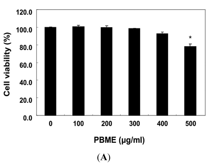
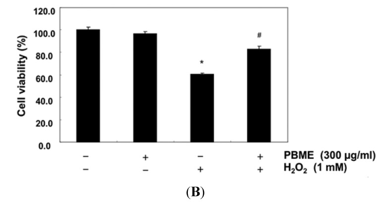
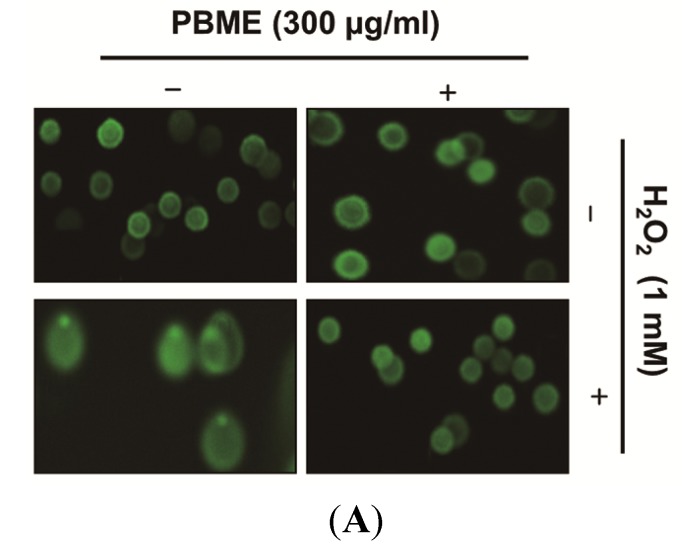
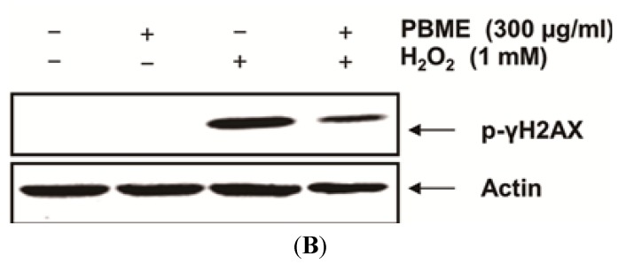
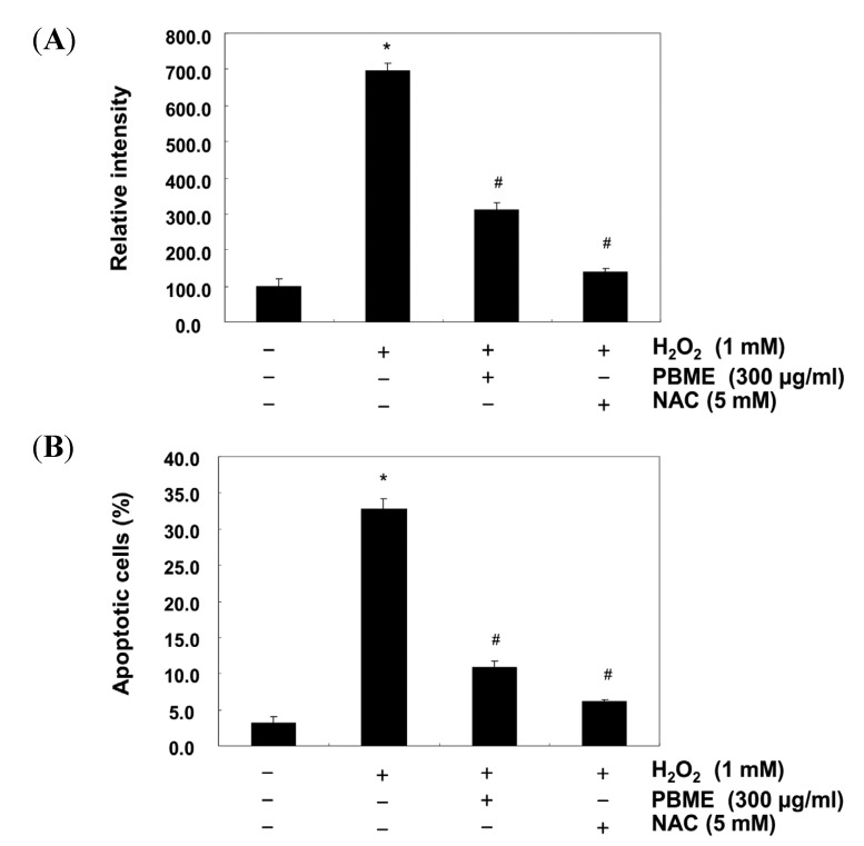
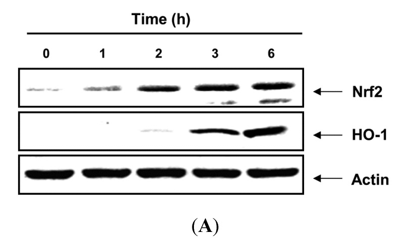
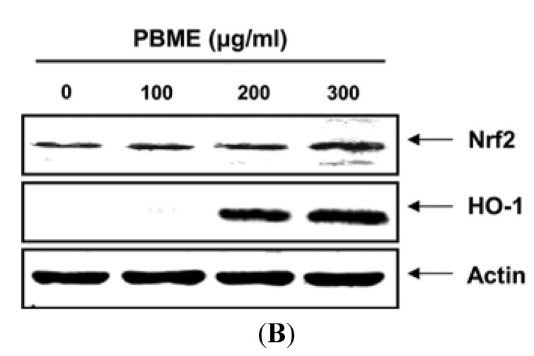
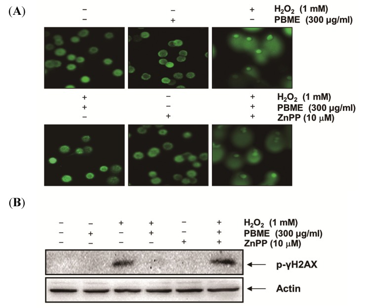
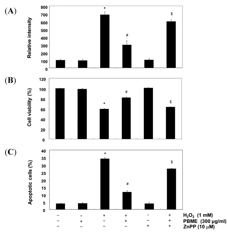
This study was designed to examine the protective effects of the marine brown algae Petalonia binghamiae against oxidative stress-induced cellular damage and to elucidate the underlying mechanisms. P. binghamiae methanol extract (PBME) prevented hydrogen peroxide (H2O2)-induced growth inhibition and exhibited scavenging activity against intracellular reactive oxygen species (ROS) induced by H2O2 in mouse-derived C2C12 myoblasts. PBME also significantly attenuated H2O2-induced comet tail formation in a comet assay, histone γH2A.X phosphorylation, and annexin V-positive cells, suggesting that PBME prevented H2O2-induced cellular DNA damage and apoptotic cell death. Furthermore, PBME increased the levels of heme oxygenase-1 (HO-1), a potent antioxidant enzyme, associated with the induction of nuclear factor-erythroid 2 related factor 2 (Nrf2). However, zinc protoporphyrin IX, a HO-1 competitive inhibitor, significantly abolished the protective effects of PBME on H2O2-induced ROS generation, growth inhibition, and apoptosis. Collectively, these results demonstrate that PBME augments the antioxidant defense capacity through activation of the Nrf2/HO-1 pathway.


Figures

Figure 1
Figure 1
Effects of P. binghamiae methanol extract (PBME) on cell viability and H2O2-induced growth inhibition in C2C12 cells. The cells were treated with various concentrations of PBME for 24 h (A) or pretreated with 300 μg/mL of PBME for 1 h and then incubated with and without 1 mM of H2O2 for 6 h (B). The cell viability was assessed with a 3-(4,5-dimethylthiazol-2-yl)-2,5-diphenyltetrazolium bromide (MTT) reduction assay. The results are presented as the mean ± standard deviation (SD) values obtained in three independent experiments (* p < 0.05 compared with the control group; # p < 0.05 compared with the H2O2-treated group).
Figure 1
Figure 1
Effects of P. binghamiae methanol extract (PBME) on cell viability and H2O2-induced growth inhibition in C2C12 cells. The cells were treated with various concentrations of PBME for 24 h (A) or pretreated with 300 μg/mL of PBME for 1 h and then incubated with and without 1 mM of H2O2 for 6 h (B). The cell viability was assessed with a 3-(4,5-dimethylthiazol-2-yl)-2,5-diphenyltetrazolium bromide (MTT) reduction assay. The results are presented as the mean ± standard deviation (SD) values obtained in three independent experiments (* p < 0.05 compared with the control group; # p < 0.05 compared with the H2O2-treated group).
Figure 2
Figure 2
Effect of PBME on H2O2-induced DNA damage in the C2C12 cells. The C2C12 cells were pretreated with 300 μg/mL of PBME for 1 h and then incubated with and without 1 mM of H2O2 for 6 h. (A) To detect cellular DNA damage, a comet assay was performed, and representative pictures of the comets were taken using a fluorescence microscope (200× original magnification); (B) The cells were lysed, and equal amounts of cell lysates were then separated on SDS-polyacrylamide gels and transferred to nitrocellulose membranes. The membranes were probed with specific antibodies against p-γH2A.X and actin, as an internal control, and the proteins were visualized using an enhanced chemiluminescence (ECL) detection system. A representative blot from three independent experiments is shown.
Figure 2
Figure 2
Effect of PBME on H2O2-induced DNA damage in the C2C12 cells. The C2C12 cells were pretreated with 300 μg/mL of PBME for 1 h and then incubated with and without 1 mM of H2O2 for 6 h. (A) To detect cellular DNA damage, a comet assay was performed, and representative pictures of the comets were taken using a fluorescence microscope (200× original magnification); (B) The cells were lysed, and equal amounts of cell lysates were then separated on SDS-polyacrylamide gels and transferred to nitrocellulose membranes. The membranes were probed with specific antibodies against p-γH2A.X and actin, as an internal control, and the proteins were visualized using an enhanced chemiluminescence (ECL) detection system. A representative blot from three independent experiments is shown.
Figure 3
Figure 3
Effect of PBME on H2O2-induced ROS generation and apoptosis in the C2C12 cells. The C2C12 cells were pretreated with 300 μg/mL of PBME for 1 h and then incubated with and without 1 mM H2O2 for 6 h. (A) To monitor the production of ROS, the cells were incubated at 37 °C in the dark for 20 min with new culture medium containing 10 μM of H2DCFDA. The generation of ROS was measured with a flow cytometer; (B) The cells were also stained with annexin V-FITC and propidium iodide (PI), and the percentages of apoptotic cells (annexin V+/PI cells) were then analyzed using flow cytometric analysis. The results are presented as the mean ± SD values obtained in three independent experiments (* p < 0.05 compared with the control group; # p < 0.05 compared with the H2O2-treated group).
Figure 4
Figure 4
Induction of nuclear factor-erythroid 2 related factor 2 (Nrf2) and heme oxygenase-1 (HO-1) expression by PBME in the C2C12 cells. The cells were incubated with 300 μg/mL of PBME for the indicated periods (A) or with various concentrations of PBME for 6 h (B). The cellular proteins were separated on SDS-polyacrylamide gels and then transferred onto nitrocellulose membranes. The membranes were probed with the specific antibodies against Nrf2 and HO-1. Actin was used as a loading control.
Figure 4
Figure 4
Induction of nuclear factor-erythroid 2 related factor 2 (Nrf2) and heme oxygenase-1 (HO-1) expression by PBME in the C2C12 cells. The cells were incubated with 300 μg/mL of PBME for the indicated periods (A) or with various concentrations of PBME for 6 h (B). The cellular proteins were separated on SDS-polyacrylamide gels and then transferred onto nitrocellulose membranes. The membranes were probed with the specific antibodies against Nrf2 and HO-1. Actin was used as a loading control.
Figure 5
Figure 5
Effects of an inhibitor of HO-1 on PBME-mediated protection of DNA damage by H2O2 in the C2C12 cells. The cells were pretreated for 1 h with 300 μg/mL of PBME and then treated for 6 h, with or without 1 mM of H2O2 in the absence or presence of 10 μM of zinc protoporphyrin IX (ZnPP). (A) A comet assay was performed, and representative pictures of the comets were taken using a fluorescence microscope at 200× original magnification; (B) Cell lysates were prepared and subjected to Western blot analysis with a specific antibody against p-γH2A.X. Actin was used as a loading control.
Figure 6
Figure 6
Effects of an inhibitor of HO-1 on PBME-mediated attenuation of ROS formation, apoptosis, and growth inhibition by H2O2 in the C2C12 cells. (A) The cells grown under the same conditions as in Figure 5 and assayed for ROS generation; (B) The degree of apoptosis was evaluated using a flow cytometer; (C) The cell viability was estimated with an MTT assay. The results are presented as the mean ± SD values obtained in three independent experiments (* p < 0.05 compared with the control group; # p < 0.05 compared with the H2O2-treated group; $ p < 0.05 compared with the H2O2- and PBME-treated group).

References

    1. Finkel T. Signal transduction by reactive oxygen species. J. Cell Biol. 2011;194:7–15. doi: 10.1083/jcb.201102095. - DOI - PMC - PubMed
    1. Kregel K.C., Zhang H.J. An integrated view of oxidative stress in aging: basic mechanisms, functional effects, and pathological considerations. Am. J. Physiol. Regul. Integr. Comp. Physiol. 2007;292:R18–R36. doi: 10.1152/ajpregu.00327.2006. - DOI - PubMed
    1. Vesely M.J., Exon D.J., Clark J.E., Foresti R., Green C.J., Motterlini R. Heme oxygenase-1 induction in skeletal muscle cells: Hemin and sodium nitroprusside are regulators in vitro. Am. J. Physiol. 1998;275:C1087–C1094. - PubMed
    1. Motohashi H., Yamamoto M. Nrf2-Keap1 defines a physiologically important stress response mechanism. Trends Mol. Med. 2004;10:549–557. doi: 10.1016/j.molmed.2004.09.003. - DOI - PubMed
    1. Surh Y.J., Kundu J.K., Na H.K. Nrf2 as a master redox switch in turning on the cellular signaling involved in the induction of cytoprotective genes by some chemopreventive phytochemicals. Planta Med. 2008;74:1526–1539. doi: 10.1055/s-0028-1088302. - DOI - PubMed

Publication types

MeSH terms

LinkOut - more resources