Observational Study
. 2015 Jul;122(7):1330-9.
doi: 10.1016/j.ophtha.2015.03.020. Epub 2015 May 1.

High-Resolution Imaging of the Optic Nerve and Retina in Optic Nerve Hypoplasia

Affiliations
Observational Study

High-Resolution Imaging of the Optic Nerve and Retina in Optic Nerve Hypoplasia

Anastasia Pilat et al. Ophthalmology. 2015 Jul.

Abstract

Purpose: To investigate the optic nerve and macular morphology in patients with optic nerve hypoplasia (ONH) using spectral-domain optical coherence tomography (SD OCT).

Design: Prospective, cross-sectional, observational study.

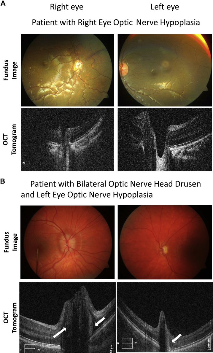
Subjects: A total of 16 participants with ONH (10 female and 6 male; mean age, 17.2 years; 6 bilateral involvement) and 32 gender-, age-, ethnicity-, and refraction-matched healthy controls.

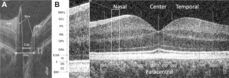
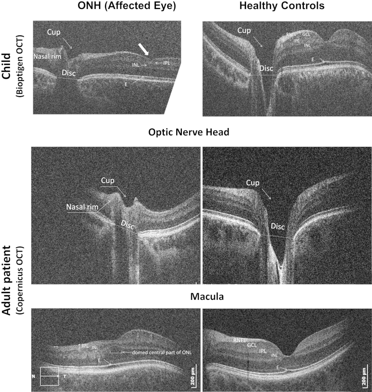
Methods: High-resolution SD OCT (Copernicus [Optopol Technology S.A., Zawiercie, Poland], 3 μm resolution) and handheld SD OCT (Bioptigen Inc [Research Triangle Park, NC], 2.6 μm resolution) devices were used to acquire horizontal scans through the center of the optic disc and macula.

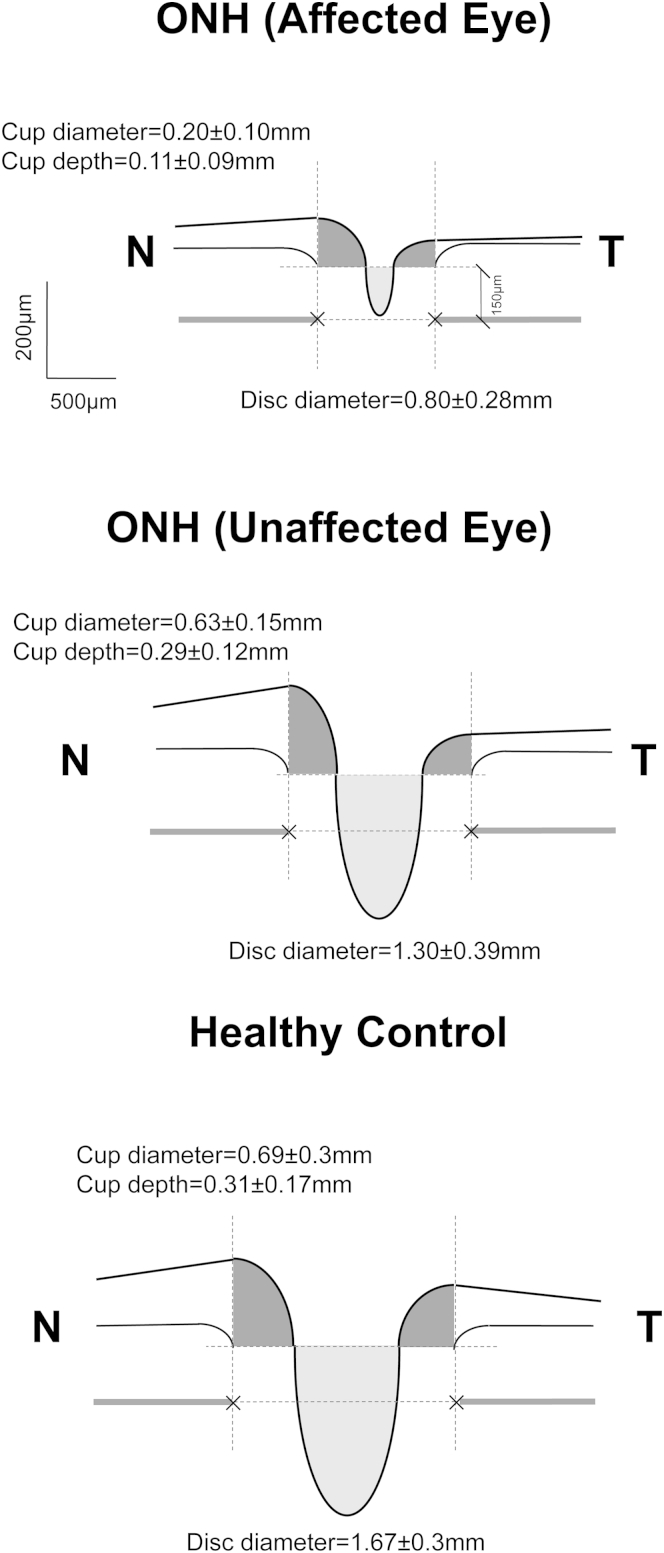
Main outcome measures: Horizontal optic disc/cup and rim diameters, cup depth, peripapillary retinal nerve fiber layer (RNFL), and thickness of individual retinal layers in participants with ONH and in controls.

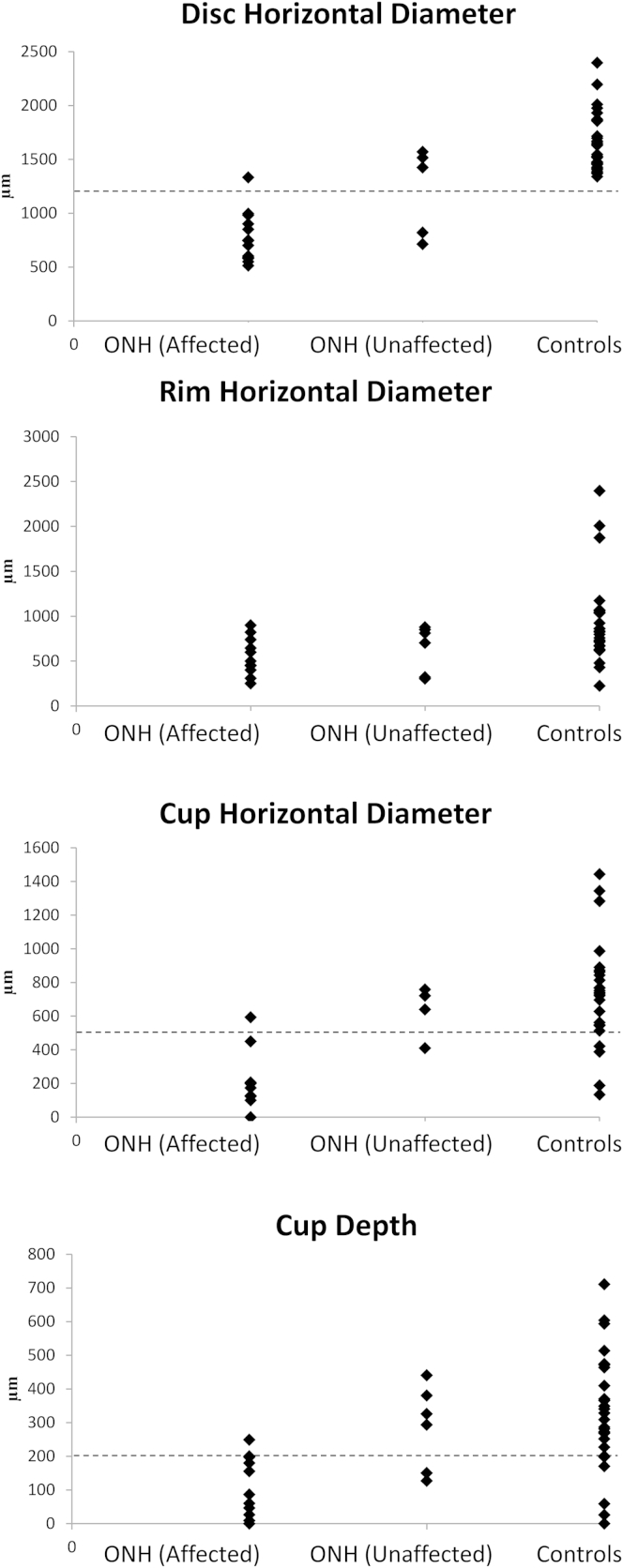
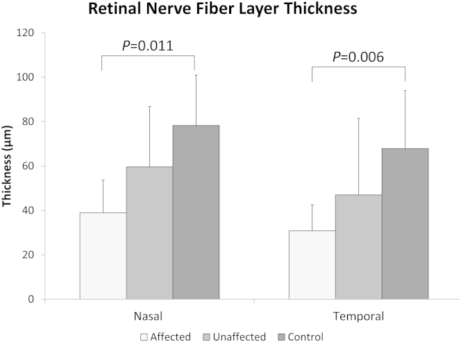
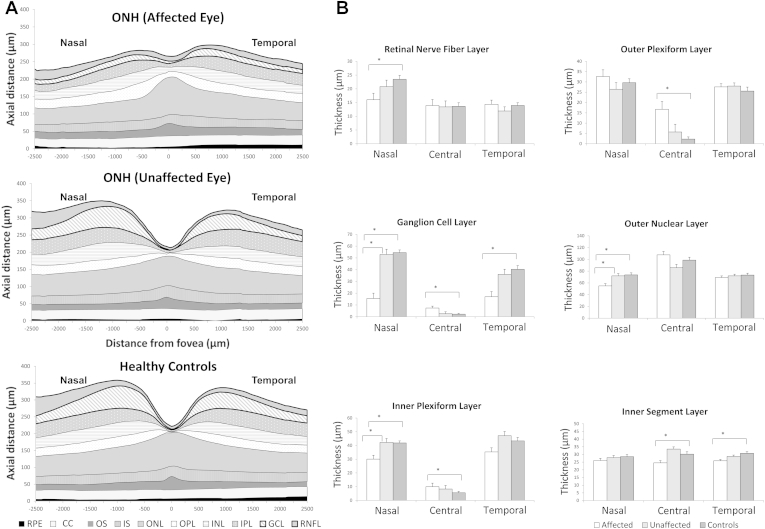
Results: Patients with ONH had significantly smaller discs (P < 0.03 and P < 0.001 compared with unaffected eye and healthy controls, respectively), horizontal cup diameter (P < 0.02 for both), and cup depth (P < 0.02 and P < 0.01, respectively). In the macula, significantly thinner RNFL (nasally), ganglion cell layer (GCL) (nasally and temporally), inner plexiform layer (IPL) (nasally), outer nuclear layer (ONL) (nasally), and inner segment (centrally and temporally) were found in patients with ONH compared with the control group (P < 0.05 for all comparisons). Continuation of significantly thicker GCL, IPL, and outer plexiform layer in the central retinal area (i.e., foveal hypoplasia) was found in more than 80% of patients with ONH. Clinically unaffected fellow eyes of patients with ONH showed mild features of underdevelopment. Visual acuity and presence of septo-optic dysplasia were associated with changes in GCL and IPL. Sensitivity and specificity for the detection of ONH based on disc and retinal optical coherence tomography (OCT) parameters were >80%.

Conclusions: Our study provides evidence of retinal changes in ONH. In addition to thinning of retina layers mainly involving the RNFL and GCL, signs reminiscent of foveal hypoplasia were observed in patients with ONH. Optic nerve and foveal parameters measured using OCT showed high sensitivity and specificity for detecting ONH, demonstrating their useful for clinical diagnosis.


Figures

Figure 1
Figure 1
Horizontal spectral-domain optical coherence tomography (SD OCT) B-scan images of the (A) optic nerve head and (B) macula area of a control subject. A, An OCT scan of the optic nerve head where the disc diameter was defined as the distance between the edges of retinal pigment epithelium (RPE); cup diameter was measured as the length of the line parallel to the disc diameter in 150 μm anteriorly to the disc and limited by the internal limiting membrane; nasal and temporal rims were calculated as the difference between the disc and cup edges; and maximal cup depth was measured as a perpendicular line between the cup diameter and the deepest point of the cup, retinal nerve fiber layer (RNFL), was delineated manually. B, The position of the different retinal layers (left) in an OCT scan of the macular area (right). The thickness of the layers was measured in the center of the fovea, in the paracentral area (from 250 μm nasally to 250 μm temporally), and nasally and temporally (from 500–2000 μm). BM = Bruch's membrane; CC = contact cylinder; E = ellipsoid; ELM = external limiting membrane; GCL = ganglion cell layer; INL = inner nuclear layer; IPL = inner plexiform layer; IS = inner segment; ONL = outer nuclear layer; OPL = outer plexiform layer; OS = outer segment.
Figure 2
Figure 2
Fundus images (top) and spectral-domain optical coherence tomography (SD OCT) horizontal B-scans (bottom) through the center of the disc in (A) a patient with right eye optic nerve hypoplasia (ONH) (patient 1, Table 1, available at www.aaojournal.org) and (B) bilateral optic nerve head drusen (ONHD) with ONH in the left eye (patient 8, Table 1, available at www.aaojournal.org). Arrows indicate the position of the drusen above the edges of the RPE. The disc size of the left eye with both ONHD and ONH is considerably smaller than in the unaffected eye.
Figure 3
Figure 3
Examples of spectral-domain optical coherence tomography (SD OCT) of affected left eyes of the patients with optic nerve hypoplasia (ONH) (left) and age-, gender-, ethnicity-, and refraction-matched healthy controls (right). The optic nerve head scans of patients with ONH demonstrated small disc and cup diameters, and cup depth. The horizontal macula scans of the patients with ONH (bottom left and top left) demonstrate a thin retina, flat foveal pits, a thin retinal nerve fiber layer (RNFL) and ganglion cell layer (GCL), continuation of the inner plexiform layer (IPL) and inner nuclear layer (INL), a domed central area of ONL, and absence of the upward deflection of the ellipsoid line in the center of the fovea in patients with ONH. E = ellipsoid.
Figure 4
Figure 4
Cross-sectional schematic diagrams representing mean values of optic nerve head parameters of patients with optic nerve hypoplasia (ONH) (affected and clinically unaffected eyes) and controls. Upper horizontal dotted lines represent horizontal offset (150 μm) used to determine cup diameters, and lower horizontal dotted lines indicate disc horizontal diameters. Vertical dotted lines show margins of rim areas. Mean (± standard error) values of parameters that were statistically significant are given. N = nasal; T = temporal.
Figure 5
Figure 5
Distribution of optic nerve head parameters (μm) in patients with optic nerve hypoplasia (ONH) in affected/clinically unaffected eyes and healthy controls. Horizontal dotted line shows the threshold for optic disc hypoplasia detection in parameters that were highly sensitive and specific.
Figure 6
Figure 6
Average thickness of the peripapillary retinal nerve fiber layer (RNFL) in patients with optic nerve hypoplasia (ONH) (affected and unaffected eyes) and healthy controls nasally and temporally from the disc center. Error bars are standard deviations.
Figure 7
Figure 7
Cross-sectional schematic diagram of individual retinal layers (A) and mean and standard error (μm) of the thicknesses of retinal layers when the difference between groups was significant (B) in patients with optic nerve hypoplasia (ONH) (affected and clinically unaffected eyes) and controls. CC = contact cylinder; GCL = ganglion cell layer; INL = inner nuclear layer; IPL = inner plexiform layer; IS = inner segment; ONL = outer nuclear layer; OPL = outer plexiform layer; OS = outer segment; RNFL = retinal nerve fiber layer; RPE = retinal pigment epithelium. *Significant difference between groups, P ≤ 0.05.

Comment in

References

    1. Saadati H.G., Hsu H.Y., Heller K.B., Sadun A.A. A histopathologic and morphometric differentiation of nerves in optic nerve hypoplasia and Leber hereditary optic neuropathy. Arch Ophthalmol. 1998;116:911–916. - PubMed
    1. Patel L., McNally R.J., Harrison E. Geographical distribution of optic nerve hypoplasia and septo-optic dysplasia in Northwest England. J Pediatr. 2006;148:85–88. - PubMed
    1. Blohme J., Bengtsson-Stigmar E., Tornqvist K. Visually impaired Swedish children. Longitudinal comparisons 1980-1999. Acta Ophthalmol Scand. 2000;78:416–420. - PubMed
    1. Dattani M.T., Robinson I.C. HESX1 and septo-optic dysplasia. Rev Endocr Metab Disord. 2002;3:289–300. - PubMed
    1. Taranova O.V., Magness S.T., Fagan B.M. SOX2 is a dose-dependent regulator of retinal neural progenitor competence. Genes Dev. 2006;20:1187–1202. - PMC - PubMed

Publication types

Supplementary concepts