Comparative Study
. 2015 Jul 9;178(1-2):31-40.
doi: 10.1016/j.vetmic.2015.04.022. Epub 2015 Apr 30.

Comparative pathogenesis of US porcine epidemic diarrhea virus (PEDV) strain PC21A in conventional 9-day-old nursing piglets vs. 26-day-old weaned pigs

Affiliations
Comparative Study

Comparative pathogenesis of US porcine epidemic diarrhea virus (PEDV) strain PC21A in conventional 9-day-old nursing piglets vs. 26-day-old weaned pigs

Kwonil Jung et al. Vet Microbiol. .

Abstract

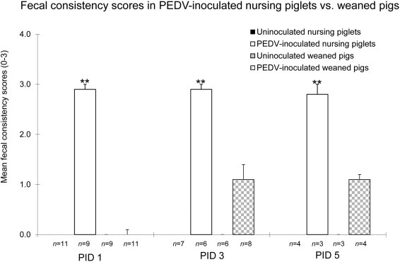
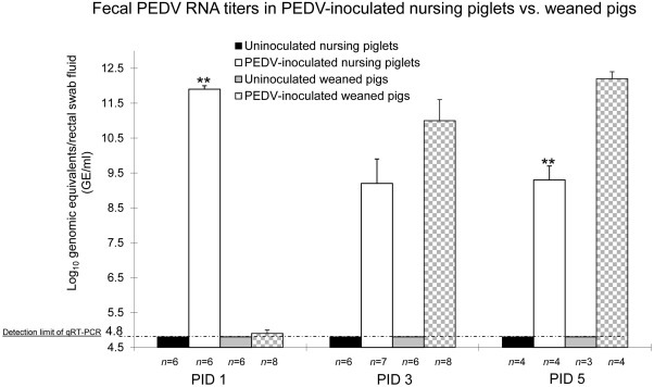
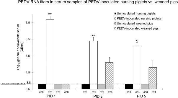
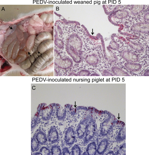
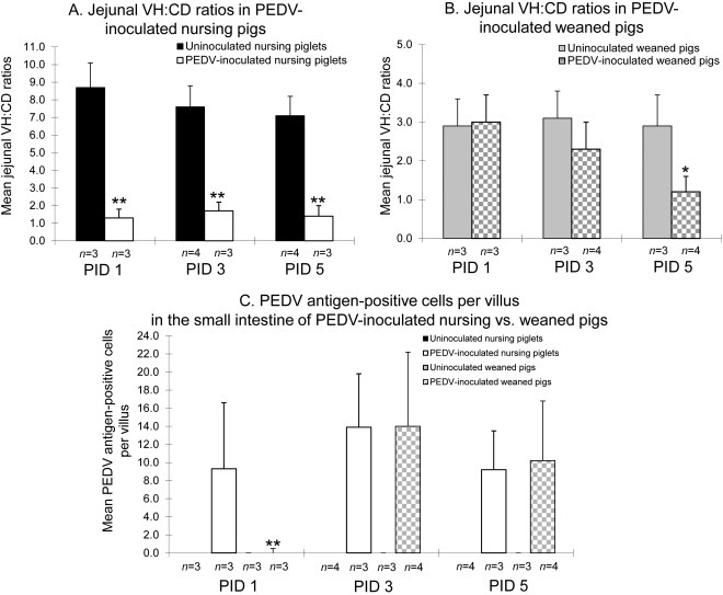
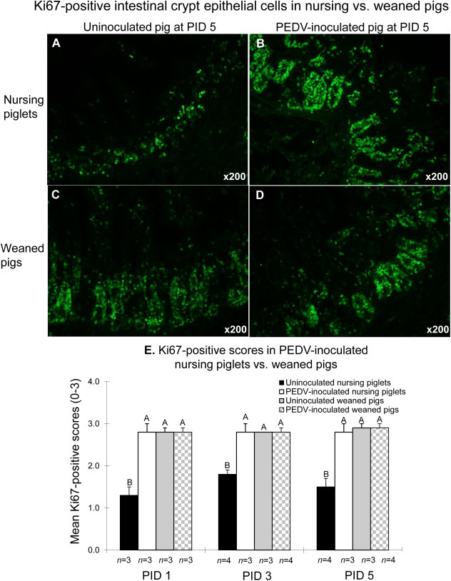
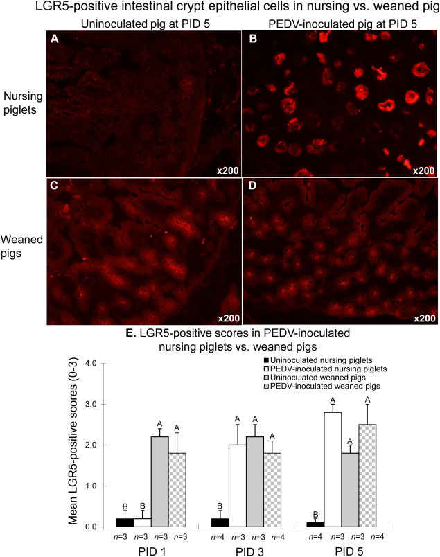
Our study demonstrated potential mechanisms by which porcine epidemic diarrhea virus (PEDV) infection induces greater disease severity of nursing vs. weaned conventional pigs. Twenty-six-day-old weaned [PEDV-inoculated (n=11); mock (n=9)] and 9-day-old nursing pigs [PEDV-inoculated (n=9); mock (n=11)] were inoculated orally [8.9 log10 genomic equivalents (GE)/pig] with PC21A strain or mock (MEM). Pigs were monitored for clinical signs and PEDV RNA titers in feces and serum. For pathology and immunofluorescence staining for Ki67 (marker for crypt proliferation) and LGR5 (marker for crypt stem cell), 3-4 pigs were euthanized at postinoculation days (PIDs) 1, 3 and 5. Severe watery diarrhea and atrophic enteritis with moderate to high PEDV RNA titers in feces (7.5-12.2 log10 GE/ml) and low viral RNA titers in serum (5.6-8.6 log10 GE/ml) were observed in all inoculated nursing piglets at PIDs 1-5. In contrast, weaned pigs did not show evidence of PEDV infection at PID 1. Pigs exhibited high fecal shedding titers at PIDs 2-5 and mild to severe atrophic enteritis at PIDs 3-5, indicating a longer incubation for PEDV infection. While uninoculated or inoculated 27-31-day-old pigs showed large numbers of Ki67- or LGR5-positive cells in the intestinal crypts, there was a lack of LGR5-positive cells and low proliferation of crypts in jejunum of uninoculated 10-14-day-old piglets, possibly causing a slower turnover of enterocytes; however, the number of LGR5-positive cells and proliferation of intestinal crypts increased remarkably at 3-5 days after inoculation. Biologic mediators that promote crypt stem cell regeneration would be targets to improve the intestinal epithelium renewal during PEDV infection.

Keywords: PEDV; Pathogenesis; Pig; Porcine epidemic diarrhea virus; Virus.


Figures

Fig. 1
Fig. 1
Significantly higher fecal consistency scores in PEDV-inoculated nursing pigs compared to PEDV-inoculated weaned pigs at PIDs 1-5. After PEDV inoculation, pigs were monitored for clinical signs 2–3 times daily until necropsy. Diarrhea was assessed by scoring fecal consistency. Fecal consistency was scored as follows: 0 = solid; 1 = pasty; 2 = semi-liquid; 3 = liquid, with scores of 2 or more considered diarrheic. Each bar represents the mean ± SEM. **, < 0.01 (statistically significant differences between the PEDV-inoculated nursing and weaned pigs by Student’s t-test).
Fig. 2
Fig. 2
Significantly higher fecal shedding PEDV RNA titers at PID 1 but lower titers at PID 5 in PEDV-inoculated nursing pigs compared to PEDV-inoculated weaned pigs. Fecal shedding PEDV RNA titers were determined by qRT-PCR. The detection limit of qRT-PCR was 10 GE per reaction, corresponding to 4.8 log10 GE/ml of rectal swab fluid. Each bar represents the mean ± SEM. **, < 0.01 (statistically significant differences between the PEDV-inoculated nursing and weaned pigs by Student’s t-test).
Fig. 3
Fig. 3
Significantly higher PEDV RNA titers in serum samples of PEDV-inoculated nursing pigs compared to PEDV-inoculated weaned pigs at PIDs 1-5. PEDV RNA titers in serum samples were determined by qRT-PCR. The detection limit of qRT-PCR was 10 GE per reaction, corresponding to 3.8 log10 GE/ml of serum sample. Each bar represents the mean ± SEM. *, < 0.05; **, < 0.01 (statistically significant differences between the PEDV-inoculated nursing and weaned pigs by Student’s t-test).
Fig. 4
Fig. 4
Gross lesions and immunohistochemistry for the detection of PEDV antigen in the intestine of PEDV-inoculated weaned (A and B) and nursing (C) pigs. (A) Intestine of a PEDV-inoculated weaned pig at PID 5, showing thin and transparent intestinal walls in the small intestine and accumulation of large amounts of fluid in the small intestinal lumen (arrows). Notice normal appearance of the large intestine and solid stools filled in the lumen (asterisk). (B) Colon of a PEDV-inoculated weaned pig at PID 5, showing low numbers of PEDV antigen-positive cells in the colonic epithelium. Original magnification × 200. (C) Colon of a PEDV-inoculated nursing pig at PID 5, showing moderate numbers of PEDV antigen-positive cells in the colonic epithelium. Original magnification × 200. Immunohistochemistry. Fast Red. Gill’s hematoxylin counterstaining.
Fig. 5
Fig. 5
Mean ratios of jejunal villous height to crypt depth (VH:CD) in PEDV-inoculated nursing (A) and weaned (B) pigs. (C) Mean PEDV antigen-positive cells per villus in the small intestine of PEDV-inoculated nursing piglets vs. weaned pigs. Eight pieces of formalin-fixed mid- to distal jejunum were taken from each virus-infected and control pigs for morphometric analysis. Only well-orientated, H&E- or IHC-stained jejunal sections were measured. Villous height, crypt depth, and number of PEDV antigen-positive cells were estimated by measuring at least 10 villi and crypts throughout the section. Each bar represents the mean ± SEM. *, < 0.05; **, < 0.01 (statistically significant differences between the PEDV-inoculated nursing and weaned pigs by Student’s t-test).
Fig. 6
Fig. 6
Detection of Ki67 protein by immunofluorescence (IF) staining in the small intestine of uninoculated (A) or PEDV-inoculated (B) nursing piglets compared to uninoculated (C) or PEDV-inoculated (D) weaned pigs at PID 5. (E) Mean Ki67-positive scores in the small intestine of PEDV-inoculated nursing piglets vs. weaned pigs. Ki67-positive scores were computed by estimating the number of IF-positive cells in the intestinal section per microscopic area, at ×200 magnification based on the following criteria: 0, no positive cells; 1, 1–29% of Ki67-positive crypt epithelial cells showed staining; 2, 30–59% of Ki67-positive crypt epithelial cells showed staining; and 3, 60–100% of Ki67-positive crypt epithelial cells showed staining. Each bar represents the mean ± SDM. Different letters denote significant differences among groups at each time-point (ANOVA test, < 0.05).
Fig. 7
Fig. 7
Detection of LGR5 protein by immunofluorescence (IF) staining in the small intestine of uninoculated (A) or PEDV-inoculated (B) nursing piglets compared to uninoculated (C) or PEDV-inoculated (D) weaned pigs at PID 5. (E) Mean LGR5-positive scores in the small intestine of PEDV-inoculated nursing piglets vs. weaned pigs. LGR5-positive scores were computed by estimating the number of IF-positive cells in the intestinal section per microscopic area, at ×200 magnification based on the following criteria: 0, no positive cells; 1, 1–29% of LGR5-positive crypt epithelial cells showed staining; 2, 30–59% of LGR5-positive crypt epithelial cells showed staining; and 3, 60–100% of LGR5-positive crypt epithelial cells showed staining. Each bar represents the mean ± SDM. Different letters denote significant differences among groups at each time-point (ANOVA test, < 0.05).

References

    1. Amimo J.O., Vlasova A.N., Saif L.J. Detection and genetic diversity of porcine group A rotaviruses in historic (2004) and recent (2011 and 2012) swine fecal samples in Ohio: predominance of the G9P[13] genotype in nursing piglets. J. Clin. Microbiol. 2013;51:1142–1151. - PMC - PubMed
    1. Amimo J.O., Vlasova A.N., Saif L.J. Prevalence and genetic heterogeneity of porcine group C rotaviruses in nursing and weaned piglets in Ohio, USA and identification of a potential new VP4 genotype. Vet. Microbiol. 2013;164:27–38. - PMC - PubMed
    1. Burkey T.E., Skjolaas K.A., Minton J.E. Board-invited review: porcine mucosal immunity of the gastrointestinal tract. J. Anim. Sci. 2009;87:1493–1501. - PubMed
    1. Chu D.K., Poon L.L., Guan Y., Peiris J.S. Novel astroviruses in insectivorous bats. J. Virol. 2008;82:9107–9114. - PMC - PubMed
    1. Chung W.B., Chan W.H., Chaung H.C., Lien Y., Wu C.C., Huang Y.L. Real-time PCR for quantitation of porcine reproductive and respiratory syndrome virus and porcine circovirus type 2 in naturally-infected and challenged pigs. J. Virol. Methods. 2005;124:11–19. - PubMed

Publication types

LinkOut - more resources