. 2015 Jun 19;290(25):15892-15903.
doi: 10.1074/jbc.M115.640607. Epub 2015 May 4.

The Function of Embryonic Stem Cell-expressed RAS (E-RAS), a Unique RAS Family Member, Correlates with Its Additional Motifs and Its Structural Properties

Affiliations

The Function of Embryonic Stem Cell-expressed RAS (E-RAS), a Unique RAS Family Member, Correlates with Its Additional Motifs and Its Structural Properties

Saeideh Nakhaei-Rad et al. J Biol Chem. .

Abstract

E-RAS is a member of the RAS family specifically expressed in embryonic stem cells, gastric tumors, and hepatic stellate cells. Unlike classical RAS isoforms (H-, N-, and K-RAS4B), E-RAS has, in addition to striking and remarkable sequence deviations, an extended 38-amino acid-long unique N-terminal region with still unknown functions. We investigated the molecular mechanism of E-RAS regulation and function with respect to its sequence and structural features. We found that N-terminal extension of E-RAS is important for E-RAS signaling activity. E-RAS protein most remarkably revealed a different mode of effector interaction as compared with H-RAS, which correlates with deviations in the effector-binding site of E-RAS. Of all these residues, tryptophan 79 (arginine 41 in H-RAS), in the interswitch region, modulates the effector selectivity of RAS proteins from H-RAS to E-RAS features.

Keywords: E-RAS; H-RAS; RAS protein; Raf kinase; effector selection; embryonic stem cell-expressed RAS; phosphatidylinositide 3-kinase (PI 3-kinase); phosphatidylinositol kinase (PI kinase); small GTPase; specificity determining residues.


Figures

FIGURE 1.
FIGURE 1.
Human E-RAS is largely associated with plasma membrane and some regions of E-RAS modulating its cellular localization. A, different E-RAS variants used in this study, including N-terminal truncated E-RASΔN (aa 39–233), palmitoylation-deficient E-RASSer-226/Ser-228 (aa 1–233), N-terminal putative PXXP motif mutant E-RASSer-7 (aa 1–233), and an N-terminal triple arginine motif variant E-RASAla-31/Ala-32/Ala-33 (aa 1–233). B and C, confocal live images of transiently transfected MDCK II cells with EYFP-tagged E-RASWT, H-RASWT, E-RASSer-226/Ser-228, E-RASΔN, E-RASSer-7, and E-RASAla-31/Ala-32/Ala-33. D, confocal imaging was performed using transiently transfected MDCK II cells with E-RAS and H-RAS. FLAG-tagged E-RAS co-localized with Na+/K+-ATPase to the plasma membrane, very similar to H-RAS, which was used as a control. Scale bar, 10 μm.
FIGURE 2.
FIGURE 2.
Different effector selection of E-RAS and H-RAS. A, effector-binding residues of H-RAS, obtained from various crystal structures, are highlighted with blue letters and yellow background, RAF1 (PDB code 1C1Y), PLCϵ (PDB code 2C5L), RalGDS (PDB code 1LFD), PI3Kγ (PDB code 1HE8), and RASSF5 (PDB code 3DDC). B, effector binding regions (in yellow and orange) of H-RAS and E-RAS were structurally analyzed on the basis of the H-RAS structure in complexes with p120RASGAP (PDB code 1WQ1). The orange amino acids indicate the sequence deviation between H-RAS and E-RAS. C, schematic view of RAS effector pathways and their cellular functions. D, E-RAS and H-RAS pulldown (PD) with various RAS effectors using COS-7 cell lysates transiently transfected with FLAG-tagged E-RASWT, H-RASWT, and H-RASVal-12 using GST-fused effector proteins, such as RAF1-RBD, RalGDS-RA, PLCϵ-RA, PI3Kα-RBD, and RASSF5-RA. RAS proteins were analysis by immunoblot using an anti-FLAG antibody. Immunoblots (IB) of total cell lysates were used as a control to detect FLAG-RAS. Exp. time stands for exposure time. RAF, rapidly accelerated fibrosarcoma; MEK, mitogen-activated protein kinase/ERK kinase; ERK, extracellular signal-regulated kinase; PLCϵ, phospholipase Cϵ; PKC, protein kinase C; RalGDS, Ral GDP dissociation stimulator; RLIP76, Ral-interacting protein 76 kDa; PI3K, phosphoinositide 3-kinase; PIP3, phosphoinositide 3,4,5-trisphosphate; MST1/2, mammalian Ste20-like kinases 1.
FIGURE 3.
FIGURE 3.
Specificity-determining residues in E-RAS-effector interaction. A, display of different effector binding mutations in E-RAS: E-RASSwI, H70Y/Q75E (Tyr-32 and Gslu-37 in H-RAS); E-RASArg-79, W79R (Arg-41 in H-RAS); E-RASSwII, A100E/I101E/H102Y/R103S (Glu-62, Glu-63, Tyr-64, and Ser-65 in H-RAS); E-RASSwI/Arg-79, H70Y/Q75E/W79R; E-RASArg-79/SwII, W79R/A100E/I101E/H102Y/R103S; RASSwI/SwII, H70Y/Q75E/A100E/I101E/H102Y/R103S, and E-RASSwI/Arg-79/SwII, H70Y/Q75E/W79R/A100E/I101E/H102Y/R103S. For details about the amino acid sequences, see supplemental Fig. S1. B, pulldown assay of FLAG-fused E-RAS variants carried out with RBD or RA domain of GST-fused effector proteins, including RAF1-RBD, RalGDS-RA, PLCϵ-RA, PI3Kα-RBD, and RASSF5-RA. The results were analyzed by immunoblot using an anti-FLAG antibody. Exp. time stands for exposure time. C, total cell lysates were used to monitor the level of phosphorylated AKT (pAKTT308 and pAKTS473), MEK1/2 (pMEK1/2), and ERK1/2 (pERK1/2) proteins.
FIGURE 4.
FIGURE 4.
Glutamate 41 function and its role in effector selection is discharged in E-RAS. A, in H-RAS-GTP, Arg-41 (Trp-79 in E-RAS) is intramolecularly stabilized by Glu-3 (Glu-41 in E-RAS), attracted by backbone oxygen of Asn-64, and repulsed by Lys-65 in RAF1. B, in E-RAS, Trp-79 is expelled from Glu-41 and cannot adopt favorable conformation because of the close presence of Asn-64 and Lys-65 of RAF1. The conformation of arginine at the place of Trp-79 in E-RASArg-79 would be restored due to its interaction with Glu-41 similarly to H-RAS, thus increasing the binding affinity of RAF1. C, PI3K does not contact E-RAS tightly in the vicinity of Trp-79 leaving enough space for proper reorientation of tryptophan side chain expelled from Glu-41 and not disfavoring the affinity of their complex. Moreover, orientation of Thr-228 enables tight hydrophobic contact with Trp-79. In E-RASArg-79, arginine attracted by Glu-41 would not contribute to the interaction with PI3K weakening its affinity to E-RASArg-79. D, selectivity-determining amino acids in RAS effectors. Multiple amino acid sequence alignments of the RBD of human RAF isoforms and the catalytic subunits of human PI3K isoforms are illustrated with major focus on the some RAS-binding residues. The corresponding sequences are RAF-1 (P04049; aa 51–131), A-RAF (P10398; aa 14–91), B-RAF (P15056; aa 105–227), PI3Kα (P42336; aa 184–276), PI3Kβ (P42338; aa 191–272), PI3Kγ (P48736; aa 214–296), and PI3Kδ (O00329; aa 184–226). X highlights residues interacting in β-β manner with switch I. # highlights additional residues interacting with switch I. Ø shows residues interacting with Tyr-64 in switch II. * shows residues close to Arg-41 in H-RAS or Trp-79 in E-RAS.
FIGURE 5.
FIGURE 5.
E-RAS signaling activities in COS-7 cells. Pulldown (PD) experiments and immunoblot (IB) analysis of total cell lysates were derived from transfected COS-7 cells with FLAG-tagged E-RAS variants H-RASWT and H-RASVal-12. A, pulldown analysis revealed that E-RAS variants like E-RASWT most strongly bind to GST-fused PI3Kα-RBD than RAF1-RBD, whereas hyperactive H-RASVal-12 mainly bound to GST-fused RAF1-RBD. In addition, PI3Kα-RBD PD showed that all E-RAS variants are in the GTP-bound state and consequently in their activated forms. Total amounts of the RAS proteins were detected as a control using anti-FLAG antibody. B, schematic view of MAPK and PI3K-AKT cascades. C, total cell lysates were analyzed for the phosphorylation level of AKT (pAKT308 and pAKT473), MEK1/2 (pMEK1/2) and ERK1/2 (pERK1/2). Total amounts of AKT, MEK1/2, and ERK1/2 were applied as loading controls.
FIGURE 6.
FIGURE 6.
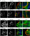
Co-localization of E-RAS with PI3Kα. Transfected MDCKII cells with FLAG-tagged E-RAS were incubated with bacterial lysates, containing GST-RBDs of PI3Kα and RAF1 proteins and stained with antibodies raised against GST and FLAG to investigate their co-localization with GTP-bound H-RAS and E-RAS proteins. E-RAS co-localized with PI3Kα. Scale bar, 10 μm.

References

    1. Vetter I. R. (2001) The guanine nucleotide-binding switch in three dimensions. Science 294, 1299–1304 - PubMed
    1. Boguski M. S., McCormick F. (1993) Proteins regulating Ras and its relatives. Nature 366, 643–654 - PubMed
    1. Ahmadian M. R., Wittinghofer A., Schmidt G. (2002) The actin filament architecture: tightly regulated by the cells, manipulated by pathogens. International Titisee Conference on the actin cytoskeleton: from signalling to bacterial pathogenesis. EMBO Rep. 3, 214–218 - PMC - PubMed
    1. Gremer L., Merbitz-Zahradnik T., Dvorsky R., Cirstea I. C., Kratz C. P., Zenker M., Wittinghofer A., Ahmadian M. R. (2011) Germ line KRAS mutations cause aberrant biochemical and physical properties leading to developmental disorders. Hum. Mutat. 32, 33–43 - PMC - PubMed
    1. Karnoub A. E., Weinberg R. A. (2008) Ras oncogenes: split personalities. Nat. Rev. Mol. Cell Biol. 9, 517–531 - PMC - PubMed

Publication types

Substances