. 2015 Jul 3;290(27):16708-22.
doi: 10.1074/jbc.M114.593228. Epub 2015 May 4.

Parvulin 17-catalyzed Tubulin Polymerization Is Regulated by Calmodulin in a Calcium-dependent Manner

Affiliations

Parvulin 17-catalyzed Tubulin Polymerization Is Regulated by Calmodulin in a Calcium-dependent Manner

Noelia Inés Burgardt et al. J Biol Chem. .

Abstract

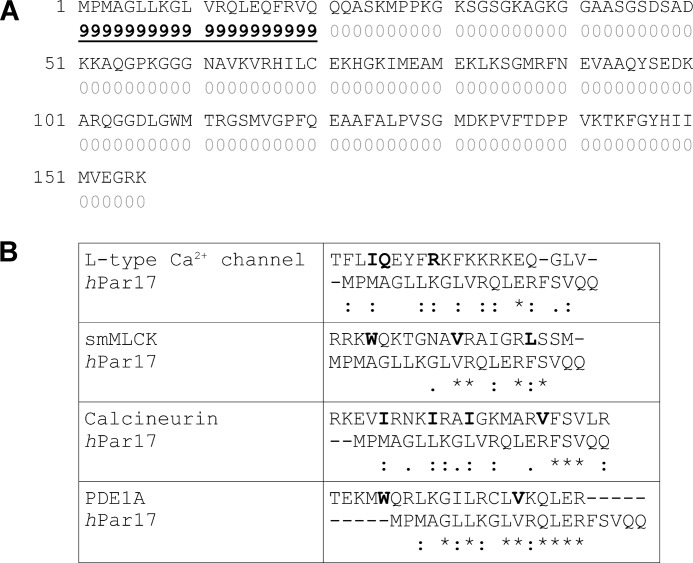
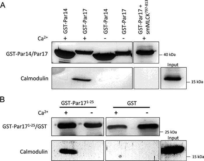
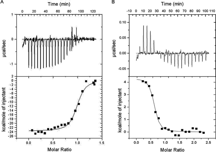
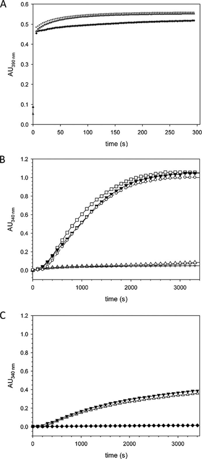
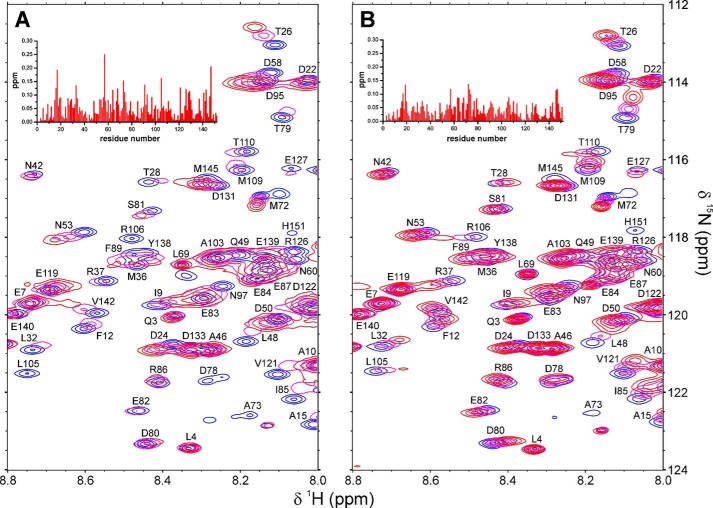
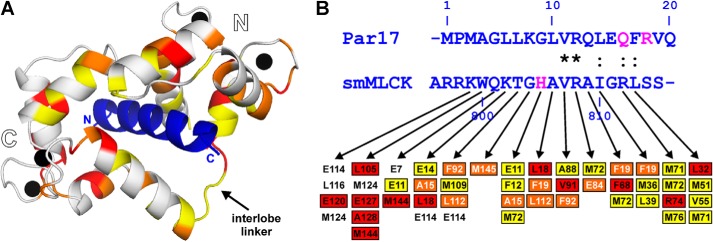
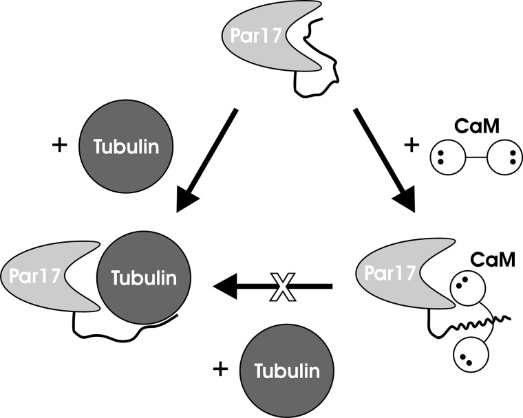
Recently we have shown that the peptidyl-prolyl cis/trans isomerase parvulin 17 (Par17) interacts with tubulin in a GTP-dependent manner, thereby promoting the formation of microtubules. Microtubule assembly is regulated by Ca(2+)-loaded calmodulin (Ca(2+)/CaM) both in the intact cell and under in vitro conditions via direct interaction with microtubule-associated proteins. Here we provide the first evidence that Ca(2+)/CaM interacts also with Par17 in a physiologically relevant way, thus preventing Par17-promoted microtubule assembly. In contrast, parvulin 14 (Par14), which lacks only the first 25 N-terminal residues of the Par17 sequence, does not interact with Ca(2+)/CaM, indicating that this interaction is exclusive for Par17. Pulldown experiments and chemical shift perturbation analysis with (15)N-labeled Par17 furthermore confirmed that calmodulin (CaM) interacts in a Ca(2+)-dependent manner with the Par17 N terminus. The reverse experiment with (15)N-labeled Ca(2+)/CaM demonstrated that the N-terminal Par17 segment binds to both CaM lobes simultaneously, indicating that Ca(2+)/CaM undergoes a conformational change to form a binding channel between its two lobes, apparently similar to the structure of the CaM-smMLCK(796-815) complex. In vitro tubulin polymerization assays furthermore showed that Ca(2+)/CaM completely suppresses Par17-promoted microtubule assembly. The results imply that Ca(2+)/CaM binding to the N-terminal segment of Par17 causes steric hindrance of the Par17 active site, thus interfering with the Par17/tubulin interaction. This Ca(2+)/CaM-mediated control of Par17-assisted microtubule assembly may provide a mechanism that couples Ca(2+) signaling with microtubule function.

Keywords: calcium; calmodulin (CaM); chemical shift perturbation (CSP) analysis; microtubule-associated protein (MAP); nuclear magnetic resonance (NMR); parvulin; protein conformation; protein-protein interaction; tubulin.


Figures

FIGURE 1.
FIGURE 1.
A, analysis of potential CaM binding sequences by the online server CaM target database. The figure shows the sequence of human Par17 (hPar17). The scores below the amino acids indicate the probability for the location of a CaM-binding site. A consecutive series of high values suggests the existence of a putative CaM-binding site. B, alignment of CaM binding motifs with the N-terminal sequence of hPar17 (*, identical; :, conserved). Key residues that classify the CaM-binding sites to a particular motif are highlighted in bold letters. The L-type Ca2+ channel displays an IQ motif, and smMLCK comprises a 1-8-14 motif, whereas calcineurin and phosphodiesterase 1A (PDE 1A) encompass a binding sequence of the 1-5-8-14 and the 1-12 class, respectively.
FIGURE 2.
FIGURE 2.
Par17 interacts with Ca2+/CaM via its N-terminal extension. For the CaM coprecipitation experiment, glutathione-Sepharose beads loaded with 20 μg of GST fusion proteins or GST alone were blocked with 3% BSA in the incubation buffer. A, Par14- and Par17-bound resins were incubated for 1 h at room temperature with 20 μm CaM in the (i) absence of Ca2+, (ii) presence of Ca2+, and (iii) presence of both Ca2+ and 20 μm smMLCK797–813 peptide. B, GST-Par171–25-loaded as well as GST-loaded GSH beads were incubated with 20 μm CaM in the absence and presence of Ca2+.
FIGURE 3.
FIGURE 3.
ITC data of Ca2+/CaM binding to the Par17 N terminus. The calorimetric trace of heat released upon titration of Ca2+/CaM into a solution containing either the Par172–22 peptide (A) or full-length Par17 (B) is shown at the top. The corresponding heat per mole of injected Ca2+/CaM is shown at the bottom. The binding isotherms were analyzed by applying a single-site binding model using the Microcal Origin software package. The analyses revealed the following thermodynamic parameters for the Par172–22 peptide (ΔH = −23.6 ± 0.4 kcal/mol, ΔS = −49.2 cal/(mol·degree), n = 0.97 ± 0.01, Kd = 149 ± 25 nm) and for the full-length Par17 protein (ΔH = 4.3 ± 0.2 kcal/mol, ΔS = 45.8 cal/(mol·degree), n = 0.59 ± 0.02, Kd = 143 ± 41 nm).
FIGURE 4.
FIGURE 4.
Ca2+/CaM inhibits Par17-catalyzed tubulin assembly in an in vitro tubulin polymerization assay. A, the PPIase activity of Par17 in the presence of Ca2+/CaM was determined using a protease-coupled assay that employs as substrate the oligopeptide succinyl-AKPF-4-nitroanilide, as previously described (20). The PPIase activity of Par17 was measured in a reaction mixture containing 2 μm GST-Par17 and 1 mm CaCl2 either with (▴) or without (●) 15 μm CaM. The uncatalyzed cis-to-trans isomerization in the absence of Par17 is shown as a control (♦). Calculation of the first-order rate constants for the cis/trans isomerization (1.47 ± 0.02 × 10⁻² in the presence and 1.5 ± 0.03 × 10⁻² in the absence of Ca2+/CaM) indicated that Ca2+/CaM does not affect Par17 PPIase activity (insensitivity of Par17 and CaM to proteolytic digestion by α-chymotrypsin was verified for the duration of the measurements). B, assembly of microtubules was initiated by dilution of ice-cold tubulin to a final concentration of 6.7 μm in 80 μl of G-PEM buffer set to 37 °C. Polymerization was measured in absence of Par17 (♦) and in presence of 16 μm GST-Par17 (▾), 16 μm GST-Par17, and 1 mm Ca2+ (○) and 16 μm GST-Par17 and 30 μm CaM (□) as well as 16 μm GST-Par17, 1 mm Ca2+, and 30 μm CaM (▵). C, tubulin polymerization was monitored in absence of GST-Par14 (♦), in the presence of 16 μm GST-Par14 (▾), and in presence of 16 μm GST-Par14, 1 mm Ca2+, and 30 μm CaM (▵). AU, absorbance units.
FIGURE 5.
FIGURE 5.
Comparison of the Par14 and Par17 backbone amide resonances. In the two upper panels, the amide signal positions in the 1H,15N HSQC spectrum of Par17 (Biological Magnetic Resonance Bank (BMRB) accession code 18615; black squares) are overlaid with those of Par14 (red circles) based on the assignment by Terada et al. (14) (A) and Sekerina et al. (Ref. ; Biological Magnetic Resonance Bank (BMRB) accession code 4768) (B). Shifted peaks are labeled with the respective Par17 residue number and in some cases connected by a line to denote the displacement. Signals belonging to the Par17 N terminus, which corresponds to the segment missing in Par14, are generally found as non-superposed black squares in the 1H resonance range between 8.5 and 8.0 ppm, thus indicating a random-coil structure. In the two lower panels, a CSP analysis for each residue within the structured PPIase domain again compares Par17 with the Par14 assignments of Terada (C) and Sekerina (D). Residues, whose backbone amide resonance assignments are missing in either Par17 or Par14 are indicated by yellow and blue bars, respectively.
FIGURE 6.
FIGURE 6.
1H,15N HSQC spectra of 0. 9 mm13C/15N-labeled Par17 collected at 18 °C and pH 6.8 in the absence of Ca2+/CaM (A) and in the presence of a 1.4-fold excess of Ca2+/CaM (B). Relative intensities of the Par17 backbone amide signals are shown in the lower panels, where the first 25 residues comprising the N-terminal extension of Par17 as well as the PPIase domain are indicated above the intensity plots by black and white boxes, respectively. In B, the side-chain NH and NH2 signals are marked accordingly; all remaining strong peaks belong to the backbone amide groups in segment Lys-29–Ala-62.
FIGURE 7.
FIGURE 7.
Ca2+/CaM binding to 15N-labeled Par17. A, central region of superposed 1H,15N TROSY spectra collected at 25 °C and pH 6.8 with 15N-labeled Par17 (0.6 mm) in the absence of Ca2+/CaM (black) and in presence of a 1.4-fold molar Ca2+/CaM excess (red). B, CSP effects in Par17 upon Ca2+/CaM binding are shown as blue and magenta bars, indicating either signal shifts or extreme line broadening, respectively. C, helical wheel representation of the predicted amphipathic α-helix at the N terminus of Par17. Hydrophobic, polar, acidic, and basic residues are highlighted in yellow, pink, red, and blue, respectively. The arrow indicates the hydrophobic moment of the helix (65); the N- and C-terminal ends of the wheel are marked by green capital letters.
FIGURE 8.
FIGURE 8.
Peptide microarray binding pattern of Ca2+/CaM. To display Ca2+/CaM binding to the microarray of Par17 15-mer peptides, relative fluorescence intensities of spots, which were corrected for the background, are plotted against the peptide number. Incubation of peptide microarrays were performed in the presence of either Ca2+ (white bars) or the Ca2+-chelator EGTA (black bars). As a control, the array was incubated only with anti-mouse-IgG (gray bars). The inset shows the amino acid sequences of peptides 30–40, comprising the dominant binding site for Ca2+/CaM in the Par17 peptide microarray.
FIGURE 9.
FIGURE 9.
Par17 binding to 15N-labeled Ca2+/CaM. A, superposed 1H,15N TROSY spectra collected at 25 °C and pH 6.8 with 15N-labeled Ca2+/CaM (0.5 mm) in the absence of Par17 (blue) and in presence of Par17 at a 5-fold (magenta) and 2-fold (red) molar excess of Ca2+/CaM. B, superposed 1H,15N TROSY spectra collected at 25 °C and pH 6.8 with 15N-labeled Ca2+/CaM (0.5 mm) without Par172–22 (blue), with Par172–22 at 1:1 molar ratio (magenta), and with Par172–22 in 3-fold molar excess (red). The CSP effects observed in Ca2+/CaM upon binding of Par17 (at 2:1 molar stoichiometry) and Par172–22 (at 1:3 molar stoichiometry) are shown as insets in panels A and B, respectively.
FIGURE 10.
FIGURE 10.
A, structural mapping of the CSP effects observed in Ca2+/CaM upon Par17 binding based on the structure of the complex (PDB ID code 1CDL) between Ca2+/CaM and the smMLCK796–815 peptide from chicken (blue ribbon). Ca2+/CaM residues that showed significant line-broadening effects are highlighted in yellow, whereas residues featuring CSP effects >0.10 ppm or >0.15 ppm are colored in orange and red, respectively. The CaM lobes and interlobe linker as well as the N- and C-terminal ends of the smMLCK796–815 peptide are marked. B, sequence comparison between the predicted CaM-binding site at the N terminus of human Par17 and the smMLCK796–815 peptide. Sequence similarities are found only in the second half of the peptides, as indicated in the row between the sequences (*, identical; :, conserved). Arrows below the smMLCK sequence denote close contacts (<4 Å) to CaM residues according to the x-ray structure (PDB ID code 1CDL). Colored boxes illustrate where these CaM residues or one of their immediate sequential neighbors showed CSP effects in the presence of Par17 (same color code as in panel A). Magenta-colored residues in the peptide sequences indicate positions of naturally occurring amino acid substitutions; (i) due to single-nucleotide polymorphism, a second human Par17 isoform exists featuring the point mutations Q16R and R18S, and (ii) in mammalian smMLCK, H805 is generally replaced by an asparagine.
FIGURE 11.
FIGURE 11.
Schematic representation of the Ca2+/CaM-dependent blocking of the Par17-promoted tubulin polymerization. In native Par17 (top), the N-terminal extension interacts with the substrate binding pocket. In the tubulin-Par17 complex (bottom left), tubulin seems to continue to interact with the Par17 N terminus after replacing it at the substrate binding pocket, thus ensuring a higher polymerization efficiency compared with Par14. Ca2+/CaM binds strongly to the Par17 N terminus (bottom right); at the same time, the close proximity of CaM to the substrate binding pocket of the PPIase domain apparently restricts the access to the active site for larger ligands such as tubulin but not for small substrates such as the oligopeptide succinyl-AKPF-4-nitroanilide.

References

    1. Galat A., Metcalfe S. M. (1995) Peptidylproline cis/trans isomerases. Prog. Biophys. Mol. Biol. 63, 67–118 - PubMed
    1. Fanghänel J., Fischer G. (2004) Insights into the catalytic mechanism of peptidyl prolyl cis/trans isomerases. Front. Biosci. 9, 3453–3478 - PubMed
    1. Yaffe M. B., Schutkowski M., Shen M., Zhou X. Z., Stukenberg P. T., Rahfeld J. U., Xu J., Kuang J., Kirschner M. W., Fischer G., Cantley L. C., Lu K. P. (1997) Sequence-specific and phosphorylation-dependent proline isomerization: a potential mitotic regulatory mechanism. Science 278, 1957–1960 - PubMed
    1. Lu K. P. (2003) Prolyl isomerase Pin1 as a molecular target for cancer diagnostics and therapeutics. Cancer Cell 4, 175–180 - PubMed
    1. Pemberton T. J., Kay J. E. (2005) Identification and comparative analysis of the peptidyl-prolyl cis/trans isomerase repertoires of H. sapiens, D. melanogaster, C. elegans, S. cerevisiae and Sz. pombe. Comp. Funct. Genomics 6, 277–300 - PMC - PubMed

Publication types

Associated data

LinkOut - more resources