. 2015 May 11;209(3):377-86.
doi: 10.1083/jcb.201412046. Epub 2015 May 4.

Axonal autophagosomes recruit dynein for retrograde transport through fusion with late endosomes

Affiliations

Axonal autophagosomes recruit dynein for retrograde transport through fusion with late endosomes

Xiu-Tang Cheng et al. J Cell Biol. .

Abstract

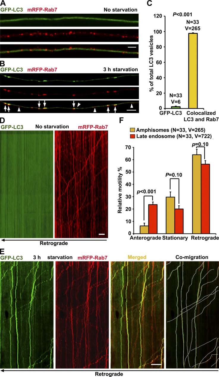
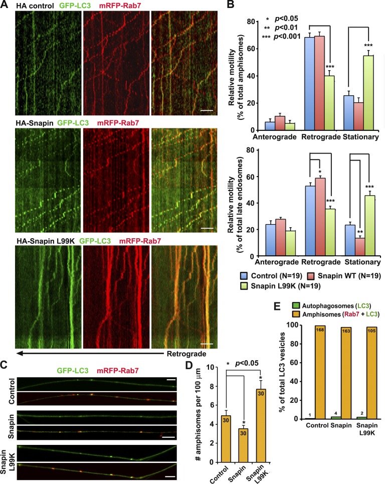
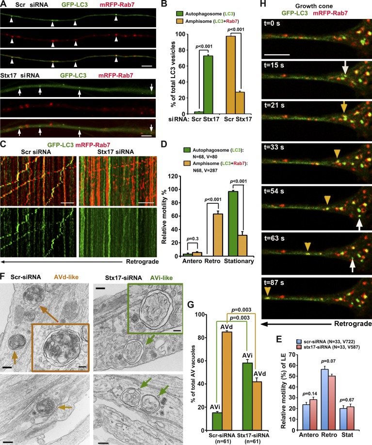
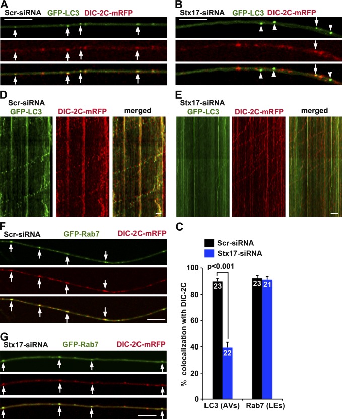
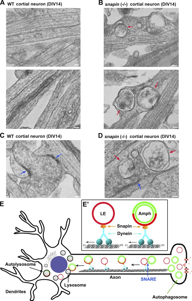
Efficient degradation of autophagic vacuoles (AVs) via lysosomes is an important cellular homeostatic process. This is particularly challenging for neurons because mature acidic lysosomes are relatively enriched in the soma. Although dynein-driven retrograde transport of AVs was suggested, a fundamental question remains how autophagosomes generated at distal axons acquire dynein motors for retrograde transport toward the soma. In this paper, we demonstrate that late endosome (LE)-loaded dynein-snapin complexes drive AV retrograde transport in axons upon fusion of autophagosomes with LEs into amphisomes. Blocking the fusion with syntaxin17 knockdown reduced recruitment of dynein motors to AVs, thus immobilizing them in axons. Deficiency in dynein-snapin coupling impaired AV transport ,: resulting in AV accumulation in neurites and synaptic terminals. Altogether, our study provides the first evidence that autophagosomes recruit dynein through fusion with LEs and reveals a new motor-adaptor sharing mechanism by which neurons may remove distal AVs engulfing aggregated proteins and dysfunctional organelles for efficient degradation in the soma.


Figures

Figure 1.
Figure 1.
Axonal amphisomes are the predominant AVs moving retrogradely. (A–C) Majority of autophagosomes in DRG axons target LEs after 3-h starvation. DRG neurons were cotransfected with GFP-LC3 and mRFP-Rab7 at DIV0 and imaged at DIV3 after incubation with serum (control; A) or serum-free medium (starvation; B) for 3 h. Images were taken from the middle segment of axons. Arrows indicate amphisomes colabeled with LC3 and Rab7, whereas arrowheads point out AV or LE alone. (D and E) Dual-channel kymographs showing comigration of GFP-LC3 and mRFP-Rab7 during 5-min time-lapse imaging. Vertical lines represent stationary organelles; slanted lines or curves to the right (negative slope) represent anterograde movement; to the left (positive slope), they indicate retrograde movement. An organelle is considered stationary if it remains immotile (displacement ≤ 10 µm). Under control conditions, GFP-LC3 was diffused, whereas LEs predominantly transported toward the soma (D). Under starvation, amphisomes (LC3 and Rab7) moved retrogradely (white lines in E). (F) Quantitative analysis showing that amphisomes and LEs share the similar predominant retrograde motility in DRG axons. Data were quantified from the total number of vesicles (V) in the total number of neurons (N) from greater than three experiments. Error bars: SEM. Student’s t test. Bars: (A and B) 5 µm; (D and E) 10 µm.
Figure 2.
Figure 2.
LE-loaded dynein–snapin complexes drive amphisome retrograde trafficking. (A and B) Kymographs (A) and quantitative analysis (B) showing impaired retrograde transport of amphisomes by disrupting DIC–snapin coupling. DRG neurons were cotransfected with GFP-LC3 and mRFP-Rab7 along with HA-snapin, HA-snapin-L99K, or HA vector at DIV0 and time-lapse imaged for 3 min at DIV3. The total number of neurons (N) examined is indicated in parentheses from greater than three experiments. (C and D) Images (C) and quantitative analysis (D) showing that disrupted snapin–DIC coupling increases the density of axonal amphisomes. The total number of neurons examined for each group is 30 from more than three experiments. (E) Disrupting snapin–DIC coupling had no significant effect on fusion between autophagosomes and LEs. Data were quantified from total number of AVs denoted in or above the bars from greater than three experiments. Mann–Whitney test (B) and Student’s t test (D and E). Error bars: SEM. Bars: (A) 10 µm; (C) 5 µm.
Figure 3.
Figure 3.
Autophagosomes acquire retrograde motility by fusion with LEs. (A and B) Stx17 knockdown blocks formation of amphisomes. (A) Imaging was performed on the middle to distal segments of axons. Arrowheads indicate amphisomes (yellow) colabeled with LC3 and Rab7, whereas arrows indicate autophagosomes (green) without Rab7 labeling. Note a robust increase in autophagosomes and decrease in amphisomes in axons expressing Stx17-siRNA (B). The number of autophagosomes or amphisomes was expressed as a percentage of the total LC3-labeled AVs. Data were quantified from 28 axons in each group and a total number of 293 AVs in greater than three experiments. (C–E) Relative motility of amphisomes, autophagosomes, and LEs. (C, right) Note that in neurons with Stx17 knockdown, autophagosomes (green) were largely stationary, whereas LEs (red) underwent predominant retrograde transport, some of which passed through stationary autophagosomes. (C and E) Depleting Stx17 had no effect on the motility of LEs. Quantification was performed from the total number of AVs or LEs (V, vesicles) from the total number of neurons (N) in greater than three experiments. (F and G) TEM analysis showing early stage AVi versus late-stage AVd in DRG neurons. In Stx17 knockdown neurites, the majority of AV-like structures are early stage autophagosomes (AVi; green box and arrows) and were easily found. In control neurons, the majority of AVs are late-stage autophagic vacuoles (AVd) containing electron-dense material and small vesicles (orange box and arrows), suggesting fusion with LEs. Data were collected from 61 micrographs (5 × 5 µm) for each condition. (H) Dynamic de novo autophagosomal biogenesis, fusion, and retrograde transport in a DRG neuron growth cone. Time-lapse imaging was taken once every 3 s with acquisition exposure time at 150 ms in a Nikon spinning-disk confocal. White arrows point to the appearance of new autophagosomes within growth cone at 15, 54, and 63 s; the yellow arrow denotes fusion events, and yellow arrowheads indicate a retrograde motile amphisome between 33 and 87 s (also see Video 1). Error bars: SEM. Unpaired Student’s t test. Bars: (A and H) 5 µm; (C) 10 µm; (F, main images) 200 nm; (F, boxes) 100 nm.
Figure 4.
Figure 4.
Blocking fusion reduces the recruitment of dynein motors to AVs. (A–C) Representative images (A and B) and quantitative analysis (C) showing that blocking fusion reduces DIC recruitment to AVs. Arrows indicate AVs containing DIC-2C (A), whereas arrowheads indicate AVs not labeled with DIC-2C (B). (D and E) AVs (green) comigrate with DIC-2C in retrograde direction in control neurons (D), whereas depleting Stx17 immobilizes AVs by reducing DIC recruitment but has no effect on DIC retrograde motility along the same axons (E). The retrograde direction is toward the left. (F and G) Blocking fusion has no observable effect on DIC recruitment to LEs by quantitative analysis (C). Arrows indicate LEs containing DIC-2C. The total neuron numbers for quantification were indicated inside the bars. Error bars: SEM. Mann–Whitney test. Bars, 10 µm.
Figure 5.
Figure 5.
Aberrant accumulation of AVs in snapin-deficient neuronal processes. (A–D) TEM showing aberrant accumulation of double-membrane AVs along neurites (B) and at presynaptic terminals (D) of snapin−/− cortical neurons at DIV14. Red arrows indicate AV-like structures, which are not readily observed in WT neurons. Blue arrows point to synaptic active zones. Images were representative from 35–50 electron micrographs from three pairs of mice. Bars, 100 nm. (E) A model of LE-loaded dynein–snapin complex driving AV for retrograde trafficking along axons. (E′) Autophagosomes (green) at distal axons acquire the dynein motor complex from LEs (red) upon their fusion into amphisomes. Therefore, dynein–snapin complexes mediate amphisomes, but not autophagosomes, for long-distance retrograde trafficking to the soma, where mature acidic lysosomes are mainly located.

References

    1. Bains M., Zaegel V., Mize-Berge J., and Heidenreich K.A.. 2011. IGF-I stimulates Rab7-RILP interaction during neuronal autophagy. Neurosci. Lett. 488:112–117 10.1016/j.neulet.2010.09.018 - DOI - PMC - PubMed
    1. Cai Q., Lu L., Tian J.-H., Zhu Y.-B., Qiao H., and Sheng Z.-H.. 2010. Snapin-regulated late endosomal transport is critical for efficient autophagy-lysosomal function in neurons. Neuron. 68:73–86 10.1016/j.neuron.2010.09.022 - DOI - PMC - PubMed
    1. Caviston J.P., Ross J.L., Antony S.M., Tokito M., and Holzbaur E.L.F.. 2007. Huntingtin facilitates dynein/dynactin-mediated vesicle transport. Proc. Natl. Acad. Sci. USA. 104:10045–10050 10.1073/pnas.0610628104 - DOI - PMC - PubMed
    1. Caviston J.P., Zajac A.L., Tokito M., and Holzbaur E.L.F.. 2011. Huntingtin coordinates the dynein-mediated dynamic positioning of endosomes and lysosomes. Mol. Biol. Cell. 22:478–492 10.1091/mbc.E10-03-0233 - DOI - PMC - PubMed
    1. Fader C.M., Sánchez D., Furlán M., and Colombo M.I.. 2008. Induction of autophagy promotes fusion of multivesicular bodies with autophagic vacuoles in k562 cells. Traffic. 9:230–250 10.1111/j.1600-0854.2007.00677.x - DOI - PubMed

Publication types