. 2015 Jun 23;43(11):5394-408.
doi: 10.1093/nar/gkv426. Epub 2015 May 4.

The SOX9 upstream region prone to chromosomal aberrations causing campomelic dysplasia contains multiple cartilage enhancers

Affiliations

The SOX9 upstream region prone to chromosomal aberrations causing campomelic dysplasia contains multiple cartilage enhancers

Baojin Yao et al. Nucleic Acids Res. .

Abstract

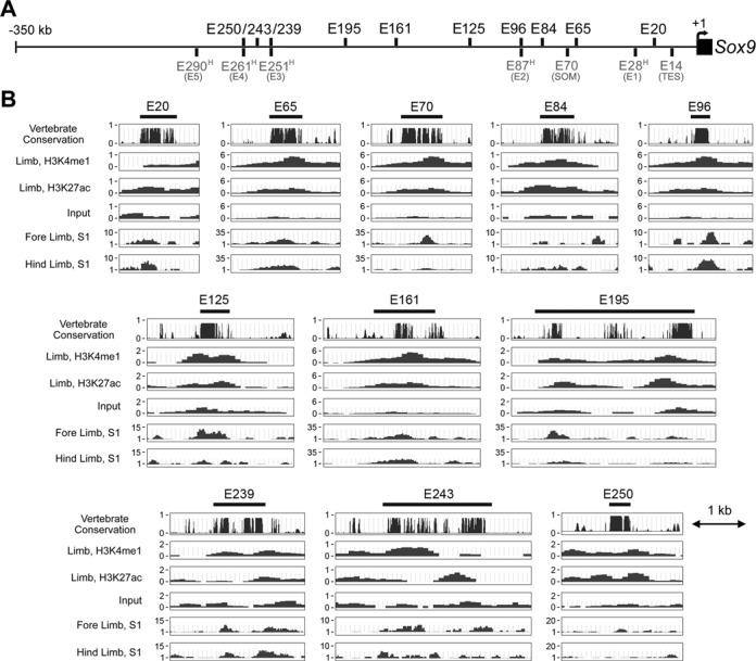
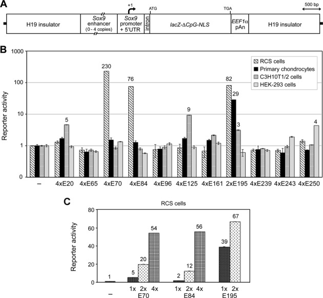
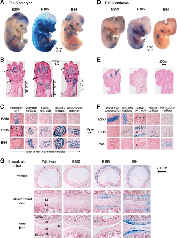
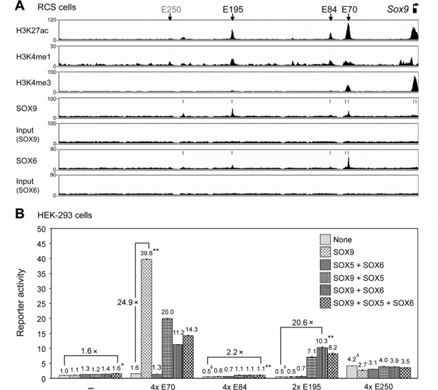
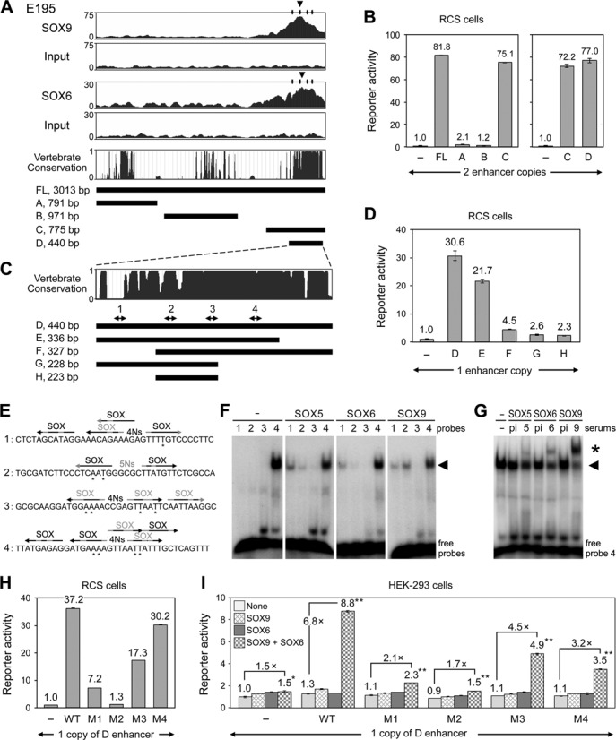
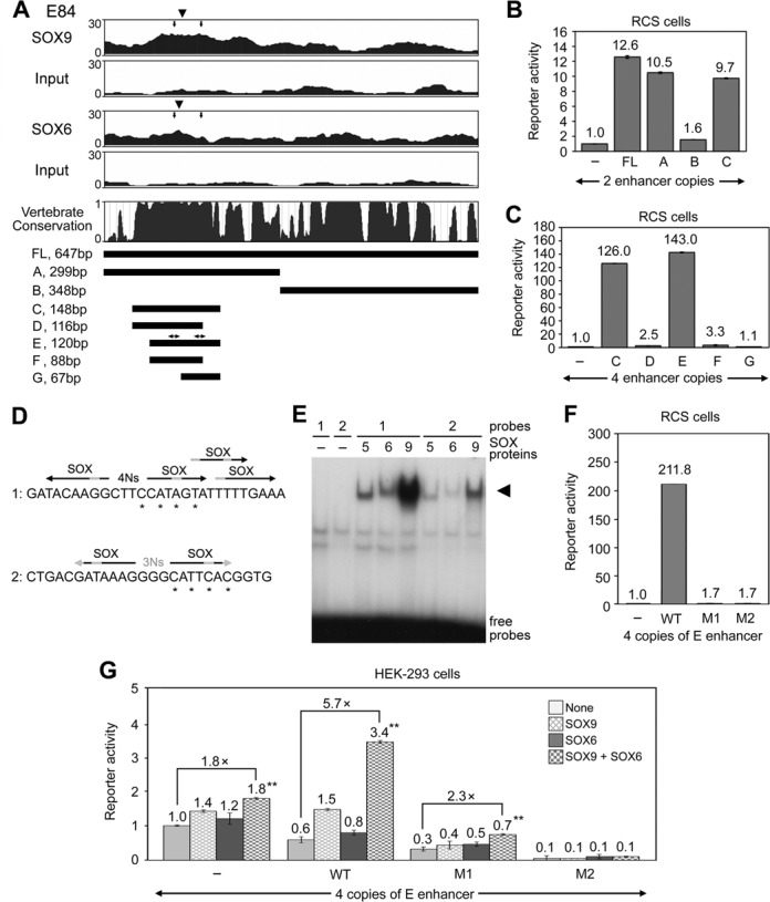
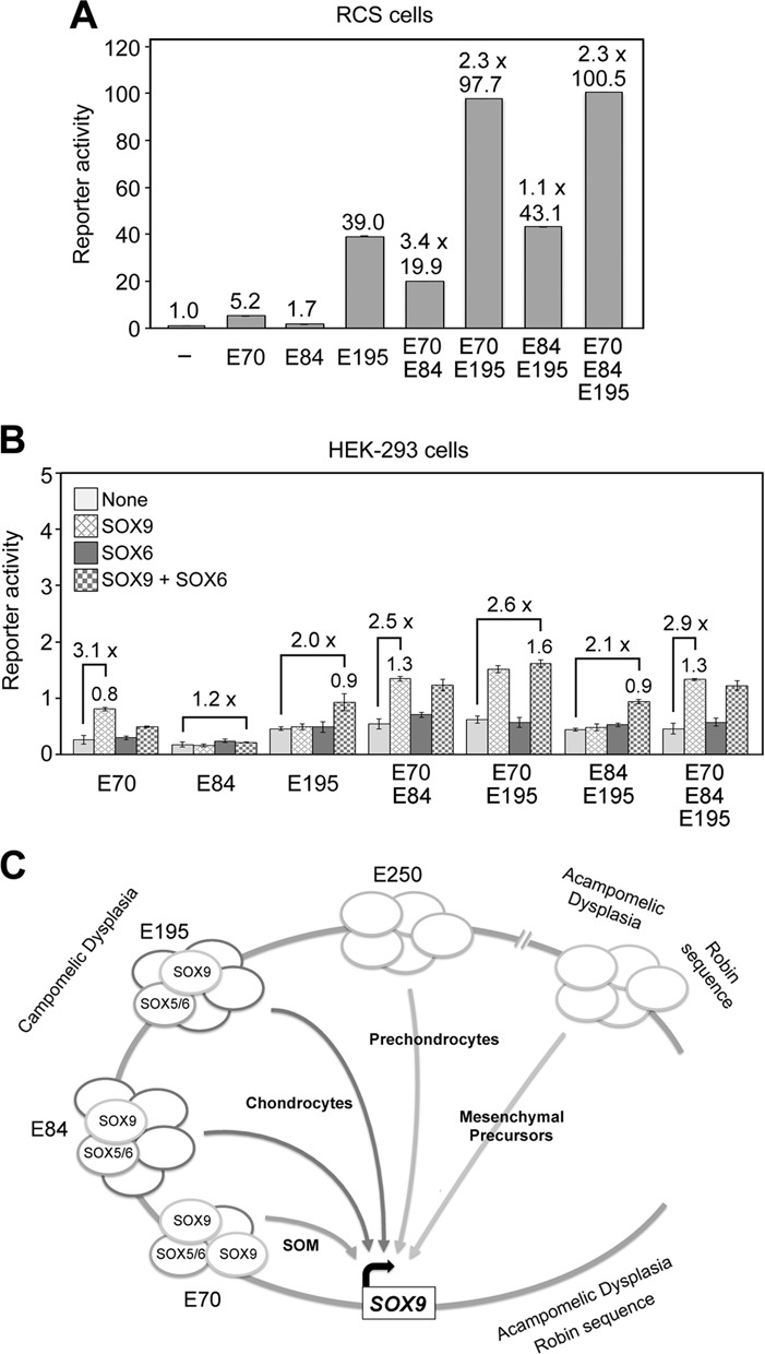
Two decades after the discovery that heterozygous mutations within and around SOX9 cause campomelic dysplasia, a generalized skeleton malformation syndrome, it is well established that SOX9 is a master transcription factor in chondrocytes. In contrast, the mechanisms whereby translocations in the --350/-50-kb region 5' of SOX9 cause severe disease and whereby SOX9 expression is specified in chondrocytes remain scarcely known. We here screen this upstream region and uncover multiple enhancers that activate Sox9-promoter transgenes in the SOX9 expression domain. Three of them are primarily active in chondrocytes. E250 (located at -250 kb) confines its activity to condensed prechondrocytes, E195 mainly targets proliferating chondrocytes, and E84 is potent in all differentiated chondrocytes. E84 and E195 synergize with E70, previously shown to be active in most Sox9-expressing somatic tissues, including cartilage. While SOX9 protein powerfully activates E70, it does not control E250. It requires its SOX5/SOX6 chondrogenic partners to robustly activate E195 and additional factors to activate E84. Altogether, these results indicate that SOX9 expression in chondrocytes relies on widely spread transcriptional modules whose synergistic and overlapping activities are driven by SOX9, SOX5/SOX6 and other factors. They help elucidate mechanisms underlying campomelic dysplasia and will likely help uncover other disease mechanisms.


Figures

Figure 1.
Figure 1.
Identification of ten putative Sox9 enhancers. (A) Schematic showing the position of newly identified enhancers (black, top) and previously identified enhancers (gray, bottom) in the –350-kb region (horizontal line) located directly upstream of the mouse Sox9 gene (black box with arrow marking the transcription start site). Enhancers identified in the human genome are presented with an ‘H’ in superscript and number corresponding to their position relative to the human SOX9 gene. Names previously given to enhancers are indicated in parentheses. (B) Specific features of E70 and ten most promising enhancers. The black bar underneath each element acronym indicates the sequence tested in reporter genes. The top graph depicts sequence conservation in 30 vertebrate genomes. The next three schematics are profiles of histone marks for active/poised enhancers (H3K4me1) and active enhancers (H3K27ac) and input background detected in E14.5 mouse embryo limbs through ChIP-seq performed by the ENCODE/LICR project. The bottom two profiles show DNase I hypersensitive sites (S1) detected in E11.5 mouse limb buds by the ENCODE/University of Washington project. See Supplementary Figure S1 for related data.
Figure 2.
Figure 2.
Schematic and activity of Sox9 enhancers in cultured cells. (A) Schematic of Sox9 enhancer-promoter-pWHERE reporters. Mouse H19 insulator sequences flank up to four copies of a Sox9 enhancer, the Sox9 307-bp proximal promoter and 364-bp 5′ untranslated region, a small intron, a modified lacZ coding sequence and the human EEF1α polyadenylation signal (pAn). The variable size of enhancers is shown with broken lines. The start of transcription (+1, angled arrow) and the start and end of translation are indicated. (B) Activities of Sox9 enhancer-promoter-pWHERE reporters in RCS cells, primary chondrocytes, C3H10T1/2 cells and HEK-293 cells. Reporters contained no enhancer (–), four tandem copies (4×) of most Sox9 enhancers, or two copies of E195. Only two copies of E195 were tested because this element is very large (3022 bp) compared to others (365–2064 bp) and more active than others. Reporter activities were normalized for transfection efficiency and calculated relative to that of the Sox9 promoter-only reporter in each cell type. Data are presented as the mean with standard deviation for technical triplicates in an experiment representative of several independent others. Activation folds ≥ 3 are indicated. (C) Activities of Sox9 enhancer-promoter-pLER reporters harboring up to four copies of E70, E84 or E195 in RCS cells.
Figure 3.
Figure 3.
Activities of E250, E195 and E84 in transgenic mice. (A) E14.5 transgenic embryos stained with X-gal. The transgenes were carrying four copies of E250, two copies of E195, or four copies of E84 upstream of the Sox9 promoter in the pWHERE reporter. Tissues expressing the reporters are seen in blue. (B) Longitudinal sections through paws and forearms. J, phalangeal joint. E, C and H stand for the epiphyseal, columnar and hypertrophic zones of cartilage growth plates, respectively. (C) High-magnification pictures of sections through various types of cartilage at different stages of development. C, cartilage. J, presumptive joint, still filled with Sox9-expressing mesenchyme. T, joint tendinous capsule. (D) E12.5 transgenic embryos stained with X-gal. (E) Paw sections showing precartilaginous digital condensations (arrow). (F) Sections through distinct cartilage tissues. C, cartilage. G, root ganglia. (G) X-gal-stained sections through distinct cartilage types from 3-week-old pups. AC, articular cartilage. B, bone. C, cartilage. GP, growth plate cartilage. M, meniscal cartilage. NP, nucleus pulposus. Bone and nucleus pulposus are not Sox9-expressing tissues. See Supplementary Figures S2 and S3 for related data.
Figure 4.
Figure 4.
Binding and activation of Sox9 enhancers by SOX9 and SOX5/SOX6. (A) ChIP-seq data obtained for histone modifications, SOX9 and SOX6, and input material for each SOX protein in the –350-kb region upstream of Sox9 in RCS cells. Vertical arrows, location of the four cartilage enhancers. Vertical bars, summits of SOX peaks identified using MACS software. (B) Activities of Sox9 enhancers in HEK-293 cells co-transfected with Sox9 reporters and expression plasmids for no protein (–) or SOX proteins, as indicated. Reporter activities were normalized for transfection efficiency and calculated relative to that of the Sox9 promoter-only reporter. Data are presented as the mean with standard deviation for technical triplicates in an experiment representative of several independent others. Δ, P <0.01 in a t-test for the difference in Sox9 promoter activity caused by an enhancer in HEK-293 cells not forced to express SOX proteins. * and **, P <0.01 and <0.001, respectively, in a t-test for the difference in a reporter activity caused by SOX protein overexpression.
Figure 5.
Figure 5.
E195 is directly activated by the SOX trio. (A) ChIP-seq data, vertebrate conservation and schematics of the full-length E195 enhancer (FL) and segments A to D. The top four graphs show ChIP-seq profiles obtained in RCS cells for SOX9 and SOX6 and for respective input material at the level of E195. Large arrowheads, summits of SOX peaks identified by MACS software. Small arrows, SOX-binding regions (see panel E). The vertebrate conservation graph was obtained by comparing 30 genomes, from lamprey to human, in the UCSC genome browser. (B) Activity of Sox9 promoter reporters harboring two enhancer copies in RCS cells. (C) Conservation and schematics of E195D to H segments. Double arrows and numbers indicate putative the location of putative SOX-binding sites (see panel E). (D) Activity of reporters harboring one copy of E195D to E195H in RCS cells. (E) Probe sequences used for EMSA. Sequences are shown for the upper strand only. Each arrow represents a putative SOX-binding site. The black and gray shades highlight nucleotides that match and do not match the SOX domain-binding consensus CT/ATTGT/AT/A, respectively. SOX is written in black for sequences presenting at least five consensus nucleotides, and in gray for others. Inverted sites separated by four nucleotides (strong SOX9 site) or five nucleotides (weak SOX9 site) are indicated. Asterisks mark nucleotides mutated in reporters used in panels H and I. (F) EMSA with probes 1–4 and nuclear extracts from COS-7 cells forced to express no protein, SOX5, SOX6 or SOX9 (Supplementary Figure S4A shows that the extracts contained similar amounts of SOX protein). Note that probe 4 forms a complex with a nonspecific protein migrating at the same level as SOX/DNA complexes (arrowhead). (G) EMSA of extracts incubated with probe 4 and preimmune serum (pi) or antiserums against SOX5 (5), SOX6 (6) or SOX9 (9). Star, antibody supershifts. (H) Reporter activities generated by one copy of wild-type and mutant E195D enhancers in RCS cells. The M1 to M4 enhancers featured point mutations, as shown in panel E and Supplementary Table S2. (I) Same experiment as in panel G, but using HEK-293 cells co-transfected with SOX expression plasmids. Reporter activities are presented as described in Figures 2B and 4B. See Supplementary Figure S4A and B for related data.
Figure 6.
Figure 6.
E84 activity involves the SOX trio. (A) ChIP-seq data, vertebrate conservation and schematics of the full-length E84 enhancer (FL) and segments A to G. The top four graphs show ChIP-seq profiles obtained in RCS cells for SOX9 and SOX6 and for respective input material at the level of E84. Large arrowheads, summits of SOX peaks identified using MACS software. Small arrows, SOX-binding regions (see panel D). The vertebrate conservation graph was obtained by comparing 30 genomes, from lamprey to human, in the UCSC genome browser. Double arrows above the E segment schematic indicate the positions of SOX-binding regions. (B) Activities of reporters harboring two copies of E84FL, A, B and C in RCS cells. (C) Activities of reporters harboring four copies of E84C to E84G in RCS cells. (D) EMSA probe sequences. Only upper strands are shown. See Figure 5E for labeling explanations. Asterisks mark nucleotides mutated in reporters tested in panels F and G. (E) EMSA with SOX protein-containing extracts from COS-7 cells and probes described in panel D. Arrowhead, SOX protein/DNA complexes. (F) Reporter activities generated by four copies of wild-type and mutant E84E enhancers in RCS cells. Mutations were as described in panel D. (G) Same experiment as in panel F, but in HEK-293 cells co-transfected with SOX expression plasmids. Reporter activities were calculated and are presented as described in Figures 2B and 4B. See Supplementary Figure S4A and C for related data.
Figure 7.
Figure 7.
Sox9 enhancer synergy and model for the activation of SOX9 in chondrocytes. (A) Activities of reporters carrying one copy of E70, E84 or E195, or tandem combinations of one copy of these enhancers in RCS cells. Synergy between enhancers (1.1 to 3.4×) was calculated as the fold activation of the Sox9 promoter obtained relative to the sum of the activities of individual enhancers. (B) Activities of the same reporters as in panel A in HEK-293 cells co-transfected with SOX expression plasmids. The best activation fold of the Sox9 promoter achieved by SOX proteins (SOX9 alone or together with SOX6) is indicated for each reporter. (C) Model. See the first paragraph of Discussion for explanations.

References

    1. Foster J.W., Dominguez-Steglich M.A., Guioli S., Kwok C., Weller P.A., Stevanovic M., Weissenbach J., Mansour S., Young I.D., Goodfellow P.N., et al. Campomelic dysplasia and autosomal sex reversal caused by mutations in an SRY-related gene. Nature. 1994;372:525–530. - PubMed
    1. Wagner T., Wirth J., Meyer J., Zabel B., Held M., Zimmer J., Pasantes J., Bricarelli F.D., Keutel J., Hustert E., et al. Autosomal sex reversal and campomelic dysplasia are caused by mutations in and around the SRY-related gene SOX9. Cell. 1994;79:1111–1120. - PubMed
    1. Unger S., Scherer G., Superti-Furga A. GeneReviews. Seattle: University of Washington; 2013. Campomelic dysplasia. - PubMed
    1. Akiyama H., Chaboissier M. C., Behringer R. R., Rowitch D. H., Schedl A., Epstein J. A., de Crombrugghe B. Essential role of Sox9 in the pathway that controls formation of cardiac valves and septa. Proc. Natl. Acad. Sci. U.S.A. 2004;101:6502–6507. - PMC - PubMed
    1. Stolt C.C., Wegner M. SoxE function in vertebrate nervous system development. Int. J. Biochem. Cell Biol. 2010;42:437–440. - PubMed

Publication types