. 2015 Jun 23;43(11):5537-49.
doi: 10.1093/nar/gkv406. Epub 2015 May 4.

The telomerase essential N-terminal domain promotes DNA synthesis by stabilizing short RNA-DNA hybrids

Affiliations

The telomerase essential N-terminal domain promotes DNA synthesis by stabilizing short RNA-DNA hybrids

Benjamin M Akiyama et al. Nucleic Acids Res. .

Abstract

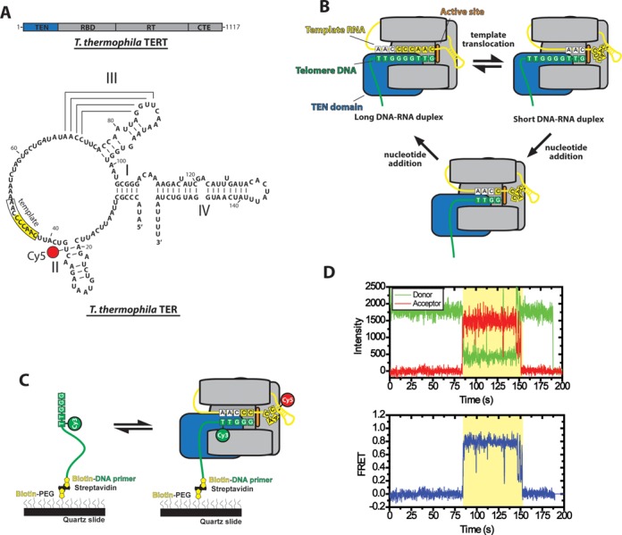
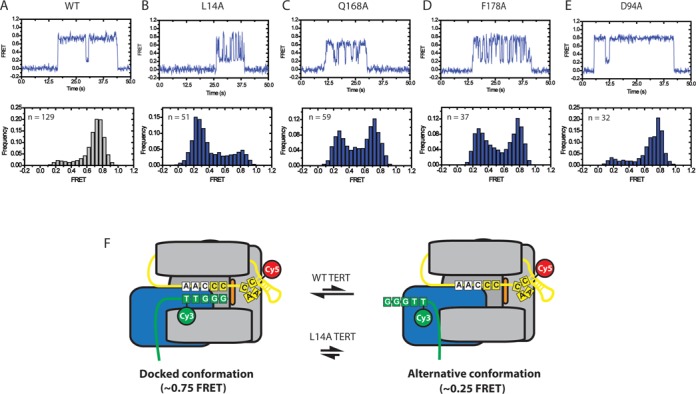
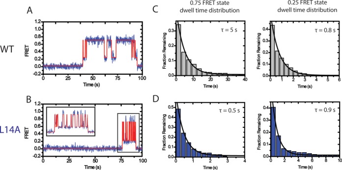
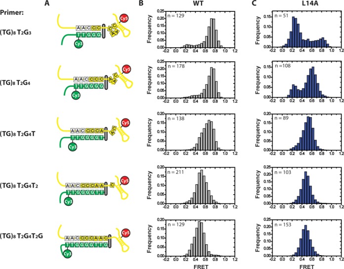
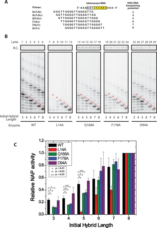
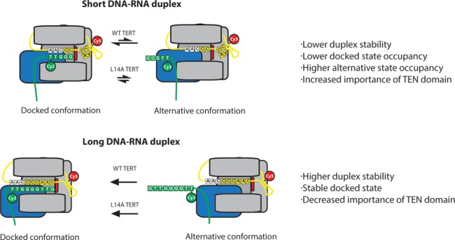
Telomerase is an enzyme that adds repetitive DNA sequences to the ends of chromosomes and consists of two main subunits: the telomerase reverse transcriptase (TERT) protein and an associated telomerase RNA (TER). The telomerase essential N-terminal (TEN) domain is a conserved region of TERT proposed to mediate DNA substrate interactions. Here, we have employed single molecule telomerase binding assays to investigate the function of the TEN domain. Our results reveal telomeric DNA substrates bound to telomerase exhibit a dynamic equilibrium between two states: a docked conformation and an alternative conformation. The relative stabilities of the docked and alternative states correlate with the number of basepairs that can be formed between the DNA substrate and the RNA template, with more basepairing favoring the docked state. The docked state is further buttressed by the TEN domain and mutations within the TEN domain substantially alter the DNA substrate structural equilibrium. We propose a model in which the TEN domain stabilizes short RNA-DNA duplexes in the active site of the enzyme, promoting the docked state to augment telomerase processivity.


Figures

Figure 1.
Figure 1.
Overview of telomerase smFRET binding assay. (A) Domain organization of Tetrahymena thermophila TERT and secondary structure of Tetrahymena thermophila TER. TERT is divided into the telomerase essential N-terminal domain (TEN, blue), the RNA binding domain (RBD), the reverse transcriptase domain (RT) and the C-terminal extension (CTE). TER contains stems I, II, III and IV as well as a conserved RNA template (boxed region). The position of the Cy5 modification used for smFRET studies at U36 is indicated. (B) Diagram of telomerase catalytic cycle. TERT is represented in gray with the TEN domain highlighted in blue and the active site in orange. The telomeric DNA substrate is represented in green and the telomerase RNA is represented in yellow. The template RNA and telomere DNA form basepairing interactions and this heteroduplex is positioned in a central channel of the enzyme adjacent to the active site (6). When the end of the template is reached, the RNA–DNA duplex is denatured and the RNA template re-anneals downstream to position the template for another round of synthesis (template translocation). The post-translocation state of the enzyme contains a short RNA–DNA duplex which must be stabilized in the active site in order to become extended by the enzyme's reverse transcriptase activity to complete the catalytic cycle. (C) Schematic diagram of smFRET telomerase binding assay. DNA primers containing telomeric DNA sequence are labeled with a donor Cy3 dye at their 5′ most alignment residue and immobilized on a quartz microscope slide by a biotin-streptavidin linkage. Telomerase labeled with Cy5 in its TER subunit is flowed onto the slide and FRET is measured on individual molecules for the duration of the binding events. (D) Example smFRET trace for a (TG)8T2G3 primer incubated with telomerase labeled at the U36 position of the TER subunit. Donor (Cy3) and acceptor (Cy5) intensity are plotted over time (Top panel). The binding event (shaded region) is marked by the onset of a FRET signal, characterized by an anti-correlated drop in donor fluorescence and rise in acceptor fluorescence. Loss of FRET signal at ∼150s occurs either due to Cy5 photobleaching or diffusion of telomerase off of the primer. Loss of Cy3 signal at ∼190s is due to a normal process of Cy3 photobleaching. Donor and acceptor intensity values from the top panel are used to calculate a FRET trace in the bottom panel. The FRET values from each point during the binding event are combined with multiple other binding events to generate smFRET histograms.
Figure 2.
Figure 2.
Representative smFRET traces and histograms for wild-type and mutant telomerase. (A) Representative smFRET trace (top) and smFRET histogram (bottom) for Cy3-labeled (TG)8T2G3 primer incubated with wild-type telomerase labeled with Cy5 at the U36 position of TER. Wild-type enzyme demonstrates a stable ∼0.75 FRET state with transient excursions to a ∼0.25 FRET state (top panel). This is also reflected in a smFRET histrogram of FRET values compiled from 129 separate binding events (bottom panel) demonstrating a predominant ∼0.75 FRET distribution with a small shoulder at ∼0.25 FRET. (BE) Representative smFRET traces (top) and smFRET histograms (bottom) for L14A, Q168A and F178A mutant telomerase respectively. (F) Model of telomerase DNA binding dynamics. smFRET data indicate that DNA associated with telomerase can exist in one of at least two conformations. In the docked state, represented by the ∼0.75 FRET population, the RNA–DNA duplex is positioned in the enzyme active site. The ∼0.25 FRET population represents an alternative state that exists in an equilibrium with the docked state. In this conformation the 3′ end of the DNA is positioned away from the enzyme active site. TEN domain residues L14, Q168 and F178 bias the internal equilibrium towards the docked conformation. Importantly, smFRET alone does not provide sufficient information to fully map the contacts present in the alternative state. Therefore, although we can confidently assert that an alternative state exists, the schematic layout presented in this figure represents only one of many possible organizations that could comprise the alternative state of the enzyme.
Figure 3.
Figure 3.
Dwell time analysis of TEN domain mutants demonstrate that the TEN domain stabilizes the docked state. (A) smFRET traces (blue) were analyzed by HaMMy (27) to generate idealized traces (red). These were used to determine the dwell time of the enzyme in each state. (B) The dwell times for the WT enzyme in the 0.75 FRET state and the 0.25 FRET state incubated with primer (TG)8T2G3 were compiled into histograms. The histograms were fit to an exponential function to identify the average dwell time. Wild-type TERT demonstrated a dwell time of τdocked = 5 s for the 0.75 FRET state and a dwell time of τalt = 0.8 s for the 0.25 FRET state. (C) Representative smFRET trace and idealized HaMMy trace for L14A TERT telomerase incubated with the (TG)8T2G3 primer. (D) Compiled histograms for L14A enzyme. L14A TERT demonstrated a dwell time of τdocked = 0.5 s for the 0.75 FRET state and a dwell time of τalt = 0.9 s for the 0.25 FRET state.
Figure 4.
Figure 4.
Effect of primer-template hybrid formation on FRET distributions. Primers capable of forming 5–9 basepairs with template RNA were tested in smFRET telomerase binding assays with U36-labeled telomerase. (A) Schematic diagram of the docked state for all six primers used in smFRET experiments, demonstrating the number of basepairs formed and the expansion of the template RNA as the RNA–DNA duplex becomes progressively longer (22). (B) smFRET histograms for wild-type enzyme. As primers contain progressively more telomeric DNA sequence, the predominant FRET distribution of the docked state shifts from ∼0.75 FRET to ∼0.5 FRET. In addition, the ∼0.25 FRET alternative state disappears. (C) smFRET histograms for L14A mutant enzyme.
Figure 5.
Figure 5.
Telomerase activity assays demonstrate TEN domain mutations affect nucleotide addition processivity of primers with short RNA–DNA duplexes. (A) Primer permutants used in in vitro extension assays. Primers were length-matched at 3 telomeric repeats (18 nts), but staggered such that they formed different initial potential RNA–DNA duplex lengths with template RNA. (B) Telomerase was reconstituted in rabbit reticulocyte lysate and telomerase activity was assayed on the six DNA primers corresponding to six potential RNA–DNA hybrid lengths. WT enzyme was compared against enzyme harboring L14A, Q168A, F178A and D94A mutations. Mutants were assayed for primer-specific NAP defects by comparing the accumulation of the first repeat addition band (red asterisks) between primers tested with the same enzyme. (C) Quantification of relative NAP activity as a function of initial primer duplex length. Telomerase activity assay gels (Figure 5B) were performed in triplicate and quantified using the program SAFA (26). The quantification of each band was then corrected for specific activity. NAP was quantified as the amount of product that was extended to the 5′ end of the RNA template (Figure 5B, red asterisks, see Materials and Methods for details). For each wild-type or mutant enzyme, the observed NAP activity for each primer variant was normalized to the (GGGGTT)3 primer, which has the potential to form eight basepairs of RNA–DNA hybrid, and displayed the maximal NAP activity. P-values indicating statistical significance are as marked on the graph, error bars indicate one standard deviation based on triplicate measurements.
Figure 6.
Figure 6.
Model demonstrating the role of the TEN domain in stabilizing short RNA–DNA duplexes. Primers corresponding to early catalytic intermediates that contain fewer RNA–DNA basepairs are in a conformational equilibrium between a docked state and an alternative state (top panel). TEN domain mutants L14A, Q168A and F178A destabilize the docked state such that the alternative state is favored, disrupting the catalytic activity of the enzyme. In primers corresponding to late catalytic intermediates, the docked state is heavily favored due to the presence of additional RNA–DNA basepairs (bottom panel). As a result, the alternative state is not observed, even in the presence of TEN domain mutants.

References

    1. Palm W., de Lange T. How shelterin protects mammalian telomeres. Annu. Rev. Genet. 2008;42:301–334. - PubMed
    1. Bodnar A.G., Ouellette M., Frolkis M., Holt S.E., Chiu C.P., Morin G.B., Harley C.B., Shay J.W., Lichtsteiner S., Wright W.E. Extension of life-span by introduction of telomerase into normal human cells. Science. 1998;279:349–352. - PubMed
    1. Vulliamy T.J., Dokal I. Dyskeratosis congenita: the diverse clinical presentation of mutations in the telomerase complex. Biochimie. 2008;90:122–130. - PubMed
    1. Kim N.W., Piatyszek M.A., Prowse K.R., Harley C.B., West M.D., Ho P.L., Coviello G.M., Wright W.E., Weinrich S.L., Shay J.W. Specific association of human telomerase activity with immortal cells and cancer. Science. 1994;266:2011–2015. - PubMed
    1. Greider C.W. Telomerase is processive. Mol. Cell. Biol. 1991;11:4572–4580. - PMC - PubMed

Publication types