. 2015 Jun 18;125(25):3905-16.
doi: 10.1182/blood-2015-01-621474. Epub 2015 May 4.

Tumor indoleamine 2,3-dioxygenase (IDO) inhibits CD19-CAR T cells and is downregulated by lymphodepleting drugs

Affiliations

Tumor indoleamine 2,3-dioxygenase (IDO) inhibits CD19-CAR T cells and is downregulated by lymphodepleting drugs

Soranobu Ninomiya et al. Blood. .

Abstract

Although T cells expressing CD19-specific chimeric antigen receptors (CARs) are a promising new therapy for B-cell malignancies, objective responses are observed at lower frequencies in patients with lymphoma than in those with acute B-cell leukemia. We postulated that the tumor microenvironment suppresses CAR-expressing T cells (CARTs) through the activity of indoleamine 2,3-dioxygenase (IDO), an intracellular enzyme that converts tryptophan into metabolites that inhibit T -: cell activity. To investigate the effects of tumor IDO on CD19-CART therapy, we used a xenograft lymphoma model expressing IDO as a transgene. CD19-CARTs inhibited IDO-negative tumor growth but had no effect on IDO-positive tumors. An IDO inhibitor (1-methyl-tryptophan) restored IDO-positive tumor control. Moreover, tryptophan metabolites inhibited interleukin (IL)-2-, IL-7-, and IL-15-dependent expansion of CARTs; diminished their proliferation, cytotoxicity, and cytokine secretion in vitro in response to CD19 recognition; and increased their apoptosis. Inhibition of CD19-CARTs was not mitigated by the incorporation of costimulatory domains, such as 4-1BB, into the CD19-CAR. Finally, we found that fludarabine and cyclophosphamide, frequently used before CART administration, downregulated IDO expression in lymphoma cells and improved the antitumor activity of CD19-CART in vivo. Because tumor IDO inhibits CD19-CARTs, antagonizing this enzyme may benefit CD19-CART therapy.


Figures

Figure 1
Figure 1
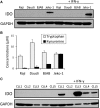
IDO in B-cell lymphoma lines and primary CLL cells. (A) Four B-cell lymphoma cell lines (Raji, Daudi, BJAB, and Jeko-1) and (C) 5 CLL patients’ PBMCs were cultured in the absence or presence of exogenous IFN-γ (50 U/mL) for 24 hours. Protein extracts were prepared for IDO western immunoblot analysis. Results are representative of 3 independent experiments. (B) IDO enzyme activity was evaluated by measuring the concentrations of l-tryptophan and l-kynurenine in the cell culture medium of the B-cell lymphoma lines using HPLC. Each bar represents the mean ± SD of the results from 3 independent experiments.
Figure 2
Figure 2
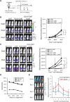
Effect of tumor-derived IDO on CD19-CARTs. (A) Schematic representation of the experiments in SCID/Beige mice comparing the antitumor effects of CD19-CARTs on Raji-control (left flank) and Raji-IDO tumor (right flank). (B) Time course of tumor bioluminescence in mice treated with nontransduced (NT) T cells or CD19-CARTs. (C) Time course of bioluminescence of Raji-control and Raji-IDO tumors treated with NT and CARTs. Data represent mean ± SD of 8 mice per group from 2 independent experiments (*P < .05).
Figure 3
Figure 3
Combination of IDO inhibitor and CD19-CART therapy on IDO-positive tumors. (A) Schematic representation of the experiments in SCID/Beige mice comparing the antitumor effect of IDO inhibitor (1-MT), CD19-CARTs, or the combination of both on IDO-positive malignancies (Raji-IDO and Jeko-1). (B) Time course of Raji-IDO tumor bioluminescence. Data represent mean ± SD of 7 mice per group from 2 independent experiments (*P < .05). (C) Time course of Jeko-1 tumor bioluminescence. Data represent mean ± SD of 8 mice per group from 2 independent experiments (*P < .05). (D) CD19-CARTs retained similar cytotoxic activity against Raji-control and Raji-IDO in a 4-hour 51Cr-release assay. (E) Luciferase-labeled CD19-CARTs were injected intravenously in mice that had been subcutaneously inoculated with nonlabeled Raji-control (left flank) and Raji-IDO (right flank). The time course of T-cell bioluminescence at Raji-control and Raji-IDO tumor site is shown. Data represent mean ± SD of 4 mice.
Figure 4
Figure 4
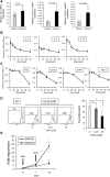
Effect of tryptophan metabolites on CD19-CART proliferation. (A) AHR, CYP1A1, and CYP1B1 mRNA expression measured by real-time PCR on CD19-CARTs treated with or without KHAA (12.5 μM) for 24 hours. Data represent mean ± SD of 4 T-cell lines generated from 4 healthy donors and normalized to GAPDH expression. (B) CD19-CARTs were cultured in media containing increasing amounts of l-kynurenine and 3-HAA (KHAA) (0, 6.25, 12.5, 25, or 50 μM) with IL-2 (50 U/mL), IL-7 (10 ng/mL), or IL-15 (5 ng/mL) for 72 hours. (C) Four B-cell lymphoma lines (Raji, Daudi, BJAB, or Jeko-1) were cultured in media containing increasing amounts of KHAA for 72 hours, without exogenous cytokines. Cell proliferation was studied by the XTT assay. One representative set of 3 experiments is shown (*P < .05). (D) NTs or CD19-CARTs labeled with CFSE were cultured with irradiated Raji cells for 3 days in the absence or presence of KHAA (12.5 or 25 μM) without any added exogenous cytokines. CFSE dilution was assayed by flow cytometry, gating on CD3-positive cells. Controls (dotted line) were cultured without Raji cells. Data represent mean ± SD of CSFE dilution in 4 T-cell lines generated from 4 healthy donors (*P < .05). (E) The number of CD19-CARTs cultured in the presence or absence of KHAA (12.5 μM) was determined at the end of each 7-day expansion cycle. Data represent mean ± SD of 5 T-cell lines generated from 5 donors (*P < .01).
Figure 5
Figure 5
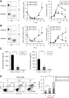
Effect of tryptophan metabolites on killing, apoptosis, and cytokine secretion by CD19-CARTs. GFP-transduced wild-type Raji cells were cocultured with CD19-CARTs at E:T ratios (A) 1:2 and (B) 1:4 in the presence or absence of KHAA (12.5 μM). The total number of tumor cells and CARTs in each culture condition was determined using flow cytometry analysis at the indicated time points. Dot plot shows representative data on day 14. Three experiments using CARTs generated from 3 different donors were analyzed and shown as the mean ± SD in the graph (*P < .05). (C) Seven days after the first expansion, CD19-CARTs (1 × 10⁶) were cultured with Raji cells (1 × 10⁶) in the presence or absence of KHAA (12.5 μM); supernatants were collected after 24 hours and analyzed for IL-2 and IFN-γ by ELISA. Data represent mean ± SD of 4 T-cell lines generated from 4 donors (*P < .05). (D) Apoptosis was determined by annexin V/7-AAD staining of CD19-CARTs after 3 days of culture in the absence or presence of KHAA (12.5 or 25 μM) and IL-2 (50 U/mL). Data represent mean ± SD of 4 T-cell lines generated from 4 donors (*P < .05).
Figure 6
Figure 6
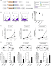
Additional costimulatory domains are not able to mitigate the inhibitory effects of tryptophan metabolites. (A) Schematic representation of recombinant retrovirus vectors encoding CD19-CAR constructs (CD19.ζ, containing the CD3ζ chain alone; CD19.28.ζ, CD3ζ chain and CD28 endodomain; CD19.28.4-1BB.ζ, CD3ζ chain and CD28 and 4-1BB endodomains) (B) CD19-CAR surface expression on T cells transduced with each construct. (C) The number of CD19-CARTs cultured in the presence or absence of KHAA (12.5 μM) was determined at the end of each 7-day expansion cycle. Data represent the mean ± SD of 5 T-cell lines generated from 5 donors (*P < .05). (D) GFP-transduced wild-type Raji cells were cocultured with each CD19-CART type at an E:T ratio of 1:2 in the presence or absence of KHAA (12.5 μM). The total number of tumor cells and CARTs in each culture condition was determined by flow cytometry. Dot plot shows representative data on day 14. Five experiments using CARTs generated from 5 different donors were analyzed and represent mean ± SD in the graph (*P < .05). In the absence and presence of KHAA, the number of residual tumor was 0.28 ± 0.28 × 10⁶ and 1.49 ± 0.84 × 10⁶, respectively, for the first-generation CAR; 0.16 ± 0.13 × 10⁶ and 0.83 ± 0.3 × 10⁶, respectively, for the second-generation CAR; and 0.99 ± 0.33 × 10⁶ and 1.79 ± 0.39 × 10⁶, respectively, for the third-generation CAR.
Figure 7
Figure 7
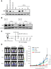
Fludarabine and cyclophosphamide downregulate IDO expression. (A) Jeko-1 cells were treated with mafosfamide (2 μg/mL), fludarabine (20 μM), or both for 24 hours. Cells were then washed and cultured with or without IFN-γ. Protein extracts were prepared for IDO immunoblot analysis. Results are representative of 3 independent experiments. (B) PBMCs from 2 CLL patients were treated with mafosfamide (2 μg/mL), fludarabine (20 μM), or both for 24 hours. Cells were then washed and incubated with IFN-γ. Twenty-four hours later, proteins were extracted, and IDO expression was revealed by immunoblot. (C) Schematic representation of the experiments in SCID/Beige mice comparing the antitumor effect of chemotherapy, CD19-CARTs, or their combination on Jeko-1 tumors. Mice were subcutaneously injected with luciferase-transduced Jeko-1 cells (3 × 10⁶). Five days later, mice were treated with 0.75 mg fludarabine (Flu) and 0.75 mg cyclophosphamide (Cy) intraperitoneally. Two days after chemotherapy, CD19-CARTs (10 × 106) were infused intravenously. (D) Time course of Jeko-1 tumor bioluminescence. Data represent mean ± SD of 7 mice per group from 2 independent experiments (*P < .05).

Similar articles

Cited by

References

    1. Cruz CR, Micklethwaite KP, Savoldo B, et al. Infusion of donor-derived CD19-redirected virus-specific T cells for B-cell malignancies relapsed after allogeneic stem cell transplant: a phase 1 study. Blood. 2013;122(17):2965–2973. - PMC - PubMed
    1. Porter DL, Levine BL, Kalos M, Bagg A, June CH. Chimeric antigen receptor-modified T cells in chronic lymphoid leukemia. N Engl J Med. 2011;365(8):725–733. - PMC - PubMed
    1. Kalos M, Levine BL, Porter DL, et al. T cells with chimeric antigen receptors have potent antitumor effects and can establish memory in patients with advanced leukemia. Sci Transl Med. 2011;3(95):95ra73. - PMC - PubMed
    1. Davila ML, Riviere I, Wang X, et al. Efficacy and toxicity management of 19-28z CAR T cell therapy in B cell acute lymphoblastic leukemia. Sci Transl Med. 2014;6(224):224ra25. - PMC - PubMed
    1. Kochenderfer JN, Dudley ME, Carpenter RO, et al. Donor-derived CD19-targeted T cells cause regression of malignancy persisting after allogeneic hematopoietic stem cell transplantation. Blood. 2013;122(25):4129–4139. - PMC - PubMed

Publication types

MeSH terms