. 2015 Aug 1;404(1):27-39.
doi: 10.1016/j.ydbio.2015.04.017. Epub 2015 May 1.

Niche signaling promotes stem cell survival in the Drosophila testis via the JAK-STAT target DIAP1

Affiliations

Niche signaling promotes stem cell survival in the Drosophila testis via the JAK-STAT target DIAP1

Salman Hasan et al. Dev Biol. .

Abstract

Tissue-specific stem cells are thought to resist environmental insults better than their differentiating progeny, but this resistance varies from one tissue to another, and the underlying mechanisms are not well-understood. Here, we use the Drosophila testis as a model system to study the regulation of cell death within an intact niche. This niche contains sperm-producing germline stem cells (GSCs) and accompanying somatic cyst stem cells (or CySCs). Although many signals are known to promote stem cell self-renewal in this tissue, including the highly conserved JAK-STAT pathway, the response of these stem cells to potential death-inducing signals, and factors promoting stem cell survival, have not been characterized. Here we find that both GSCs and CySCs resist cell death better than their differentiating progeny, under normal laboratory conditions and in response to potential death-inducing stimuli such as irradiation or starvation. To ask what might be promoting stem cell survival, we characterized the role of the anti-apoptotic gene Drosophila inhibitor of apoptosis 1 (diap1) in testis stem cells. DIAP1 protein is enriched in the GSCs and CySCs and is a JAK-STAT target. diap1 is necessary for survival of both GSCs and CySCs, and ectopic up-regulation of DIAP1 in somatic cyst cells is sufficient to non-autonomously rescue stress-induced cell death in adjacent differentiating germ cells (spermatogonia). Altogether, our results show that niche signals can promote stem cell survival by up-regulation of highly conserved anti-apoptotic proteins, and suggest that this strategy may underlie the ability of stem cells to resist death more generally.

Keywords: DIAP1; Drosophila spermatogenesis; JAK–STAT signaling; Stem cell niche.


Figures

Fig 1
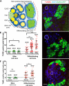
Fig 1. Stem cells in the testis niche are more resistant to radiation and starvation induced cell death than differentiating cells
(A) Illustration of the Drosophila adult testis niche. GSCs (yellow) and CySCs (dark blue) are adjacent to the hub at the testis apex. GSCs divide to give rise to gonialblasts, which continue to divide, forming spermatogonial cysts. CySCs divide to form cyst cells, which envelop gonialblasts and their progeny. TUNEL-positive cells were quantified in two adjacent regions of the testis 1), A “Stem cell zone” comprised of the first two tiers of cells adjacent to the hub which includes all stem cells (GSCs and CySCs) and an occasional gonialblast and/or early cyst cell. 2), A “Differentiating cell zone”, which extends from the boundary of the stem cell zone through the Bam-GFP positive spermatogonial cysts (green). (B, C, D) Confocal sections of testes expressing Bam-GFP fusion protein and immuno-stained with anti-GFP antisera (Bam expression pattern- spermatogonial cysts, green), TUNEL (apoptotic cells, red) and DAPI (DNA, blue) under control conditions (B), after 3 hours of exposure to 25 Gy γ radiation (C) and after 19.5 hours of protein-starvation (D). TUNEL-positive spots (arrows) are typically seen within and adjacent to Bam-positive spermatogonial cysts (C, D). Scale bar 10 μm. (E, F) Scatter plots showing the number of TUNEL-positive spots per testis apex in the stem cell and differentiating cell zones. (E) Upon 25Gy of irradiation, there is no significant difference in TUNEL-positive spots in stem cell zone compared to un-irradiated controls. In contrast, 50 Gy of IR causes significantly more stem cells to die. In the differentiating cell zone, significantly more cells die under both 25Gy and 50Gy doses compared to un-irradiated controls. (*, p<0.05), (**, p<0.01). 2-tailed Kruskal-Wallis test with multiple comparisons. Bars indicate mean and S.D. (F) There is no significant difference in TUNEL-positive spots in the stem cell zone after starvation. There is a significant increase in TUNEL-positive spots in differentiating cell zone after starvation. (*, p<0.05). 2-tailed Mann Whitney test.
Fig 2
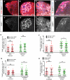
Fig 2. DIAP1 is enriched in the stem cells and is a target of JAK-STAT signaling
(A-C) Confocal sections through the apex of testes stained with anti-DIAP1 (white), anti-Zfh1 (CySCs and cyst cells, red), and DAPI (DNA, blue). (A’-C’) DIAP1 channel alone. In control testes, DIAP1 protein is enriched around the hub (white outline) and co-localizes with CySCs (Zfh1 positive cells near the hub; one indicated, arrowhead) and GSCs (Zfh1- negative cells contacting the hub; one indicated, arrow). (B) DIAP1 levels are reduced in testes with reduced JAK-STAT signaling (genotype: statF/stat06436) after 24h at restrictive temperature (see methods). (C) DIAP1 levels are increased in both stem cell populations and in differentiating cells in testes with increased JAK-STAT signaling (hs-upd testes after 45 minutes of induction). (D, E) Bar graph showing quantification of DIAP1 levels in stem cells normalized to DAPI levels in the same cells in control (green) and experimental (red) flies when JAK-STAT is reduced (D) or up-regulated (E).(***, p<0.001), (****, p<0.0001). 2-tailed Mann Whitney test. Error Bars indicate mean and S.E.M.
Figure 3
Figure 3. DIAP1 is required for stem cell survival
(A, B) Confocal sections through the testis apex show control (A) or DIAP1 null (thread5, B) clones 2 days after clone induction (ACI). Hubs are outlined in white (thick line). Negatively marked clones, identified by the absence of GFP (green) staining, are outlined in white (thin line). Clones positive for Tj (red) are CySC lineage clones (arrowheads), and those negative for Tj are germ line clones (arrows). Both control (A, A’) and DIAP1 null (B, B’) clones can be seen next to the hub. (C) Line diagram showing the percentage of testes with DIAP1 null (thread5, Exptl) or control (Cntrl) GSC or CySC clones after clone induction. Very few thread5 CySC clones (blue solid line) and thread5 GSC clones (red solid line), are seen compared to control CySC (blue dashed line) or GSC (red dashed line) clones. (D-H) Confocal sections through the apex of testes stained with TUNEL (red) and DAPI (blue). Hubs are outlined in white. (D’-H’), TUNEL channel alone. (D, D’) TUNEL positive cells next to the hub were rarely seen in control testes after induction of GFP RNAi. (E, E’) After heat shock-mediated induction of DIAP1-RNAi (E), TUNEL positive cells were seen adjacent to the hub (arrow in E, E’). (F) Scatter plot showing quantification of data from D and E. The number of dying cells in the stem cell zone (see methods) increased significantly upon induction of DIAP1 RNAi compared to induction of GFP RNAi in controls (*, p<0.05). No significant difference in cell death was observed without induction of RNAi or with induction of GFP RNAi. Statitistical test used is Kruskal – Wallis test of multiple comparisons. (G, G’) Irradiation of control testes (genotype- statF / TM3, after 24h at restrictive temperature) at 25 Gy did not result in TUNEL positive cells next to the hub (stem cell zone). (H, H’) Irradiation of testes with reduced JAK-STAT signaling (genotype: statF/stat06436, after 24 h at restrictive temperature) at 25 Gy resulted in increased number of TUNEL positive cells next to the hub (arrows in G, G’) (stem cell zone). Scale bars 10 μm. (I) Scatter plot shows quantification of data from G and H. The number of TUNEL positive cells in the stem cell zone increased significantly upon irradiation (25 Gy) after reduction of JAK-STAT signaling compared to irradiated control testes (****, p<0.0001). 2 tailed Mann-Whitney test. Bars (F, I) indicate mean and S.D.
Figure 4
Figure 4. DIAP1 over-expression in CySCs and their daughters rescues differentiating germline cells from stress induced cell death
(A -D) Confocal sections through the apex of testes stained with anti-DIAP1 (white), anti-Vasa (A-C, germ cells, red) or anti-Tj (D, somatic cells, red), and DAPI. (A’-D’) DIAP1 channel alone. Testes were imaged at the same gain to show different levels of DIAP1 protein in (A, A’) a control testis or in testes overexpressing DIAP1 in (B, B’) GSCs and their daughters using Nanos Gal4; (C, C’) in both germ cells and somatic cyst cells using Tubulin Gal4; (D, D’) in CySCs and their daughters using c587 Gal4. Hubs are outlined in white. Scale bar, 10 μm. (E-H) Scatter plots showing quantification of TUNEL-positive spermatogonial cell clusters (see text) under normal conditions or under stress from protein starvation (E-F) or irradiation (G-H). Over expression of DIAP1 in GSCs and their daughters (E) under normal or protein starvation conditions does not significantly alter the number of TUNEL positive spermatogonial cell clusters compared to controls without DIAP1 over expression. However, there is a decrease in TUNEL positive spermatogonial cell clusters under both protein starvation and irradiation conditions in testes over expressing DIAP1 in CySCs and their daughters (F, G). Simultaneous over expression of DIAP1 in both germ cells and somatic cyst cells also results in a decrease in TUNEL positive spermatogonial cell clusters under irradiation conditions (H). (*, p<0.05), (**, p<0.01). 2-tailed Mann-Whitney test. In all graphs, bars indicate mean and S.D.; sample size for each treatment set indicated under the respective plot.
Figure 5
Figure 5. Cyst cells die before the enclosed germ cell clusters under stress condition
(A –A”) Confocal section through the apex of a testis expressing Bam-GFP fusion protein and stained with anti-TJ (somatic cells, white, A’), anti- cleaved Dcp-1 (Death caspase-1: effector caspase activity, red, A”) or anti-GFP (Bam expression pattern: germ cell clusters, green), and DAPI. (A) A dying cyst cells (TJ positive) with cleaved Dcp-1 (white arrow) was seen adjacent to a living (cleaved Dcp-1 negative) germ cell cluster (Bam expression) after irradiation (25 Gy). Scale bar 10 µm. (B) Scatter plots showing quantification of dying cyst cell (TJ positive, cleaved Dcp-1 positive)-living germ cell cluster (Bam expression) pair in a spermatogonial cyst under control or irradiation conditions. Dying cyst cell-living germ cell cluster pairs are seen in control as well as irradiated testes. Significantly more dying cyst cell-living germ cell cluster pairs are seen under irradiation conditions (*, p<0.05). 1-tailed Student's T-test. In all graphs, bars indicate mean and S.D., sample size for each treatment set indicated under the respective plot.

References

    1. Abdullah LN, Chow EK-H. Mechanisms of chemoresistance in cancer stem cells. Clin. Transl. Med. 2013;2:3. doi:10.1186/2001-1326-2-3. - PMC - PubMed
    1. Allan DJ, Harmon BV, Roberts SA. Spermatogonial apoptosis has three morphologically recognizable phases and shows no circadian rhythm during normal spermatogenesis in the rat. Cell Prolif. 1992;25:241–250. - PubMed
    1. Betz A, Ryoo HD, Steller H, Darnell JE. STAT92E is a positive regulator of Drosophila inhibitor of apoptosis 1 (DIAP/1) and protects against radiation-induced apoptosis. Proc. Natl. Acad. Sci. U. S. A. 2008;105:13805–10. doi:10.1073/pnas.0806291105. - PMC - PubMed
    1. Blanpain C, Mohrin M, Sotiropoulou P. a, Passegué E. DNA-damage response in tissue-specific and cancer stem cells. Cell Stem Cell. 2011;8:16–29. doi:10.1016/j.stem.2010.12.012. - PubMed
    1. Brawley C, Matunis E. Regeneration of male germline stem cells by spermatogonial dedifferentiation in vivo. Science. 2004;304:1331–1334. doi:10.1126/science.1097676. - PubMed

Publication types

Substances

LinkOut - more resources