. 2015 Jun 9;112(23):7249-54.
doi: 10.1073/pnas.1424236112. Epub 2015 May 4.

Myocardial pressure overload induces systemic inflammation through endothelial cell IL-33

Affiliations

Myocardial pressure overload induces systemic inflammation through endothelial cell IL-33

Wei-Yu Chen et al. Proc Natl Acad Sci U S A. .

Abstract

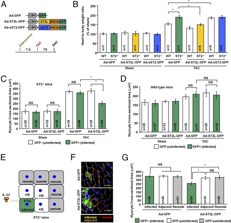
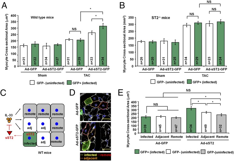
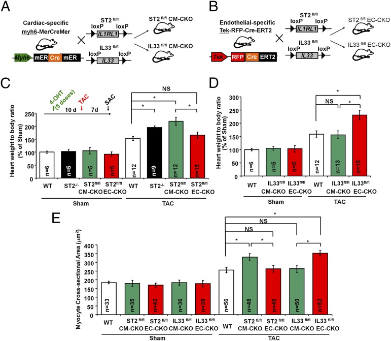
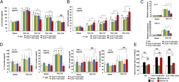
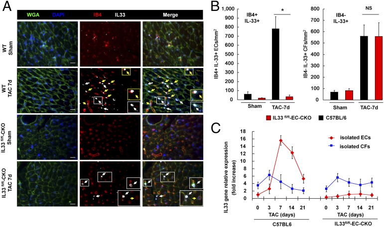
Hypertension increases the pressure load on the heart and is associated with a poorly understood chronic systemic inflammatory state. Interleukin 33 (IL-33) binds to membrane-bound ST2 (ST2L) and has antihypertrophic and antifibrotic effects in the myocardium. In contrast, soluble ST2 appears to act as a decoy receptor for IL-33, blocking myocardial and vascular benefits, and is a prognostic biomarker in patients with cardiovascular diseases. Here we report that a highly local intramyocardial IL-33/ST2 conversation regulates the heart's response to pressure overload. Either endothelial-specific deletion of IL33 or cardiomyocyte-specific deletion of ST2 exacerbated cardiac hypertrophy with pressure overload. Furthermore, pressure overload induced systemic circulating IL-33 as well as systemic circulating IL-13 and TGF-beta1; this was abolished by endothelial-specific deletion of IL33 but not by cardiomyocyte-specific deletion of IL33. Our study reveals that endothelial cell secretion of IL-33 is crucial for translating myocardial pressure overload into a selective systemic inflammatory response.

Keywords: cardiac hypertrophy; endothelial cells; inflammation; interleukin-33.


Conflict of interest statement

Conflict of interest statement: Brigham and Women’s Hospital holds patents on ST2, listing R.T.L. as inventor.

Figures

Fig. 1.
Fig. 1.
ST2L controls pressure overload-induced hypertrophy in individual cardiomyocytes. (A) Adenovirus gene constructs and experiment design. SAC, sacrifice; TAC, transverse aortic constriction. (B) Adenoviral vectors (Ad-GFP, Ad-ST2L-GFP, or Ad-sST2-GFP) were injected into the left ventricular free wall in mice. Seven days after adenovirus gene transfer, the mice were subjected to Sham or TAC surgery. (C) Adenoviral transfer of ST2L to ST2−/− mice leads to reduced CSA in infected cardiomyocytes. (D) Adenoviral transfer of ST2L to WT mice had no effect on the CSAs between infected and uninfected cardiomyocytes. (E) Schematic representation of ST2L-transferred cardiomyocyte responses to local IL-33. Cardiomyocytes were grouped as infected or uninfected on the basis of GFP fluorescence. Adj, adjacent; infected, adenovirus infected cardiomyocyte; remote, remote cardiomyocyte. (F) Representative image showed the immunofluorescence staining of GFP and selection of the cells for CSA analysis. (Scale bar, 50 μm.) (G) ST2L-transferred cardiomyocytes develop lower cell size after TAC-induced hypertrophy. Only myocytes that had both a visible nucleus in the center and an intact cellular membrane were included in these studies. CSA was measured in ≥10 distinct microscope fields for each slide. Three hearts from each group were analyzed. Data are shown as mean ± SD. Statistical analysis was performed with one-way ANOVA. *P < 0.05. NS, no significance.
Fig. 2.
Fig. 2.
sST2 exacerbates pressure overload-induced hypertrophy locally in myocardium. (A) Adenoviral transfer of sST2 to WT mice leads to greater CSA in infected cardiomyocytes after TAC. (B) Gene transfer of the sST2 gene to ST2−/− mice had no differences in CSAs between infected and uninfected cardiomyocytes. (C) Schematic representation of sST2-expressing cardiomyocytes affecting nearby cells by altering local IL-33 signaling via sST2. (D) Representative image showed the immunofluorescence staining of GFP and selection of the cells for CSA analysis. (Scale bar, 50 μm.) (E) sST2-expressing cardiomyocytes develop more hypertrophy and affect adjacent but not remote cardiomyocytes after TAC-induced hypertrophy. CSA was measured in ≥10 distinct microscope fields for each slide. Three hearts from each group were analyzed.
Fig. 3.
Fig. 3.
Endothelial-specific IL33 and cardiomyocyte-specific ST2 deficiency exacerbate pressure overload-induced hypertrophy. Mice with (A) cardiomyocyte-specific deletion (CM-CKO) or (B) endothelial-specific deletion (EC-CKO) of the ST2 or IL33 gene were generated by cross-breeding the mice with floxed alleles (ST2fl/fl or IL33fl/fl) and the mice with tissue-specific Cre recombinase (myh6-mER-Cre-mER or Tek-RFP-CreERT2). (C) Mice were injected with 4-OH tamoxifen (five doses) to induce Cre recombination. After 10 d the mice were subjected to Sham or TAC surgery. ST2fl/fl-CM-CKO mice develop greater cardiac hypertrophy after TAC. (D) IL33fl/fl-EC-CKO but not IL33fl/fl-CM-CKO mice developed more hypertrophy after TAC. (E) TAC induces greater myocardial cellular hypertrophy in ST2fl/fl-CM-CKO and IL33fl/fl-EC-CKO mice. CSA was measured in ≥10 distinct microscope fields for each slide. Four hearts from each group were analyzed. Data are shown as mean ± SD. Statistical analysis was performed with one-way ANOVA. *P < 0.05. NS, no significance.
Fig. 4.
Fig. 4.
EC IL-33 induces a selective systemic inflammatory response after myocardial pressure overload. (A) Serum IL-33 and (B) sST2 protein levels from the mice were analyzed by using ELISA (n = 6 in each group). #P < 0.05 compared with same genotype Sham controls. (C) mRNA expression of the IL13 and TGFβ1 gene in left ventricular tissues. (D) Serum levels of IL-13, TGF-β1, TNF-α, and IL-6 from the mice with TAC and Sham controls. (E) Treatment with IL-33 reduced hypertrophy only in WT and IL33fl/fl-EC-CKO but not ST2fl/fl-CM-CKO mice (n = 6 in each group). IL-33 caused no significant change under nonstress conditions in vivo. Data are shown as mean ± SD. Statistical analysis was performed with one-way ANOVA. *P < 0.05 and #P < 0.05 compared with same genotype Sham control group. NS, no significance.
Fig. 5.
Fig. 5.
Myocardial pressure overload induces IL-33 in cardiac ECs. (A) Expression of IL-33 in IB4+ cells (yellow arrowheads) and IB4– cells (white arrowheads) was analyzed using immunofluorescence staining. Endothelial IL-33 was not detected in cardiac tissues from IL33fl/fl-EC-CKO mice. Wheat germ agglutinin (WGA) staining was used for cellular membrane (green); DAPI staining was used for the nucleus (blue); IB4 staining for was used for ECs (red); IL-33 staining was detected in the nucleus (gray). (Scale bar, 50 μm.) (B) Quantification of IB4+IL-33+ and IB4–IL-33+ cells in cardiac tissues from mice with Sham or TAC (n = 3 mice from each group). Data are shown as mean ± SD. Statistical analysis was performed with one-way ANOVA. *P < 0.05. NS, no significance. (C) IL33 transcript levels in isolated cardiac ECs and CFs from mice with TAC surgery. Relative expression was normalized to the IL33 levels in ECs isolated from mice prior to TAC (day 0).

Comment in

  • Stressed hearts inflame the body (in a good way).
    Hashimoto T, Kass DA. Hashimoto T, et al. Proc Natl Acad Sci U S A. 2015 Jun 9;112(23):7113-4. doi: 10.1073/pnas.1507821112. Epub 2015 May 21. Proc Natl Acad Sci U S A. 2015. PMID: 25997444 Free PMC article. No abstract available.

References

    1. Kearney PM, et al. Global burden of hypertension: Analysis of worldwide data. Lancet. 2005;365(9455):217–223. - PubMed
    1. Harrison DG, et al. Inflammation, immunity, and hypertension. Hypertension. 2011;57(2):132–140. - PMC - PubMed
    1. Libby P. Inflammation in atherosclerosis. Arterioscler Thromb Vasc Biol. 2012;32(9):2045–2051. - PMC - PubMed
    1. Ky B, et al. High-sensitivity ST2 for prediction of adverse outcomes in chronic heart failure. Circ Heart Fail. 2011;4(2):180–187. - PMC - PubMed
    1. Shimpo M, et al. Serum levels of the interleukin-1 receptor family member ST2 predict mortality and clinical outcome in acute myocardial infarction. Circulation. 2004;109(18):2186–2190. - PubMed

Publication types

MeSH terms