. 2015 May 19;112(20):E2575-84.
doi: 10.1073/pnas.1420115112. Epub 2015 May 4.

Organization and dynamics of the nonhomologous end-joining machinery during DNA double-strand break repair

Affiliations

Organization and dynamics of the nonhomologous end-joining machinery during DNA double-strand break repair

Dylan A Reid et al. Proc Natl Acad Sci U S A. .

Abstract

Nonhomologous end-joining (NHEJ) is a major repair pathway for DNA double-strand breaks (DSBs), involving synapsis and ligation of the broken strands. We describe the use of in vivo and in vitro single-molecule methods to define the organization and interaction of NHEJ repair proteins at DSB ends. Super-resolution fluorescence microscopy allowed the precise visualization of XRCC4, XLF, and DNA ligase IV filaments adjacent to DSBs, which bridge the broken chromosome and direct rejoining. We show, by single-molecule FRET analysis of the Ku/XRCC4/XLF/DNA ligase IV NHEJ ligation complex, that end-to-end synapsis involves a dynamic positioning of the two ends relative to one another. Our observations form the basis of a new model for NHEJ that describes the mechanism whereby filament-forming proteins bridge DNA DSBs in vivo. In this scheme, the filaments at either end of the DSB interact dynamically to achieve optimal configuration and end-to-end positioning and ligation.

Keywords: DNA repair; genomic integrity; nonhomologous end-joining; single-molecule FRET; super-resolution microscopy.


Conflict of interest statement

The authors declare no conflict of interest.

Figures

Fig. 1.
Fig. 1.
Organization of NHEJ proteins in response to DNA damage. (A) Representative nuclei (dashed yellow line) stained for Ku/TUNEL displayed with conventional resolution microscopy (TIRF) and reconstructed SR microscopy. (Inset) Zoomed region in which Ku associates with a DNA break marked by TUNEL. (Scale bars: 5 µm and 500 nm, respectively.) (B) Quantification of DNA breaks measured over the course of 6 h by observing the normalized stained area of TUNEL foci (area of TUNEL particles/nuclear area). The amount of TUNEL staining increases rapidly after DNA damage, but decreases as repairs occur. Number of cells, n = 39, 14, 14, and 16, respectively. (C) Quantification of the association between various NHEJ proteins at the basal level and after bleomycin treatment. The number of interactions/nuclei increases after damage. Number of cells, n = 63/12, 27/17, and 20/19, respectively. (D) Repair structures (Caterpillars) from cells in which NHEJ filament proteins interact with a DSB. LigIV/TUNEL shows a long filament capped by a DSB (orange arrows). These structures are observable using Ku and XRCC4/XLF/LigIV. (Scale bar: 250 nm.) (E) Repair structures (Butterflies) from cells showing NHEJ filament proteins interacting with a DSB. LigIV/TUNEL shows DSB (orange arrow) roughly near the filament center of mass. These structures are observable using Ku and XRCC4/XLF/LigIV. In this class of structure, we identified two characteristic subtypes; in the first, gapped filaments are separated by a cluster of Ku, whereas in the second, we found continuous filaments with Ku at their center. (Scale bar: 250 nm.) Error bars represent SEM. **P < 0.01; ***P < 0.001; ****P < 0.0001.
Fig. 2.
Fig. 2.
NHEJ proteins form structures in vitro. (A) (Upper) Cartoon showing the assembly procedure used to reconstitute NHEJ repair structures for in vitro SR imaging. DNA, protein, and DNA with proteins were reacted for 30 min and then cross-linked. Subsequently, they were bound to a coverslip, immunofluorescently labeled, and imaged as in our cellular SR assay. (Lower) Comparison of the regular diffraction limited microscopy image showing blurred features and a reconstructed SR image, in which the nanoscale organization and features of NHEJ DDR intermediates are clearly shown. (Scale bar: 250 nm.) (B) Observation of LX filaments formed in the absence of DNA. NHEJ proteins (6 μM Ku, LX, and XLF) were incubated in the absence of DNA and stained for LigIV and Ku. (Scale bar: 250 nm.) (C and D) In the presence of DNA, NHEJ proteins (6 μM Ku/LX/XLF) formed two characteristic structures. The first type of structure (Caterpillars) is shown in C, where Ku is localized at the end of LX filaments. The second type of structure (Butterflies) is shown in D, in which Ku is localized near the center of the LX filaments. Orange arrows illuminate Ku locations. (Scale bar: 250 nm.) (E) Quantification of the frequency of Caterpillar and Butterfly structures in our reconstituted reactions, showing that the formation of the observed structures is highly reliant on DNA. The addition of 100 μM SCR7 resulted in a decrease in Butterfly structures. The abundance of each structure is normalized to the amount present in the DNA, Ku, LX, and XLF reactions. Error bars represent SEM. ns, P value not significant; *P < 0.05; **P < 0.001.
Fig. 3.
Fig. 3.
Particle averaging of repair intermediates and kinetic analysis. (A–C) Representative average particle obtained for each of the three categories: Caterpillar (n = 42) (A), gapped Butterfly (n = 42) (B), and continuous Butterfly (n = 20) (C). (Scale bar: 250 nm.) Illustrations of the three types of observed structures are below the particle average image. (D) Quantification of the relative abundance of LigIV Caterpillar and Butterfly structures in untreated cells (basal), following bleomycin treatment, taken between 0–60 min (bleomycin), and a further recovery time point at 360 min (recovery). Structures were normalized to the total number of interacting particles in each cell. Number of cells, n = 27, 100, and 21, respectively. (E) Kinetics of Butterfly structures during repair. The two different Butterfly structures were monitored after 5 min of bleomycin-induced damage. Gapped Butterflies (blue squares) and continuous Butterflies (black squares) are shown as a function of DSB recovery time. These populations were strongly anticorrelated in time, with gapped filaments decreasing sharply within 15 min of recovery time and continuous filaments increasing within 15 min. This analysis shows that the gapped structures are most abundant immediately after DSB induction. With increased recovery time, the predominant population becomes continuous Butterflies. Number of cells, n = 13, 17, 8, 19, 27, and 16, respectively, with n = 119 structures examined. Error bars represent SEM. ns, P value not significant; *P < 0.05; ***P < 0.001.
Fig. 4.
Fig. 4.
smFRET of NHEJ synapsis and ligation. (A) Diagram illustrating our NHEJ smFRET dsDNA capture assay. (1) dsDNA with four nucleotides of ssDNA and an acceptor dye bound to the surface. (2) Solution containing various NHEJ proteins and a dsDNA with a complementary four-nucleotide overhang labeled with a donor dye is added into the chamber. (3) Pairing between the two dsDNAs occurs, and FRET is observed. (B) Images from the smFRET synapsis reaction showing donor/acceptor channels for different combinations of NHEJ factors. Spots represent individual pairs of DNA molecules. Ku/LX/XLF showed the most abundant pairing, whereas reactions containing DNA-PKcs resulted in formation of large aggregates of the solution DNA strand. (C) Quantification of synapsis as a function of NHEJ proteins added using an smFRET dsDNA capture assay. Although some stable synapsis is observed for LX/XLF, synapsis improves with the addition of Ku, as seen in Ku/XLF and Ku/LX/XLF. Pairing efficiency is normalized to the reaction containing Ku/LX/XLF. Number of observed FRET pairs, n >1,000 molecules. Error bars represent SEM.
Fig. 5.
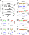
Fig. 5.
Kinetic analysis of NHEJ dynamics with smFRET. (A) FRET histograms of the synaptic complex (50 nM Ku/LX/XLF) during ligation reactions showing a broad distribution of FRET values. The Reaction panel is a substrate with a 5′ phosphate capable of undergoing ligation. The Ligation panel is the remaining population from the Reaction panel following a 1 M NaCl wash. (Inset) Comparison of the normalized number of molecules (Reaction) and following the salt wash (Ligation), in which the effective yield of the ligation was calculated to be ∼38% of FRET pairs seen in the reaction. The Distal panel shows a substrate in which the acceptor was placed ∼60 bp from the DNA end. The ddC panel shows a substrate in which ligation is blocked by dideoxy nucleotides on the 3′ ends. The no Phosphate panel shows a substrate that lacks 5′ phosphate and is unable to complete ligation. Each pair of substrates has complementary four nucleotide overhangs. All histograms show broad distributions of FRET values. Number of observed FRET pairs, n = 200, 200, 68, 200, and 100 molecules, respectively. (B) Two representative smFRET trajectories showing the initial NHEJ pairing of the two dsDNA strands at a high-FRET state (Left) and a low-FRET state (Right), demonstrating that initial pairing occurs at both end-to-end and adjacent configurations, as illustrated in the cartoons on the right. The reaction was carried out with 50 nM Ku/LX/XLF. (C) Two representative smFRET trajectories for surface dsDNA with distal acceptor dye. Initial pairing of the two dsDNA strands occurs at either a high-FRET state (Left) or a low-FRET state (Right), further demonstrating that initial pairings occur at both end-to-end and adjacent configurations, as illustrated in the cartoons on the right. The reactions were carried out with 50 nM Ku/LX/XLF. The trajectories exhibit well-defined FRET values and limited fluctuations, consistent with stably ligated dsDNA. (D) Representative smFRET trajectories showing repetitive transitions of a paired synaptic complex between adjacent and end-to-end configurations. (E) Representative smFRET trajectories showing fast transitions during synapsis for distal acceptor surface dsDNA. These trajectories resembles those in which the dye is placed close to the accessible DNA end showing repetitive transitions between adjacent configurations and end-to-end configurations. (F) Representative smFRET trajectories showing fast transitions during synapsis for ddC substrates. These trajectories resemble those in which the dye is placed close to the accessible DNA end (as in Fig. 5E), showing repetitive transitions between adjacent configurations and end-to-end configurations. (G) Representative smFRET trajectories showing fast transitions during synapsis for substrates with no phosphate. These trajectories resemble those in which the dye is placed close to the accessible DNA end (as in Fig. 5E), showing repetitive transitions between adjacent and end-to-end configurations. Error bars represent SEM.
Fig. 6.
Fig. 6.
Model for DSB repair via NHEJ. After Ku loading, XRCC4/XLF/LigIV filaments are recruited, creating Caterpillar structures. Synapsis between two Caterpillar structures commences, such that the structures can align end-to-end, as seen in our SR images. When the two DNA ends are in an end-to-end configuration in which the ends are compatible for ligation, the filament will merge over the two ends to initiate end ligation.

References

    1. Lieber MR. The mechanism of double-strand DNA break repair by the nonhomologous DNA end-joining pathway. Annu Rev Biochem. 2010;79:181–211. - PMC - PubMed
    1. Ramsden DA, Weed BD, Reddy YV. V(D)J recombination: Born to be wild. Semin Cancer Biol. 2010;20(4):254–260. - PMC - PubMed
    1. Weinfeld M, Lees-Miller SP. DNA double-strand break repair by non-homologous end joining and its clinical relevance. In: Kelley MR, editor. DNA Repair in Cancer Therapy: Molecular Targets and Clinical Applications. 2012. pp. 161–189.
    1. Lees-Miller SP, Meek K. Repair of DNA double-strand breaks by non-homologous end-joining. Biochimie. 2003;85(11):1161–1173. - PubMed
    1. Limp-Foster M, Kelley MR. DNA repair and gene therapy: Implications for translational uses. Environ Mol Mutagen. 2000;35(2):71–81. - PubMed

Publication types