. 2015 Apr 20:9:63.
doi: 10.3389/fnsys.2015.00063. eCollection 2015.

Rebuilding a realistic corticostriatal "social network" from dissociated cells

Affiliations

Rebuilding a realistic corticostriatal "social network" from dissociated cells

Marianela Garcia-Munoz et al. Front Syst Neurosci. .

Abstract

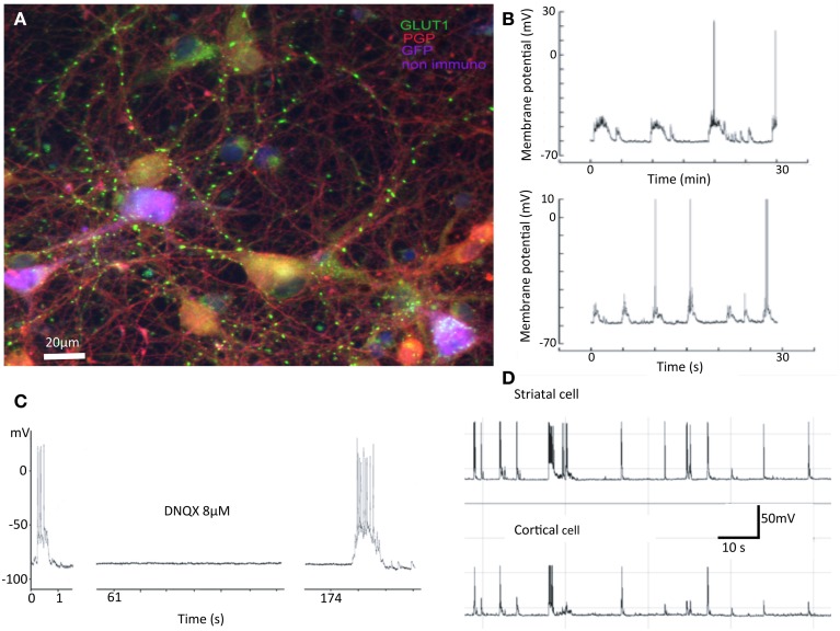
Many of the methods available for the study of cortical influences on striatal neurons have serious problems. In vivo the connectivity is so complex that the study of input from an individual cortical neuron to a single striatal cell is nearly impossible. Mixed corticostriatal cultures develop many connections from striatal cells to cortical cells, in striking contrast to the fact that only connections from cortical cells to striatal cells are present in vivo. Furthermore, interneuron populations are over-represented in organotypic cultures. For these reasons, we have developed a method for growing cortical and striatal neurons in separated compartments that allows cortical neurons to innervate striatal cells in culture. The method works equally well for acutely dissociated or cryopreserved neurons and allows a number of manipulations that are not otherwise possible. Either cortical or striatal compartments can be transfected with channel rhodopsins. The activity of both areas can be recorded in multielectrode arrays or individual patch recordings from pairs of cells. Finally, corticostriatal connections can be severed acutely. This procedure enables determination of the importance of corticostriatal interaction in the resting pattern of activity. These cultures also facilitate development of sensitive analytical network methods to track connectivity.

Keywords: cortical neurons; interneurons; mutual information; neuronal cultures; striatal neurons; synaptic connections.


Figures

Figure 1
Figure 1
Summary of previous results. Properties of mixed cultures that are also seen in separated cultures include: (A) Synapses are formed between neurons from cortex and striatum. In cultures cortical and striatal cells do not have the obviously different morphologies expected from in vivo investigations. In this false color image, cortical cells are purple, striatal gold, and inhibitory interneurons red. The green dots are glutamatergic boutons stained with antibodies against vGluT1. (B) Spontaneous activity in striatal cells shows “UP” states with action potentials riding on them. (C) Such “UP” states are abolished by application of AMPA receptor antagonist DNQX. (D) Activity of cortical and striatal cells recorded simultaneously are closely related in time (Randall et al., 2011).
Figure 2
Figure 2
Optical stimulation of ChR2 expressing striatal cells on cortical neurons plated in separate compartments. Single cortical (A) and striatal (B) neurons both depolarized with 80 nA intracellular current. (C) Striatal cell (seen in B) expressing ChR2 followed every light stimulus (at red dots) for extended periods. (D) Optical stimulation of striatal neurons (trains of 5 light pulses repeated 5 times -at the red dots) had no effect on cortical cells. (E) Optical stimulation of cortical neurons (same parameters as in D) drove EPSPs in striatal cells.
Figure 3
Figure 3
Cortically induced long-term changes in striatal synaptic responses observed in separated cortical and striatal cultures. (A,B) Optical stimulation of cortical neurons expressing ChR2: Single pulses (blue traces) and theta-like rhythm (6 trains of 5 pulses at 4 Hz at 10 s intervals red traces) stimulation. (C) Summary of EPSP amplitudes before and after theta burst stimulation (N = 14).
Figure 4
Figure 4
MEA recordings of a representative segregated culture. Top four rows contain cortical and lower three rows striatal neurons in three experimental conditions. Area of cut is indicated by white dots and clear shading.
Figure 5
Figure 5
Correlations of regionally averaged potentials for all plates and the mutual information between electrodes for a representative experiment. (A) Average correlation between electrode activity averaged over the cortical region and electrode activity averaged over the striatal region (see Figure 4). (B) Only half of the electrode-electrode mutual information values are displayed since this quantity is symmetric. In medium there are many different z-scores across electrodes. In medium there are many different z-scores across electrodes. Left hand panel (Medium): lower left quadrant displays mutual information between cortical electrodes; upper right quadrant striatal electrode mutual information; lower right quadrant mutual information shared between the compartments. Right-hand panel (Cut) displays the dramatic fall in information within the striatum and its complete absence between regions. The slight increase in the cortical region is likely due to the intervening NMDA application (see panel A) rather than a direct consequence of the cut, but its source remains to be examined in further work. Details of similar analyses in other plates in this study are illustrated in Supplementary Figure 1.

References

    1. Arbuthnott G. W., Poulter M., Staines W. A. (2005). Corticostriatal co-cultures: a minimal system for ‘up’ states in striatal cells?, in British Neuroscience Association Abstracts (Bristol: ).
    1. Beggs J. M., Plenz D. (2003). Neuronal avalanches in neocortical circuits. J. Neurosci. 23, 11167–11177. - PMC - PubMed
    1. Bologna L. L., Nieus T., Tedesco M., Chiappalone M., Benfenati F., Martinoia S. (2010). Low-frequency stimulation enhances burst activity in cortical cultures during development. Neuroscience 165, 692–704. 10.1016/j.neuroscience.2009.11.018 - DOI - PubMed
    1. Burns B. D. (1950). Some properties of the cat's isolated cerebral cortex. J. Physiol. 111, 50–68. - PMC - PubMed
    1. Burns B. D. (1951). Some properties of the isolated cerebral cortex in the unanaesthetized cat. J. Physiol. 112, 156–175. - PMC - PubMed

LinkOut - more resources