Review
. 2015 Apr 20:6:82.
doi: 10.3389/fphar.2015.00082. eCollection 2015.

Stabilization of G protein-coupled receptors by point mutations

Affiliations
Review

Stabilization of G protein-coupled receptors by point mutations

Franziska M Heydenreich et al. Front Pharmacol. .

Abstract

G protein-coupled receptors (GPCRs) are flexible integral membrane proteins involved in transmembrane signaling. Their involvement in many physiological processes makes them interesting targets for drug development. Determination of the structure of these receptors will help to design more specific drugs, however, their structural characterization has so far been hampered by the low expression and their inherent instability in detergents which made protein engineering indispensable for structural and biophysical characterization. Several approaches to stabilize the receptors in a particular conformation have led to breakthroughs in GPCR structure determination. These include truncations of the flexible regions, stabilization by antibodies and nanobodies, fusion partners, high affinity and covalently bound ligands as well as conformational stabilization by mutagenesis. In this review we focus on stabilization of GPCRs by insertion of point mutations, which lead to increased conformational and thermal stability as well as improved expression levels. We summarize existing mutagenesis strategies with different coverage of GPCR sequence space and depth of information, design and transferability of mutations and the molecular basis for stabilization. We also discuss whether mutations alter the structure and pharmacological properties of GPCRs.

Keywords: G protein-coupled receptors; GPCRs; alanine scanning; conformational thermostabilization; pharmacology; protein engineering; stabilizing mutations.


Figures
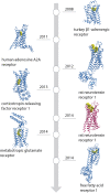
Figure 1
Figure 1
Timeline of GPCR structures based on conformational thermostabilization by alanine scanning (blue) or directed evolution (red) with their ligands (yellow). PDB IDs: 2VT4 (turkey β1-adrenergic receptor), 2YDO (human adenosine A2A receptor), 4GRV (neurotensin recetor 1, conformationally stabilized), 4K5Y (corticotropin-releasing factor receptor 1), 4BV0 (neurotensin receptor 1, directed evolution), 4OR2 (metabotropic glutamate receptor) and 4PHU (free fatty-acid receptor 1).
Figure 2
Figure 2
Favorable mutations identified by alanine scanning (A) and directed evolution (B) in presence of agonist (green), antagonist (red) and in absence of ligand (yellow). Alanine scanning identified mutations which increase the thermostability of receptors while retaining a minimal expression level. Directed evolution detected mutations that increased expression or thermostability, or both. Position of mutations is indicated by Ballesteros-Weinstein numbering. Mutations found in multiple receptors are indicated by double dots, those with high expression levels (=125% of wild-type expression level, alanine scanning only) are shown as hexagons. ECL and ICL stand for extracellular and intracellular loops, respectively.
Figure 3
Figure 3
Molecular basis for stabilization by mutation of residues involved in activation. Residues directly involved in GPCR activation (shown as magenta sticks with Ballersteros-Weinstein numbers and the backbone shown in gray), were identified by comparison of inactive and active state structures. They form a continuous path between the ligand (yellow spheres) and the effector binding site, mapped on the crystal structure of the complex between human β2adrenergic receptor and the heterotrimeric Gs (cyan, PBD ID 3SN6, Rasmussen et al., 2011b).
Figure 4
Figure 4
Molecular basis of stabilization by mutation of the 3.41 position to tryptophan. The 3.41W mutation stabilizes 5-HT1B (green), 5-HT2B (light pink) and CXCR4 (blue) through its interaction with the proline in position 5.50 and the carbonyl of the amino acid in position 5.45.
Figure 5
Figure 5
Comparison of GPCRs containing either a fusion protein or thermostabilizing mutations. Structures of neurotensin receptor 1 (A) and adenosine A2A receptor in the inactive (B) and active (C) state solved using fusion-protein or point-mutagenesis approaches show high similarity [overall RMSD = 0.95 Å (A) and 0.5 Å (B,C)]. The differences in helices five and six in the two neurotensin receptor structures may be due to mutation of an amino acid involved in activation, R1673.50L, in one receptor and the T4 lysozyme fusion in the other receptor. PDB IDs: 3ZEV (NTR1, directed evolution), 4GRV (NTR1, fusion with T4 lysozyme), 3PWH (A2A, antagonist-bound, with thermostabilizing mutations), 3EML (A2A, antagonist-bound, T4 lysozyme fusion), 3QAK (A2A, with agonist UK-432097 and T4 lysozyme) and 2YDO (with agonist adenosine and thermostabilizing mutations).

Similar articles

Cited by

References

    1. Ahern T. J., Casal J. I., Petsko G. A., Klibanov A. M. (1987). Control of oligomeric enzyme thermostability by protein engineering. Proc. Natl. Acad. Sci. U.S.A. 84, 675–679. 10.1073/pnas.84.3.675 - DOI - PMC - PubMed
    1. Alexandrov A. I., Mileni M., Chien E. Y. T., Hanson M. A., Stevens R. C. (2008). Microscale fluorescent thermal stability assay for membrane proteins. Structure 16, 351–359. 10.1016/j.str.2008.02.004 - DOI - PubMed
    1. Amin N., Liu A. D., Ramer S., Aehle W., Meijer D., Metin M., et al. . (2004). Construction of stabilized proteins by combinatorial consensus mutagenesis. Protein Eng. Des. Sel. 17, 787–793. 10.1093/protein/gzh091 - DOI - PubMed
    1. Arase A., Yomo T., Urabe I., Hata Y., Katsube Y., Okada H. (1993). Stabilization of xylanase by random mutagenesis. FEBS Lett. 316, 123–127. 10.1016/0014-5793(93)81199-A - DOI - PubMed
    1. Baker J. G., Proudman R. G. W., Tate C. G. (2011). The pharmacological effects of the thermostabilising (m23) mutations and intra and extracellular (β36) deletions essential for crystallisation of the turkey β-adrenoceptor. Naunyn. Schmiedebergs. Arch. Pharmacol. 384, 71–91. 10.1007/s00210-011-0648-4 - DOI - PMC - PubMed