. 2015 May 5;10(5):e0125875.
doi: 10.1371/journal.pone.0125875. eCollection 2015.

Structure and assembly of group B streptococcus pilus 2b backbone protein

Affiliations

Structure and assembly of group B streptococcus pilus 2b backbone protein

Roberta Cozzi et al. PLoS One. .

Abstract

Group B Streptococcus (GBS) is a major cause of invasive disease in infants. Like other Gram-positive bacteria, GBS uses a sortase C-catalyzed transpeptidation mechanism to generate cell surface pili from backbone and ancillary pilin precursor substrates. The three pilus types identified in GBS contain structural subunits that are highly immunogenic and are promising candidates for the development of a broadly-protective vaccine. Here we report the X-ray crystal structure of the backbone protein of pilus 2b (BP-2b) at 1.06Å resolution. The structure reveals a classical IgG-like fold typical of the pilin subunits of other Gram-positive bacteria. The crystallized portion of the protein (residues 185-468) encompasses domains D2 and D3 that together confer high stability to the protein due to the presence of an internal isopeptide bond within each domain. The D2+D3 region, lacking the N-terminal D1 domain, was as potent as the entire protein in conferring protection against GBS challenge in a well-established mouse model. By site-directed mutagenesis and complementation studies in GBS knock-out strains we identified the residues and motives essential for assembly of the BP-2b monomers into high-molecular weight complexes, thus providing new insights into pilus 2b polymerization.


Conflict of interest statement

Competing Interests: Most of the authors are affiliated to Novartis Vaccines and Diagnostics; RC, EM, MJB, IM, DM and CDR are employees of Novartis Vaccines. There is a pending patent application belonging to the institution, but the authors will not benefit from it (Maione D., Cozzi R., Rinaudo D., Lazzarin M., Zerbini F., Margarit Y Ros I. 2013. Pilus proteins and compositions. WO2013/124473(PCT/EP2013/053644), filed February 23, 2013 and issued August 29, 2013). There are no products in development or marketed products to declare. This does not alter the authors’ adherence to all the PLOS ONE policies on sharing data and materials, as detailed online in the guide for authors.

Figures

Fig 1
Fig 1. Overall structure and topology of the backbone pilus protein BP-2b.
(A) The structure of BP-2b is depicted with light blue and orange cartoons for domains D2 and D3. Intradomain isopeptide bonds in D2 and D3 are shown with sticks, while green spheres show calcium ions located on the surface. The red dashed lines indicate the only residues that could not be modelled due to the lack of electron density. (B) Cartoon representation of D2 and D3 of BP-2b, with the two domains reoriented with respect to the view in A), and colored with a gradient according to the topology diagram on the right. (C) Topology diagram for D2 and D3 of BP-2b, with their core β-strands labeled A—G in rainbow style colored as in panel B. The strands linked by isopeptide bonds in D2 and D3 are marked by thick horizontal black lines.
Fig 2
Fig 2. Isopeptide bonds of BP-2b.
Domains D2 and D3 are colored as in Fig 1 in blue and orange, respectively. Isopeptide bonds between Asn330 and Lys 187 for D2, and between Asn462 and Lys358 in D3 are shown with blue and orange sticks, and 1σ 2Fo-Fc electron density map around this region is shown as blue mesh (carve = 1.1). The magenta colored regions in (A) and (B) show the location of hydrophobic residues surrounding the isopeptide bonds. Hydrogen-bonds between the isopeptide bonds and the nearby Asp (225, D2) and Glu (423, D3) are shown with black dashed lines.
Fig 3
Fig 3. Structural comparisons of BP-2bD2+D3 with other pilin backbone proteins.
(A) BP-2b (blue cartoon) is shown overlaid onto: the pilus backbone protein RrgB (pdb 2x9x, red cartoon, left), the major pilin protein GBS80 (pdb 3pf2, green cartoon, middle), and on the major pilin protein BP-2a (pdb 2xtl, pink cartoon, right). (B) Domain architecture of GBS backbone proteins from pilus 1 (BP-1), pilus 2a (BP-2a) and pilus 2b (BP-2b). The proteins are comprised of a signal peptide (SP) at the N-terminus and a C-terminal LPXTG-like motif (in red) linked to the transmembrane domain (TM). BP-1 and BP-2b contain three domains, while BP-2a four domains. The pilin motif involved in pilus polymerization is located near the D1–D2 domain linker while the E-box is located close to the sorting signal. Residues involved in isopeptide bonds are indicated by black bars. Domains present in the crystal structures are included into the box outlined with dashed lines.
Fig 4
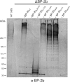
Fig 4. Lys 175, Glu 423 and the sorting motif LPSTG are involved in BP-2b polymerization in GBS.
Immunoblot analysis of total protein extracts from GBS mutant strain lacking the pilus 2b backbone protein gene (ΔBP-2b) complemented with plasmids expressing the wild-type BP-2b protein (WT) or BP-2b mutants carrying a deletion of the C-terminal sorting signal (BP-2bΔLPXTG), alanine substitutions of the putative pilin motif lysine (BP-2bK175A, BP-2bK118A BP-2bK82A) or of the E-box E423 residue (BP-2bE423A). Nitrocellulose membrane was probed with a mouse antiserum raised against the recombinant BP-2b protein (α-BP–2b).
Fig 5
Fig 5. Biochemical characterization of different BP-2b constructs.
(A) Time course of the trypsin-proteolysis reactions at 37°C of BP-2b full length and fragments, analyzed by SDS-PAGE. Different digestion patterns can be observed for the different constructs. Asterisks indicate the not-digested proteins. (B) Differential Scanning Fluorimetry (DSF) analysis of BP-2b proteins (D1+D2+D3, D2+D3 and single domains D1, D2, D3) in presence of Sypro orange showed different thermal stabilities. Graph shows the fluorescence intensity vs. the temperature for the unfolding different BP-2b constructs. (C) Correlation of BP-2b melting temperature with the concentration of Ca2+.

References

    1. Heath PT, Schuchat A. Perinatal group B streptococcal disease. Best Pract Res Clin Obstet Gynaecol. 2007;21(3):411–24. - PubMed
    1. Skoff TH, Farley MM, Petit S, Craig AS, Schaffner W, Gershman K, et al. Increasing burden of invasive group B streptococcal disease in nonpregnant adults, 1990–2007. Clin Infect Dis. 2009;49(1):85–92. 10.1086/599369 - DOI - PubMed
    1. Baker CJ. The spectrum of perinatal group B streptococcal disease. Vaccine. 2013;31 Suppl 4:D3–6. 10.1016/j.vaccine.2013.02.030 - DOI - PubMed
    1. Telford JL, Barocchi MA, Margarit I, Rappuoli R, Grandi G. Pili in gram-positive pathogens. Nat Rev Microbiol. 2006;4(7):509–19. - PubMed
    1. Maione D, Margarit I, Rinaudo CD, Masignani V, Mora M, Scarselli M, et al. Identification of a universal Group B streptococcus vaccine by multiple genome screen. Science. 2005;309(5731):148–50. - PMC - PubMed

Publication types

MeSH terms