Case Reports
. 2015 Jun-Jul;115(2-3):128-140.
doi: 10.1016/j.ymgme.2015.04.007. Epub 2015 May 1.

Expanding the clinical and molecular characteristics of PIGT-CDG, a disorder of glycosylphosphatidylinositol anchors

Affiliations
Case Reports

Expanding the clinical and molecular characteristics of PIGT-CDG, a disorder of glycosylphosphatidylinositol anchors

Christina Lam et al. Mol Genet Metab. 2015 Jun-Jul.

Abstract

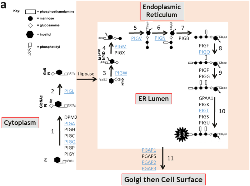
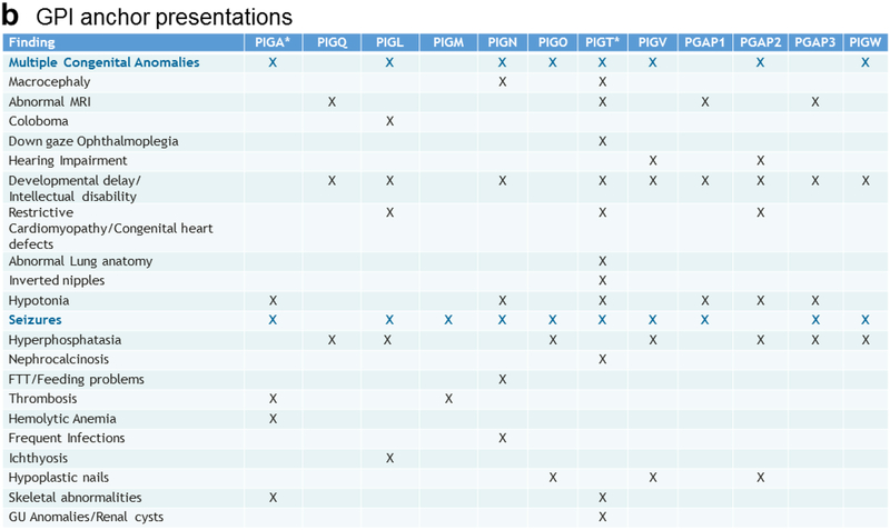
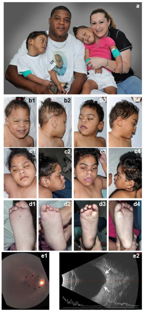
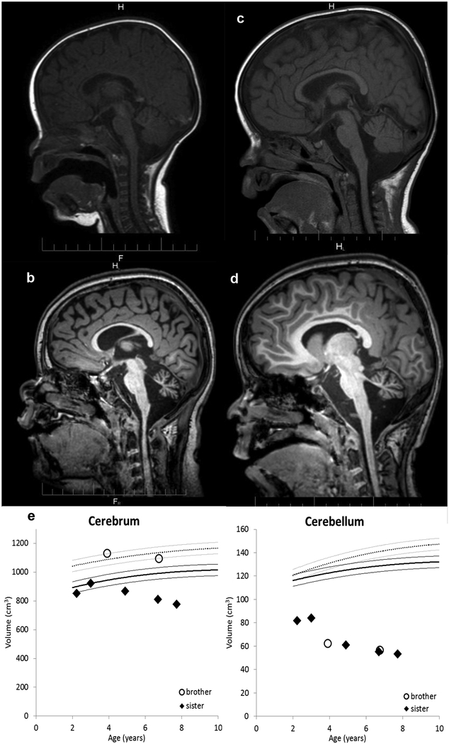
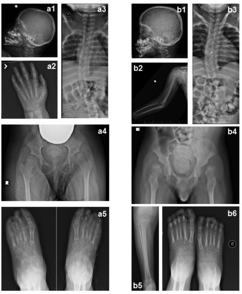
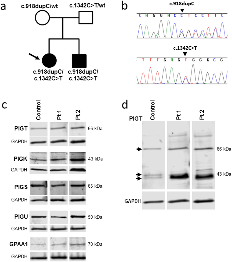
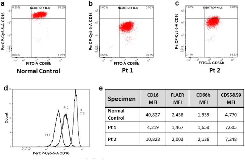
PIGT-CDG, an autosomal recessive syndromic intellectual disability disorder of glycosylphosphatidylinositol (GPI) anchors, was recently described in two independent kindreds [Multiple Congenital Anomalies-Hypotonia-Seizures Syndrome 3 (OMIM, #615398)]. PIGT encodes phosphatidylinositol-glycan biosynthesis class T, a subunit of the heteropentameric transamidase complex that facilitates the transfer of GPI to proteins. GPI facilitates attachment (anchoring) of proteins to cell membranes. We describe, at ages 7 and 6 years, two children of non-consanguineous parents; they had hypotonia, severe global developmental delay, and intractable seizures along with endocrine, ophthalmologic, skeletal, hearing, and cardiac anomalies. Exome sequencing revealed that both siblings had compound heterozygous variants in PIGT (NM_015937.5), i.e., c.918dupC, a novel duplication leading to a frameshift, and c.1342C > T encoding a previously described missense variant. Flow cytometry studies showed decreased surface expression of GPI-anchored proteins on granulocytes, consistent with findings in previous cases. These siblings further delineate the clinical spectrum of PIGT-CDG, reemphasize the neuro-ophthalmologic presentation, clarify the endocrine features, and add hypermobility, low CSF albumin quotient, and hearing loss to the phenotypic spectrum. Our results emphasize that GPI anchor-related congenital disorders of glycosylation (CDGs) should be considered in subjects with early onset severe seizure disorders and dysmorphic facial features, even in the presence of a normal carbohydrate-deficient transferrin pattern and N-glycan profiling. Currently available screening for CDGs will not reliably detect this family of disorders, and our case reaffirms that the use of flow cytometry and genetic testing is essential for diagnosis in this group of disorders.

Keywords: Congenital disorder of glycosylation; Exome; Flow cytometry; Glycosylphosphatidylinositol anchor; PIGT-CDG; Phenotype.


Conflict of interest statement

Conflict of interest

The authors declare no conflict of interest.

Figures

Fig. 1.
Fig. 1.
GPI anchor pathway and phenotypic findings of GPI anchor disorders. (a) The 11 steps of mammalian GPI anchor biosynthesis and protein attachment. The genes associated with each step are labeled. Underlined genes have been associated with congenital human disease. Note PIGT in step 10. (b) Summary of the clinical features described in cases of congenital GPI anchor biosynthesis disorders. The most common features are highlighted in blue. * Note that somatic mutations in PIGA and PIGT in hematopoietic stem cells can cause paroxysmal nocturnal hemoglobinuria manifesting with hemolytic anemia, bone marrow failure, thrombosis, and smooth muscle dystonia. This is distinct from the clinical entity caused by germline mutations in these two genes. Additionally, acquired mutations in the GPI transamidase complex subunits have been implicated in human cancers.
Fig. 1.
Fig. 1.
GPI anchor pathway and phenotypic findings of GPI anchor disorders. (a) The 11 steps of mammalian GPI anchor biosynthesis and protein attachment. The genes associated with each step are labeled. Underlined genes have been associated with congenital human disease. Note PIGT in step 10. (b) Summary of the clinical features described in cases of congenital GPI anchor biosynthesis disorders. The most common features are highlighted in blue. * Note that somatic mutations in PIGA and PIGT in hematopoietic stem cells can cause paroxysmal nocturnal hemoglobinuria manifesting with hemolytic anemia, bone marrow failure, thrombosis, and smooth muscle dystonia. This is distinct from the clinical entity caused by germline mutations in these two genes. Additionally, acquired mutations in the GPI transamidase complex subunits have been implicated in human cancers.
Fig. 2.
Fig. 2.
Photographs of the family displaying dysmorphic features in the affected siblings. For familial comparison, parents are depicted with both siblings in (a). Additional photographs are shown of each of the siblings when Patient 1 was 36 months old (b1, b2) and Patient 2 was 24 months old (b3, b4), and when Patient1 was 91 months old (c1, c2; d1, d2) and Patient 2 was 79 months old (c3, c4; d3, d4). Among the dysmorphic features, note the depressed nasal bridge, high forehead, bitemporal narrowing, rounded nasal tip that was correlated to very soft cartilage, large ears also with soft cartilage, overlapping toes, and deep plantar creases in both affected siblings. RetCAM 3 fundus image of the right eye of Patient 2 is shown (e1). The staphyloma noted on exam is located between the optic nerve head and the nasal edge of the macula (arrow-heads). This finding was confirmed in a B-scan ultrasound documenting the protrusion of the staphylomatous area (e2).
Fig. 3.
Fig. 3.
T1 sagittal brain magnetic resonance imaging (MRI) of the two affected patients. MRI scans of Patient 1 were taken at 6 months (a), and 91 months (b). MRI scans of Patient 2 were taken at 12 months (c), and 79 months (d). Brain volume measurements on the Patient 1 (diamonds) and Patient 2 (open circles) are displayed along with published normal population means [27] (dotted curve = boys, solid curve = girls, lighter lines = 95% CI) (e). Progressing volume loss is evident in both the cerebrum and cerebellum for both patients. Atrophy is more profound in the cerebellum than in the cerebrum. Atrophy in the cerebellum proceeded at about the same rate for both patients, whereas in the cerebrum, atrophy seemed to progress more rapidly in the sister.
Fig. 4.
Fig. 4.
Skeletal radiographs. Skeletal survey of Pt1(a), and Pt2 (b) performed at 91 months and 79 months of age, respectively. a1 and b1 illustrate brachycephaly, a2 shows advanced bone age (bone age 10 years +/− 20 months) of Patient 1, b2 and b5 illustrates the slender osteopenic long bones that both patients have, b2 also shows dislocation of elbow and subluxation of shoulder, a3 and b3 illustrate S-shaped scoliosis, a4 and b4 show bilateral dislocated hips with severe coxa valga-and shallow acetabulum, and a5 is unusual for pointy distal phalanxes bilaterally especially on the first digit that is not seen in b6.
Fig. 5.
Fig. 5.
Molecular data and protein expression results. (a) Pedigree of the family with identified PIGT variants in each individual indicated. (b) Sanger sequencing validation of the PIGT (NM_015937.5) variants c.918dupC and c.1342C>T. (c) Immunoblots of fibroblast lysates from control, Pt1 and Pt2 were probed with antibodies to the GPI transamidase complex subunits and GAPDH (loading control). The bands corresponding to the major isoform of each GPI transamidase subunit are shown. The predicted molecular weight of each band, calculated by gel imaging software (Image studio Software, Li-cor Inc.) is indicated in the Figure. The calculated molecular weight of each subunit based on amino acid sequence are listed in Supplemental Table 1, and full images of the immunoblots are in Supplemental Fig. 2. (d) Image of the entire PIGT antibody-probed immunoblot from (c). The indicated molecular weights are calculated by gelimaging software. The major protein bands are indicated by arrowheads. Note the protein band with increased intensity in both patients, compared to control around ~40 kDa, which corresponds to the predicted molecular weight of the truncated protein resulting from NM_015937.5:c.918dupC.
Fig. 6.
Fig. 6.
Flow cytometry results of GPI-anchored proteins in patients’ granulocytes. Scatter plots on logarithmic scale of fluorescent intensity of granulocytes tagged by antibody markers for CD66b on x axis and CD16 on y axis for normal control (a), Patient1 (b), and Patient 2 (c). Histogram overlays of normal control, Patient 1 and Patient 2 (d). The mean fluorescent intensity (MFI) determined by flow cytometry on granulocytes that were tagged with antibodies to fluorochrome conjugated aerolysin (FLAER) or antibodies to CD16, CD66b, and CD55 & 59 (e).
Fig. 7.
Fig. 7.
MRS results. Measurements on the Patient1 (diamonds) and on Patient 2 (open circles) are displayed in separate plots for each metabolite. The metabolite levels are plotted as a Z-score relative to the distribution of the reference group; the horizontal lines on the plots denote the 10th, 50th, and 90th percentiles. The metabolites that are produced by healthy neurons (NAA, creatine, glutamine, glutamate, and GABA) are generally lower than normal and decline on follow-up. The metabolites that are elevated by inflammation (choline and myo-inositol) are generally high and rising in the parietal gray matter and centrum semiovale, relatively normal in the pons and thalamus, and low and falling in the superior cerebellar vermis. The spatial and temporal patterns suggest that the cerebellum is affected earlier in the disease than is the cerebrum, and that there is a transient inflammatory phase that precedes the loss of neurons.

References

    1. Paulick MG, Bertozzi CR, The glycosylphosphatidylinositol anchor: a complex membrane-anchoring structure for proteins, Biochemistry 47 (2008) 6991–7000. - PMC - PubMed
    1. Nosjean O, Briolay A, Roux B, Mammalian GPI proteins: sorting, membrane residence and functions, Biochim. Biophys. Acta 1331 (1997) 153–186. - PubMed
    1. Ferguson MAJ, Kinoshita T, Hart GW, Glycosylphosphatidylinositol Anchors, in: Varki A, Cummings RD, Esko JD, Freeze HH, Stanley P, Bertozzi CR, Hart GW, Etzler ME (Eds), Essentials of Glycobiology, 2nd edition, Cold Spring Harbor (NY), Cold Spring Harbor Laboratory Press, 2009, Chapter 11. - PubMed
    1. Kinoshita T, Fujita M, Maeda Y, Biosynthesis, remodelling and functions of mammalian GPI-anchored proteins: recent progress, J. Biochem 144 (2008) 287–294. - PubMed
    1. Johnston JJ, Gropman AL, Sapp JC, Teer JK, Martin JM, Liu CF, Yuan X, Ye Z, Cheng L, Brodsky RA, Biesecker LG, The phenotype of a germline mutation in PIGA: the gene somatically mutated in paroxysmal nocturnal hemoglobinuria, Am. J. Hum. Genet 90 (2012) 295–300. - PMC - PubMed

Publication types

Substances