Review
. 2015 Jun;129(6):809-27.
doi: 10.1007/s00401-015-1424-1. Epub 2015 May 6.

Oligodendroglioma: pathology, molecular mechanisms and markers

Affiliations
Review

Oligodendroglioma: pathology, molecular mechanisms and markers

Pieter Wesseling et al. Acta Neuropathol. 2015 Jun.

Abstract

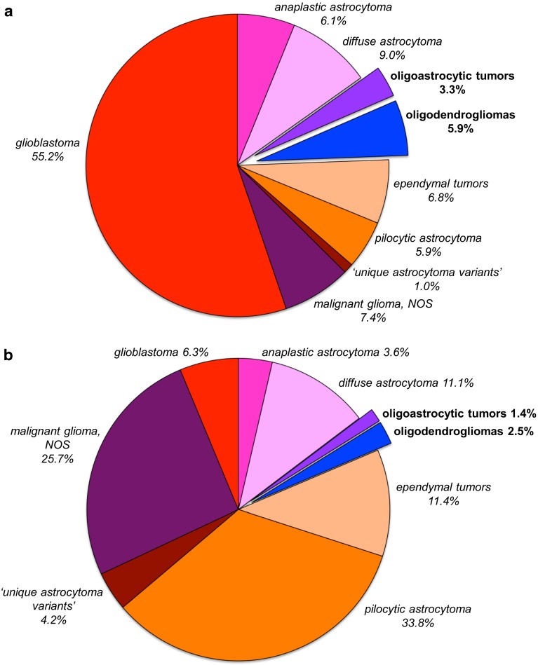
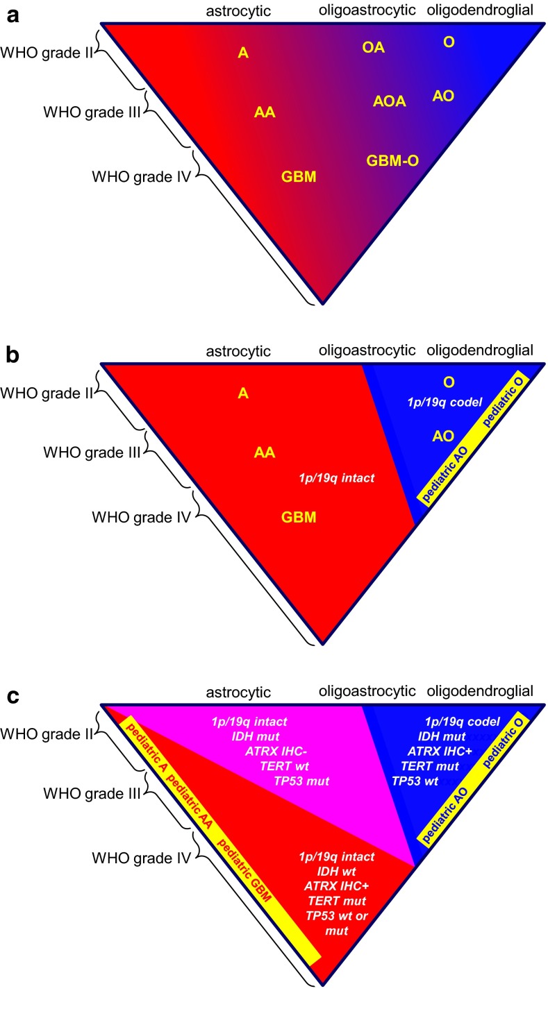
For nearly a century, the diagnosis and grading of oligodendrogliomas and oligoastrocytomas has been based on histopathology alone. Roughly 20 years ago, the first glioma-associated molecular signature was found with complete chromosome 1p and 19q codeletion being particularly common in histologically classic oligodendrogliomas. Subsequently, this codeletion appeared to not only carry diagnostic, but also prognostic and predictive information, the latter aspect only recently resolved after carefully constructed clinical trials with very long follow-up times. More recently described biomarkers, including the non-balanced translocation leading to 1p/19q codeletion, promoter hypermethylation of the MGMT gene, mutations of the IDH1 or IDH2 gene, and mutations of FUBP1 (on 1p) or CIC (on 19q), have greatly enhanced our understanding of oligodendroglioma biology, although their diagnostic, prognostic, and predictive roles are less clear. It has therefore been suggested that complete 1p/19q codeletion be required for the diagnosis of 'canonical oligodendroglioma'. This transition to an integrated morphological and molecular diagnosis may result in the disappearance of oligoastrocytoma as an entity, but brings new challenges as well. For instance it needs to be sorted out how (histopathological) criteria for grading of 'canonical oligodendrogliomas' should be adapted, how pediatric oligodendrogliomas (known to lack codeletions) should be defined, which platforms and cut-off levels should ideally be used for demonstration of particular molecular aberrations, and how the diagnosis of oligodendroglioma should be made in centers/countries where molecular diagnostics is not available. Meanwhile, smart integration of morphological and molecular information will lead to recognition of biologically much more uniform groups within the spectrum of diffuse gliomas and thereby facilitate tailored treatments for individual patients.


Figures

Fig. 1
Fig. 1
Relative frequency of histopathologically diagnosed oligodendroglial and oligoastrocytic tumors in the spectrum of glial tumors of the CNS: a in 95,564 patients of all age groups; b in 10,274 children and adolescents (0–19 years). Information extracted from CBTRUS statistical report: NPCR and SEER, 2007–2011 [85]
Fig. 2
Fig. 2
Common histopathologic patterns encountered in classic (IDH mutant, 1p19q codeleted) oligodendrogliomas include: a a honeycomb-like arrangement of evenly spaced tumor cells with uniformly rounded nuclei and clear haloes imparting a “fried egg” appearance; additionally, there are branching capillaries (arrow) resembling chicken-wire; b mucin-filled microcystic spaces; c, d minigemistocytes with small rounded bellies of eosinophilic cytoplasm and classic nuclear cytology including rounded countours, delicate “salt and pepper” chromatin, sharp nuclear membranes, and small nucleoli (c intra-operative smear; d tissue section); e foci of anaplastic transformation with increased cellularity and enlarged cell size (left) in comparison to the low-grade precursor cells (right). Anaplastic examples often feature: f enlarged epithelioid cells with increased pleomorphism, vesicular chromatin, macronucleoli, but a maintained general sense of nuclear roundness; g increased mitotic index and occasional “red crunchy” cells with brightly eosinophilic lysosomal inclusions (arrow); h microvascular proliferation; and i foci of tumor necrosis, more often consisting of collections of apoptotic cells rather than the pattern of pseudopalisading necrosis seen in glioblastomas. Immunohistochemically, some cases are completely GFAP negative, with only reactive astrocytes staining (j), while others (k) show strong expression in gliofibrillary oligodendrocytes (perinuclear rim and variable tadpole-like tail) or minigemistocytes (not shown). l A stain for neurofilament protein often highlights the infiltrative growth pattern by showing entrapped neurons and axons. m Tumoral synaptophysin positivity is not uncommon and can include a paranuclear dot-like pattern. n A stain for IDH1-R132H mutant protein highlights perineuronal satellitosis in this positive tumor with cortical involvement. Unlike diffuse astrocytomas, most oligodendrogliomas show only limited p53 immunoreactivity (o) and retained ATRX expression (p)
Fig. 3
Fig. 3
Less common and sometimes diagnostically confusing histopathologic patterns encountered in classic (IDH mutant, 1p19q codeleted) oligodendrogliomas include: ae a lobulated or nodular growth pattern can simulate a neuroendocrine tumor, with immunohistochemical pitfalls including CD56 (b) and synaptophysin (c) positivity; however, demonstration of IDH1 R132H mutant protein (d) and 1p19q codeletion confirmed the diagnosis in this case; e focal spindled cytology was also encountered, simulating a more astrocytoma-like pattern. Rare examples demonstrate more overt neuronal differentiation, including neurocytic (f) and ganglioglioma-like (g, h). Neurocytic foci often feature slightly smaller and more hyperchromatic nuclei surrounding central collections of delicate pink neuropil (i.e., neurocytic rosettes). In contrast, the case with ganglioglioma-like maturation featured areas of classic anaplastic oligodendroglioma (g note also the “red crunchy” cells); FISH studies (not shown) revealed 1p19q codeletions not only in this component, but in neoplastic ganglion cells (h) as well
Fig. 4
Fig. 4
Diffuse gliomas: from histopathologically to molecularly defined entities. Diffuse gliomas histopathologically form a spectrum, both with regard to cell type (astrocytic, oligodendroglial, mixed) and malignancy grade. Especially, delineation of oligoastrocytomas from (more) pure astrocytic and oligodendroglial tumors is poorly reproducible. In practice, (neuro)pathologists who readily accept the existence of mixed gliomas will more liberally diagnose oligoastrocytomas, while those who are skeptical that this entity exists will designate the vast majority of diffuse gliomas as either astrocytic or oligodendroglial (a). Introducing 1p/19q codeletion as a defining feature for oligodendrogliomas will in most cases allow for a clear distinction from astrocytic neoplasms and can be expected to drastically reduce the fraction of neoplasms diagnosed as mixed/oligoastrocytic (b). Meanwhile, especially in children, CNS tumors resembling classic oligodendrogliomas often lack 1p/19q codeletion; the best position for these ‘pediatric oligodendrogliomas’ in an improved taxonomy of CNS tumors requires further study. c Molecular markers helpful in daily clinical practice for state-of-the-art classification of diffuse gliomas. A recently published algorithm proposes immunohistochemistry (IHC) as a first step, allowing for correct classification of esp. the IDH1 R132H mutant protein positive, ATRX negative (and thus ATRX mutated) astrocytomas [96]. Ideally, in other tumors, subsequent molecular testing for 1p/19q codeletion status and/or other IDH1 and IDH2 mutations is performed. Variable testing for other markers can be helpful as well: in IDH mutated (mut) diffuse gliomas demonstration of TERT mutation indicates oligodendroglioma and of TP53 mutation astrocytoma; demonstration of TERT mutation in IDH wild type (wt) diffuse gliomas as well as of EGFR amplification/EGFRvIII strongly indicates high-grade malignant astrocytic tumor/glioblastoma; a KIAA-BRAF fusion gene is typically found in pilocytic astrocytoma, while mutations in the histone genes H3F3A, HIST1H3B/C indicate pediatric type high-grade gliomas (which may also harbor ATRX mutations and show loss of ATRX immunoreactivity). A, O, OA low-grade astrocytoma, oligodendroglioma, oligoastrocytic/mixed glioma, resp.; AA, AO, AOA anaplastic astrocytoma, oligodendroglioma, oligoastrocytic/mixed glioma, resp.; GBM glioblastoma, GBM-O glioblastoma with oligodendroglial component. See also text in this article and the other reviews in this cluster
Fig. 5
Fig. 5
Example of how ‘molecular reclassification’ may affect tumor grade. This tumor, previously diagnosed as GBM-O, WHO grade IV featured mostly cells resembling astrocytoma (a, c) and included necrosis (a) and microvascular proliferation (b). Cells resembling oligodendroglioma were seen only focally (d). The expression of IDH1-R132H mutant protein suggested that this represents either a secondary glioblastoma or an anaplastic oligodendroglioma, whereas the PTEN deletion identified by FISH is more common in glioblastoma (f; centromere 10 probe in green and PTEN probe in orange). Given the combined deletions of chromosome 1p (g; 1p probe in orange and 1q probe in green) and 19q (19p probe in green and 19q probe in orange), however, this case would be reclassified as an anaplastic oligodendroglioma, WHO grade III based on the most current recommendations
Fig. 6
Fig. 6
Example where ‘molecular reclassification’ does not affect tumor grade. This previously diagnosed oligoastrocytoma, WHO grade II (a) with IDH1 R132H mutant protein (b) would now be reclassified as a diffuse astrocytoma, WHO grade II based on strong p53 expression (c correlating well albeit imperfectly with TP53 mutation) and loss of ATRX expression (d note positive expression in non-neoplastic nuclei serving as a positive internal control)
Fig. 7
Fig. 7
For unequivocal assessment of 1p/19q codeletion in oligodendroglial tumors, detection of whole-arm losses is key. In order to avoid detection of false-positive cases with partial 1p and/or 19q loss, ideally a test is used that allows for analysis of multiple loci along each chromosome arm. a Example of an oligodendroglioma of which formalin-fixed, paraffin-embedded tissue was analyzed for 1p/19q status by multiplex ligation-dependent probe amplification (MLPA). With this platform, probes for 15 loci on chromosome 1p and 8 loci on 19q are used to interrogate these chromosome arms for loss or gain [the rest of the probes (3 on 1q, 2 on 19p, 15 on other chromosomes) serve as copy number references]. The test is performed in duplicate (hence the two lines) and demonstrates a clear loss for all 1p and 19q probes in this case (see [49] for technical details). b Massively parallel sequencing (MPS) allows for much more detailed analysis of copy number aberrations in the (tumor) genome. This is an example of a low-grade oligodendroglioma of which four different regions were separately analyzed. Interestingly, while complete losses of chromosome arms 1p (indicated by the arrow) and 19q (arrowhead) were uniformly present in the different regions, other copy number aberrations were only detected in some regions (most strikingly in area I, see region marked by red box). As the deletions for 1p and 19q loss in the different regions are at a similar level, this heterogeneity for copy number aberrations of other chromosomes cannot simply be explained by a difference in percentage of tumor cells in the different areas and must be interpreted as intratumoral genetic heterogeneity. Image b is modified from [122]

References

    1. Agnihotri S, Aldape KD, Zadeh G. Isocitrate dehydrogenase status and molecular subclasses of glioma and glioblastoma. Neurosurg Focus. 2014;37:E13. - PubMed
    1. Aldape K, Burger PC, Perry A (2007) Clinicopathologic aspects of 1p/19q loss and the diagnosis of oligodendroglioma. Arch Pathol Lab Med 131:242–251. doi:10.1043/1543-2165(2007)131[242:CAOQLA]2.0.CO;2 - PubMed
    1. Appin CL, Brat DJ. Molecular pathways in gliomagenesis and their relevance to neuropathologic diagnosis. Adv Anat Pathol. 2015;22:50–58. - PubMed
    1. Appin CL, Gao J, Chisolm C, Torian M, Alexis D, Vincentelli C, Schniederjan MJ, Hadjipanayis C, Olson JJ, Hunter S, et al. Glioblastoma with oligodendroglioma component (GBM-O): molecular genetic and clinical characteristics. Brain Pathol. 2013;23:454–461. - PMC - PubMed
    1. Arita H, Narita Y, Fukushima S, Tateishi K, Matsushita Y, Yoshida A, Miyakita Y, Ohno M, Collins VP, Kawahara N, et al. Upregulating mutations in the TERT promoter commonly occur in adult malignant gliomas and are strongly associated with total 1p19q loss. Acta Neuropathol. 2013;126:267–276. - PubMed

MeSH terms

Substances